Isolated coronary artery fistula is a rare congenital cardiac abnormality with an estimated incidence between 0.2 and 0.6%.Reference Effler, Sheldon, Turner and Groves 1 , Reference Liberthson, Sagar, Berkoben, Weintraub and Levine 2 Coronary artery fistulas originate most frequently from the right coronary artery and drain into the right heart in 90% of the cases. Symptoms related to coronary artery fistulas may occur at extremes of life, either in the neonatal period or in adults. Congestive cardiac failure may occur in neonates as a result of a haemodynamically significant left-to-right shunt. Symptoms in adults may include angina, dyspnoea, or arrhythmias, some of which occur as a result of possible coronary artery “steal” phenomenon. Occasionally, thrombosis of the coronary artery fistula may occur with subsequent acute myocardial infarction, and rarely an aneurysmal fistula may rupture causing haemo-pericardium.Reference Liberthson, Sagar, Berkoben, Weintraub and Levine 2 , Reference Valente, Lock and Gauvreau 3 Coronary artery fistulas can be detected prenatally by cross-sectional and colour Doppler echocardiography.Reference Schumacher, Roithmaier and Lorenz 4 After birth, echocardiography and additional investigations, such as MRI or CT scanning, may help confirm the diagnosis. Coronary angiography can also confirm the diagnosis, but is usually performed at the time of an interventional procedure. All of these investigations may provide detailed information about the anatomy of the fistula as well as, in particular, the size, the origin, the course, the presence of any stenoses or aneurysms, and the drainage site.
It is unusual for coronary artery fistulas to produce symptoms in the neonatal period or early infancy. Coronary artery fistulas can be diagnosed accurately in the fetus, but it is unclear whether these fetuses are at higher risk for developing symptoms earlier after birth. We report the natural history, progression, and outcome in five cases diagnosed during fetal life; one additional pregnancy where there was associated fetal hydrops resulted in termination.
Methods
We performed a retrospective study of all fetuses seen between January, 2000 and December, 2013, at a tertiary referral centre for fetal cardiology, to identify all the cases with a prenatal diagnosis of isolated coronary artery fistula in order to determine their progression and outcomes.
Study approval
This study was performed as an audit for service evaluation and was independently reviewed and approved in accordance with our institutional clinical audit programme policy. As this study was approved as an audit, separate ethical approval was not deemed necessary.
Study population
During the study period, 25,295 pregnant women, who were considered to be at an increased risk of having a baby with CHD, were referred to our unit for fetal cardiac assessment. Fetal echocardiography was carried out using a variety of ultrasound platforms: Toshiba Xario and Toshiba Aplio ultrasound system (Toshiba Medical Systems, Crawley, United Kingdom), Hewlett Packard Sonos 5500 ultrasound system (Philips Inc., Andover, Massachusetts, United States of America), and Voluson E8 (General Electric Company, Schenectady, New York, United States of America). A diagnosis of structural CHD was made in 2645 fetuses. In six (0.2%) of these, a diagnosis of coronary artery fistula was made in an otherwise structurally normal heart at the time of the initial fetal diagnosis. In one case, the pregnancy was terminated, and this case is excluded from the study, as it was not possible to monitor progression or postnatal outcome. This fetus had hydrops associated with the coronary artery fistula. The study group consisted of the remaining five fetuses.
Data collection
Prenatal
The patient records and all the fetal echocardiograms of the five fetuses were reviewed retrospectively to assess the progression and changes in the fetal cardiac findings during pregnancy. The reason for the referral and the gestational age at presentation and at the time of subsequent studies were noted. The origin and the site of drainage of the fistula were noted for each case. Measurements of the cardiothoracic ratio and the size of the fistula, at its origin, were made at each study. The flow pattern in the aortic arch was recorded as forward flow only or as to-and-fro flow.
Postnatal
In all liveborns, the medical records and the echocardiograms of the patients were reviewed. MRI scans and angiograms were also reviewed, when available. Measurements of the coronary artery fistulas were made using the postnatal echocardiograms. The details of the timing and the method of interventions and the outcome of the transcatheter closure of the fistulas were recorded.
Results
Prenatal findings
In five fetuses, a diagnosis of an isolated coronary artery fistula was made and the pregnancy continued to term; four of the five mothers were referred for fetal echocardiography, because of suspicion of CHD during the obstetric anomaly scan. The fifth case was referred because of a family history of CHD, as the previous child had hypoplastic left heart syndrome.
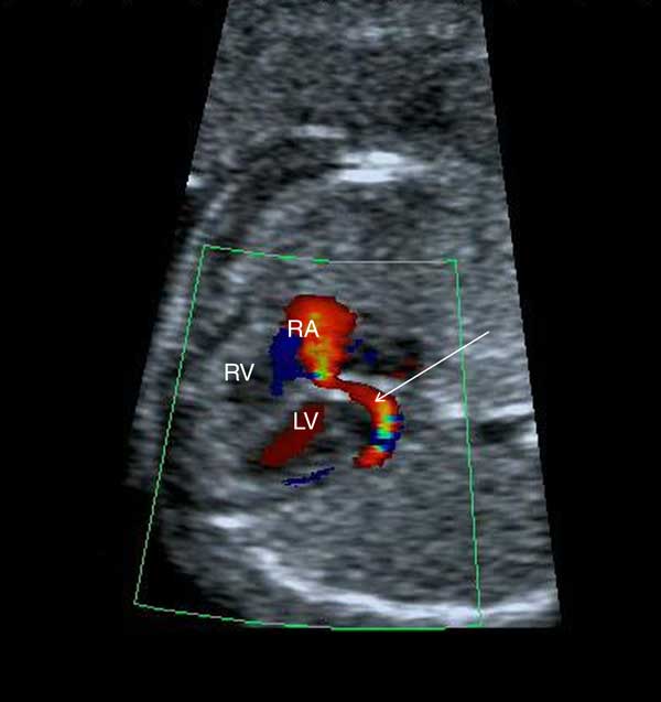
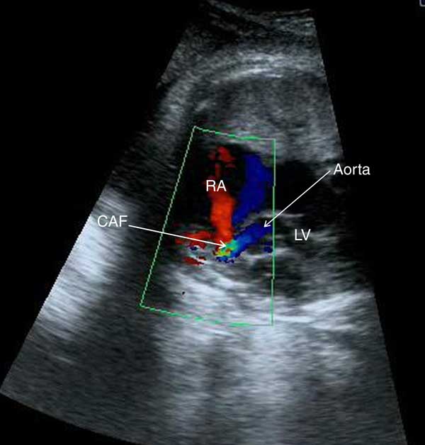
Diagnosis of coronary artery fistula was made in all five cases between 19 and 22 weeks of gestation. The diagnosis was suspected when an abnormal jet of blood was seen entering either an atrium or a ventricle. In the majority, the affected feeding coronary artery was dilated and was easily seen, so that the origin and the course of the fistula could be mapped (Figs 1 and 2 and Supplementary movies 1 and 2). The fistulas originated from the right coronary artery in four cases and from the left in one. The fistulas drained into the right atrium in four cases and to the left ventricular outflow tract in one case. Cardiomegaly with an increased cardiothoracic ratio was present in the cases with a large left-to-right shunt. The ratio was greater than 0.5 at the initial scan in two of the cases. To-and-fro flow in the aortic arch was also a feature in three cases (cases 1, 2, and 5) with a large shunt.
A coronary artery fistula from the left coronary artery to the right atrium. The arrow points to the fistula. LV=left ventricle, RA=right atrium, RV=right ventricle.

A coronary artery fistula from the right coronary artery to the right atrium. There is cardiomegaly with an increased cardiothoracic ratio. CAF=coronary artery fistula, RA=right atrium, LV=left ventricle.

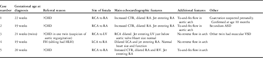
A summary of the features observed during fetal life, the referral reasons, and the gestational age at diagnosis are shown in Table 1.
Summary of fetal demographic and echocardiographic features.

?CHD=suspected congenital heart disease during obstetric screening; ASD=atrial septal defect; CTR=cardiothoracic ratio; FH=family history; HLH=hypoplastic left heart syndrome; LCA=left coronary artery; LV=left ventricle; RA=right atrium; RCA=right coronary artery; RV=right ventricle; VSD=ventricular septal defect.
Progression during fetal life
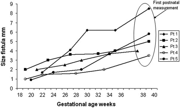
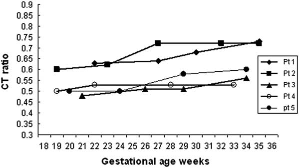
The progression of the coronary artery fistula on echocardiography is shown in Figure 3. In four of the cases, the fistulas increased significantly in size during pregnancy, whereas there was only slight progression in the size of the fistula between the left circumflex coronary artery and the right atrium in one case (case 4). The cardiothoracic ratio increased in four of the five cases, whereas in one (case 4) there was no increase in the ratio (Fig 4).
Sizes of coronary artery fistulas at different gestational ages during pregnancy in all five patients. The measurements shown within the eclipse are the first postnatal measurements and are shown at the gestational age at which delivery took place. Pt=patient.

Cardiothoracic ratios measured at different gestational ages during pregnancy in all five patients. Pt=patient.

Postnatal course
All the five babies were born between 37 and 39 weeks of gestation, with a birth weight ranging between 3.0 and 3.8 kg; four of the five babies were female. The karyotypes were normal in all five babies. Among all, two patients had additional cardiac defects; one of them had a secundum atrial septal defect, which has not been closed so far. The other was suspected to have an aortic coarctation prenatally, but this diagnosis was only confirmed at the age of 10 months, and balloon dilation was performed.
Moreover, two babies (cases 1 and 2), both with coronary artery fistulas between the right coronary artery and the right atrium, developed congestive cardiac failure soon after birth. There was no response to medical treatment with diuretics and captopril, and therefore catheter closure of the fistulas was performed at 5 and 17 days of age, respectively. During the procedure, trial occlusion of the fistula with a balloon catheter did not reveal evidence of myocardial ischaemia. In one case, a 4-mm Amplatzer Muscular Ventricular Septal Defect device (St Jude Medical Inc., St Paul, Minnesota, United States of America) was used to close the fistula. In the second case, an 8/6 Amplatzer Duct Occluder I (St Jude Medical Inc.) was used. In both cases, complete occlusion of the fistulas was achieved; one patient had transient ST segment depression 7 hours after the procedure and was maintained on warfarin for 1 year. The other patient was maintained on aspirin indefinitely.
The follow-up has ranged between 2 and 14 years; three babies were asymptomatic after birth and were managed conservatively. During the course of follow-up, one case had catheter closure of a coronary artery fistula between the left circumflex coronary artery and the right atrium at 4 years of age, because of progressive increase in the size of the feeding coronary artery. The fistula was closed with an 8/6 Amplatzer Duct Occluder I, and at follow-up 1 year later the patient has remained asymptomatic. Another patient had catheter closure of a right coronary artery-to-right atrium fistula at the age of 20 months, also using an 8/6 Amplatzer Duct Occluder I, with an uncomplicated course during a follow-up of 7 months. Both these patients have been maintained on aspirin and clopidogrel. The third patient with a fistula between the right coronary artery and the left ventricular outflow tract remained asymptomatic, but with progressive increase in the size of the feeding vessel. This fistula was closed with a 16-mm Amplatzer Vascular Plug II at the age of 7 years and the child has been maintained on aspirin and clopidogrel.
Discussion
Isolated coronary artery fistulas are a rare abnormality with an incidence of ∼1:50000 live births;Reference Effler, Sheldon, Turner and Groves 1 , Reference Liberthson, Sagar, Berkoben, Weintraub and Levine 2 , Reference Schumacher, Roithmaier and Lorenz 4 however, they may be associated with other cardiac malformations such as ventriculo-coronary arterial communications in patients with hypoplastic left heart syndrome and pulmonary atresia with intact ventricular septum.Reference Patel, Lane, Spector, Smith and Waight 5 – Reference Freedom, Anderson and Perrin 7 Many patients with coronary artery fistulas may become symptomatic after the 2nd or 3rd decade of life, and some small fistulas may close spontaneously.Reference Schleich, Rey, Gewillig and Bozio 8 , Reference Malcic, Belina, Gitter, Viljevac, Kniewald and Krmelk 9 Spontaneous closure of a prenatally detected fistula has been reported in one patient, in whom the fistula from the left anterior descending coronary artery to the right ventricle closed by 1 year of age.Reference Cotton 10 Occasionally, fetal hydrops may develop in association with a coronary artery fistula and the outcome is likely to be worse than those without hydrops.Reference Oztunc, Gokalp, Yuksel, Imamoglu and Madazil 11 The report from Oztunc et al highlights a case of a fistula between the left coronary artery and the right ventricle, diagnosed at 28 weeks gestation, and where hydrops developed during pregnancy and the baby died on the day of delivery. In our unit, only one fetus with a coronary artery fistula had fetal hydrops; this baby was excluded from the study as the pregnancy was terminated. In all other published reports of prenatally detected fistulas, congestive cardiac failure or significant cardiomegaly occurred after birth. These patients had attempted catheter or surgical closure in the newborn period or early infancy.Reference Hayashi, Inuzuka, Ono and Katori 12 – Reference Khan, Qureshi, Rosenthal and Sharland 16 The optimal timing for transcatheter or surgical closure of a coronary artery fistula in asymptomatic patients is unclear.Reference Armsby, Keane, Sherwood, Forbess, Perry and Lock 17 Occasionally, complications may be encountered even after transcatheter closure.Reference Kharouf, Cao and Hijazi 18 – Reference Gowda, Forbes and Singh 20 Therefore, the treatment options and the timing merit serious consideration. It is preferable to perform transcatheter closure in patients above 10 kg if they are asymptomatic, as bigger the patient the easier the procedure is technically, but closure may need to be performed in patients under 10 kg when they have intractable symptoms such as congestive cardiac failure.
Prenatal diagnosis of isolated coronary artery fistula is rare, with only few case reports published thus far.Reference Oztunc, Gokalp, Yuksel, Imamoglu and Madazil 11 – Reference Khan, Qureshi, Rosenthal and Sharland 16 We first reported the prenatal detection and progression of a right coronary artery-to-right ventricular fistula in one case in 1996.Reference Sharland, Tynan and Qureshi 21 The diagnosis can be made in fetal life by cross-sectional and colour Doppler echocardiography. The signs on colour Doppler echocardiography include the detection of an abnormal jet draining into one of the cardiac chambers, dilation of the feeding coronary artery, cardiomegaly, and possible reversal of flow in the ascending aorta and transverse arch.
Noticeable progression of the coronary artery fistula occurred in four out of five of our cases during pregnancy. In all except one of these four cases, drainage of the fistulas was to the right atrium, and in one case it was to the left ventricular outflow tract. In the latter patient, the fistula was closed at 7 years of age because of progressive increase in the size of the feeding vessel. In the remaining case, in whom there was a fistula between the left circumflex coronary artery and the right atrium, there was only slight progression during fetal life. None of the fistulas closed spontaneously during fetal life.
Comparison with postnatal measurements on MRI scans or angiography at the time of intervention has its limitations because these measurements are taken at different phases of the cardiac cycle. Prenatal presence of cardiomegaly and to-and-fro flow in the aorta seem to be good predictors for developing congestive cardiac failure after birth; two out of five cases developed congestive cardiac failure soon after birth and underwent transcatheter closure of the fistula with occlusion devices. In these two, the cardiothoracic ratio was increased at the time of the diagnosis and increased further as pregnancy advanced. The other three cases remained asymptomatic after birth. They had cardiothoracic ratios within the normal range at the time of the diagnosis, and in one of them there was a small increase as pregnancy advanced. These three cases were managed conservatively until there was some evidence of an increase in the left-to-right shunt or the size of the feeding coronary artery; one among them underwent catheter closure at 4 years, one at 20 months, and one at 7 years of age. From our small experience, it appears that patients with a short connection between the main coronary artery or a branch and the right atrium seem to develop congestive cardiac failure early after birth, compared with those in whom the connection is of a longer distance, tortuous, and involving a side branch of the coronary artery. Prenatally, it was not possible to determine whether the feeding vessel to the fistula was a side branch or the main coronary artery. This may be clinically important, as in the latter group it may be possible to delay catheter closure until the patient is of optimal weight to undergo catheter closure safely.
It is possible to detect coronary artery fistulas prenatally, which may indicate fistulas with the largest left-to-right shunts, and thus may predispose the patients to the early onset of congestive cardiac failure and the need for early intervention. If asymptomatic, patients can be managed conservatively until they are old enough to undergo a lower-risk procedure after achieving the optimal weight. Catheter closure of such fistulas is challenging technically in older children, and this is especially so in the neonatal period. We have shown that it is possible to detect coronary artery fistulas prenatally and have documented their course during the pregnancy and after birth.
Acknowledgement
The authors would like to acknowledge all staff at Evelina Children’s Hospital who were involved in the clinical care of the patients in this study.
Financial Support
This research received no specific grant from any funding agency, commercial, or not-for-profit sectors.
Conflicts of Interest
None.
Supplementary Material
To view supplementary material for this article, please visit http://dx.doi.org/10.1017/S1047951115001535