Introduction
Carcinoma of the upper aerodigestive tract is one of the major health problems across the globe. In 2018, cancers of various sites of the upper aerodigestive tract were among the top 36 cancers in incidence worldwide, ranking ninth globally when added together.Reference Bray, Ferlay, Soerjomataram, Siegel, Torre and Jemal1 In India, head and neck cancers account for 30 per cent of all cancers in males and 11–16 per cent in females.Reference Kulkarni2 Most of these cancers are squamous cell carcinoma.
Definitive radiotherapy (RT) is the standard of care in many of these cancers, particularly in the early stages. Finding a suspicious residual or recurrent lesion in a symptomatic (or asymptomatic) patient beyond 12 weeks post-RT puts the clinician in a difficult situation. Although early confirmation of local treatment failure is of paramount importance for successful salvage therapy, it is not easy. Clinical examination is difficult in such cases because of changes in mucosa, tissue oedema and fibrosis caused by RT.
Although non-invasive modalities like computerised tomography (CT)Reference Hermans, Pameijer, Mancuso, Parsons and Mendenhall3 and fluoro-deoxy-glucose positron emission tomography combined with CTReference Helsen, Roothans, Van Den Heuvel, Van den Wyngaert, Van den Weyngaert and Carp4 have been found to be useful in detecting post-RT local treatment failure, a tissue diagnosis obtained by biopsy is still needed to institute any oncological therapy. However, interpretation of histopathological examination of biopsied tissue is also difficult after RT. On many occasions the biopsy report is inconclusive, and a repeat biopsy is warranted, which increases morbidity, more so in irradiated tissues. Therefore, there is a need for a non-invasive technique that can provide tissue diagnosis reliably. Contact endoscopy is a technique that can show the cytological features of tissues in vivo and identify malignant tissue.
Earlier studies, performed on non-irradiated tissues, have shown that contact endoscopy can scan large areas of mucosa and detect malignancy with high accuracy.Reference Warnecke, Averbeck, Leinung, Soudah, Wenzel and Kreipe5,Reference Pelucchi, Bianchini, Travagli and Pastore6 However, the role of contact endoscopy in post-RT cases has not been well studied. We found only two studies in the literature that have evaluated contact endoscopy in post-RT cases.Reference Pak, To, Leung and van Hasselt7,Reference Pak, Vlantis, Chow and van Hasselt8 Both of these studies evaluated only nasopharyngeal cancer. We could not find any study evaluating contact endoscopy for post-RT residual or recurrent squamous cell carcinoma in other areas of the upper aerodigestive tract. This study was undertaken to fill this gap by evaluating the role of contact endoscopy in post-RT failures across various sites of the upper aerodigestive tract.
Materials and methods
This study was conducted at a tertiary care centre between January 2017 and June 2019. Ethical clearance was obtained from the Institutional Ethics Committee of the Army College of Medical Sciences and Base Hospital, Delhi Cantt, India, and the study complied with the Helsinki Declaration of 1975. Study approval was granted by the Scientific Review Committee of our hospital.
We included consecutive consenting patients planned for biopsy because of suspicion of local treatment failure following definitive RT for squamous cell carcinoma of upper aerodigestive tract. Non-consenting patients were excluded.
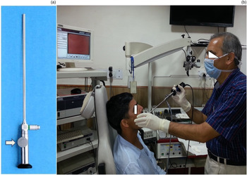
To perform contact endoscopy, we used: Andrea-Dias Contact Micro Laryngoscope with HOPKINS Straight Forward Telescope 0° and 30°, with diameter 5.5 mm, length 23 cm, magnification 60 × and 150×; a 3 chip camera (Tricam SL II); a Xenon 175 watt light source and a video recording system (AIDA), all manufactured by Karl storz, Tuttlingen, Germany (Figure 1).
Images showing equipment and set up for contact endoscopy: (a): contact micro-laryngoscope with magnification changer (arrow); and (b) contact endoscopy of the oral cavity in the out-patient department.

When performing the contact endoscopy procedure, no anaesthesia was required in the oral cavity. For nasopharynx and oropharynx procedures, 4 per cent lignocaine topical spray was used whereas general anaesthesia was required for larynx and hypopharynx procedures.
We stained the lesion by placing cottonoids soaked in 1 per cent methylene blue against it for five minutes (or longer if required for adequate staining). Then we placed the contact micro-laryngoscope (0 degree on plane surface; 30 degree on sloping surfaces) gently over the mucosa and studied the whole area by moving it slowly from one area to another area. The procedure was performed at × 60 magnification first and repeated with × 150 magnification. Cellular architecture was carefully examined. Nuclei were identified by their dark blue staining and the cytoplasm was light blue.
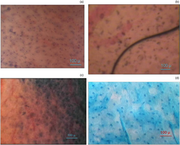
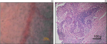
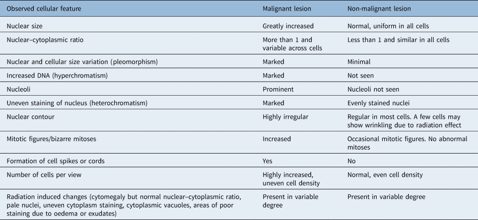
Contact endoscopy was performed by the most senior otolaryngologist in the team, who also performed digital capture and recording of videos and images of the procedure independently. The images were then presented to the other three otolaryngologists who had different amounts of experience (10 years, 4 years and 1 year) in interpreting contact endoscopy. These otolaryngologists analysed the images independently, without having prior knowledge of the clinical and radiological findings of the patients. The presence of malignancy on contact endoscopy was decided based on cytological characteristics adapted from reports of previous studies.Reference Pelucchi, Bianchini, Travagli and Pastore6,Reference Andrea, Dias and Santos9–Reference Wardrop, Sim and McLaren12 These are summarised in Table 1. The result was recorded as ‘malignant’ or ‘non-malignant’. Representative images of the tissues seen on contact endoscopy are given in Figures 2 and 3.
Contact endoscopy images of the tonsils showing: (a) normal mucosa with cells that are uniform in size and shape with the nuclear–cytoplasmic ratio maintained; (b) malignant lesion with marked nuclear pleomorphism, irregular contours and increased nuclear–cytoplasmic ratio; (c) tonsil at three months post-radiotherapy with residual cancer showing large areas of acellular necrotic material and exudate (pink), irregular nuclear contour, increased nuclear-cytoplasmic ratio and increased cells per view; and (d) tonsil at three months post-radiotherapy with no residual malignancy showing areas of oedema (unstained areas), fairly homogenous distribution of cells, normal nuclear–cytoplasmic ratio with regular contour of nuclei. (methylene blue; ×150)

Image of (a) contact endoscopy (methylene blue; ×150) and (b) histopathology image (H&E; ×200) of carcinoma of the hypopharynx. Irregular distribution of nuclei with increased nuclear–cytoplasmic ratio is evident in both images.

Interpretation of cellular features on contact endoscopy

After performing contact endoscopy, the stain was washed away by saline irrigation and the most suspicious area was infiltrated with 2 per cent lignocaine containing 1 in 100 000 adrenaline for local anaesthesia and decongestion. Surgical biopsy was then obtained, and the specimen was sent to the laboratory for histopathological examination, which was performed by an onco-pathologist who was blinded to the contact endoscopy findings.
Two weeks later, the contact endoscopy images were again reviewed by the three otolaryngologists and the result recorded as malignant or non-malignant. When the histopathological examination report was available, comparison of contact endoscopy diagnoses with histopathological examination findings was made by another otolaryngologist who had neither performed contact endoscopy nor histopathological examination. For the purpose of comparison, lesions reported as well, moderately or poorly differentiated squamous cell carcinoma and carcinoma in situ on histopathological examination were grouped as ‘malignant’ whereas dysplasia, squamous metaplasia, radiation change, and benign tissue or normal mucosa were grouped as ‘non-malignant’. The scores of correct diagnoses of each otolaryngologist were compared with each other and also with their own scores on the two occasions.
Statistical analysis
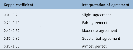
The categorical variables were compared using the chi-square test. Sensitivity, specificity, negative predictive value, positive predictive value and accuracy of contact endoscopy in various regions of the upper aerodigestive tract and also over all (combining all sites) were calculated, considering the findings of the most experienced otolaryngologist as being the correct diagnosis following contact endoscopy. Fleiss’ kappa statistic was calculated for inter-observer agreement and Cohen's kappa was used for intra-observer variability. The strengths of the kappa coefficients were interpreted as per the guidelines of Landis and Koch (Table 2). A p-value of less than 0.05 was considered significant. SPSS® statistical software (version 21) was used for analysis.

Results
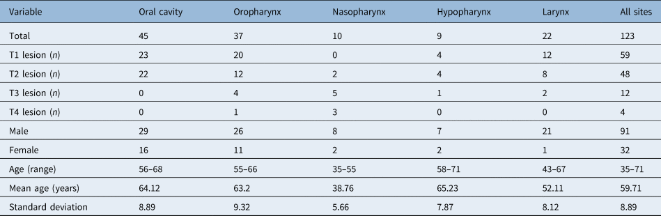
We considered 135 patients for the study initially. All had histopathologically proven squamous cell carcinoma of upper aerodigestive tract and had undergone definitive RT in the past. Demographic and descriptive data of these patients are summarised in Table 3. Of the patients, 96 (71.11 per cent) had presented with recurrent or persistent pain, dysphagia, dysphonia, aural fullness or foreign body sensation at 12-weeks post-RT follow up. There were 39 patients (28.89 per cent) who had no symptoms but who had a lesion detected on clinical examination during routine follow up. All had received 60–70 Gy of RT, depending on the tumour characteristics. The time interval between completion of RT and suspicion of local failure of treatment ranged from 3 to 21 months (mean 14.26 +/– 2.36 months).
Demographic and descriptive data

Contact endoscopy could not be performed in 7 patients (5.19 per cent) because of: inability to negotiate the rigid scope to the intended site, especially in the roof of nasophaynx behind the fossa of Rosenmüller and floor of vallecula; bleeding from the lesion on touching with the scope; failure to stain the irradiated tissue; and excessive precipitation of the stain. In 5 patients (3.70 per cent), histopathological examination was inconclusive. Both of these categories of patients were excluded from the final analysis. Thus, we were left with 123 patients for whom contact endoscopy analysis and the histopathological examination report was available for comparison.
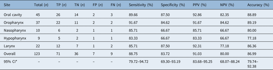
Out of these 123 patients, malignancy was reported in 80 cases (65.04 per cent) and no malignancy was seen in 43 cases (34.96 per cent) on histopathological examination. These results were accepted as a true representation of disease status and contact endoscopy findings were evaluated in comparison to these values. On contact endoscopy, 78 cases (63.41 per cent) were reported as malignant and 45 (36.59 per cent) as non-malignant by the most experienced otolaryngologist's interpretation of contact endoscopy, and these results were taken as representative of contact endoscopy diagnosis. Considering these values, the overall sensitivity, specificity and accuracy of contact endoscopy were 88.75 per cent, 88.72 per cent and 86.99 per cent, respectively (Table 4). There was no significant difference in these indices among various sites studied by us (chi-square test, p = 0.99).
Comparison of contact endoscopy with histopathology report

*95 per cent confidence interval (CI) for overall indices of contact endoscopy. TP = true positive; TN = true negative; FP = false positive; FN = false negative; PPV = positive predictive value; NPV = negative predictive value
The proportion of correct diagnoses made by the 3 otolaryngologists with an experience of 10 years, 4 years and 1 year in interpreting contact endoscopy was 91.87 per cent, 85.37 per cent and 80.49 per cent, respectively, which appeared to reflect the length of experience with contact endoscopy. However, the difference was not statistically significant (p = 0.35, chi-square test).
Overall agreement among the three observers was 92.95 per cent and the kappa value was 0.86 (95 per cent confidence interval (CI) = 0.79–0.93). Intra-observer agreement in the diagnoses made on the two occasions was 96.75 per cent for the first observer and 97.56 per cent for the second as well as the third observer. Kappa value was 0.93 (95 per cent CI = 0.87–1.00) for the first observer and 0.95 (95 per cent CI = 0.90–1.00) for the second and third observers.
There were no major complications because of the dye or the procedure. We were able to perform contact endoscopy in the out-patient department as well as in the operating theatre. As mentioned, there was difficulty in negotiating the scope to the desired site in the roof of nasopharynx behind the fossa of Rosenmüller and in the floor of vallecula. There was also some difficulty as a result of bleeding on touching friable lesions, excessive precipitation of stain in the tissues and the need for re-staining in some cases.
Discussion
Our study has demonstrated the high sensitivity, specificity and accuracy of contact endoscopy in detecting residual or recurrent malignancy in irradiated patients with squamous cell carcinoma of the upper aerodigestive tract. These results were comparable across various sites of the upper aerodigestive tract and were also in line with the findings of contact endoscopy in non-irradiated cases studied by other researchers in the head and neck region in generalReference Szeto, Wehrli, Whelan, Franklin, Nichols and Yoo13 and in specific areas like the larynx,Reference Warnecke, Averbeck, Leinung, Soudah, Wenzel and Kreipe5 lower lip,Reference Ramos, Tavares, Dedivitis, Pedruzzi and Oliveira11 oral cavity and oropharynx.Reference Pelucchi, Bianchini, Travagli and Pastore6,Reference Saeki, Tsuzuki, Negoro, Nin, Sagawa and Uwa14–Reference Dowthwaite, Szeto, Wehrli, Daley, Whelan and Franklin16 However, the sensitivity and specificity of contact endoscopy in our series was lower than in the study by Pak et al. Reference Pak, To, Leung and van Hasselt7 who studied the nasopharynx post-RT and reported 100 per cent sensitivity and specificity. The difference may not be statistically significant or, if statistically significant, may be attributable to our lack of experience with the technique. Dowthwaite et al. Reference Dowthwaite, Szeto, Wehrli, Daley, Whelan and Franklin16 also found high sensitivity and specificity of contact endoscopy in diagnosing malignant lesions (89 per cent and 100 per cent, respectively) but only 50 per cent in benign lesions. They also reported a high number of uninterpretable images (17.6 per cent).
• The role of contact endoscopy in picking up residual or recurrent cancer after radiotherapy has not been well studied
• The present study evaluated contact endoscopy in detecting post-radiotherapy failures in various sites of the upper aerodigestive tract
• The sensitivity, specificity and accuracy of contact endoscopy in these cases were 88.75, 88.72 and 86.99 per cent, respectively
• These indices matched the results in non-irradiated tissues and were also similar across various sites of the upper aerodigestive tract
• Contact endoscopy showed low inter- and intra-observer variability
Compared to non-irradiated tissues, additional problems are encountered in interpreting contact endoscopy images in irradiated cases. Oedematous and necrotic tissues do not take up the stain well. Due to RT the cells undergo generalised cytomegaly and the nuclear membrane may show wrinkling. These changes may be confused with signs of malignancy. However, a careful look at the nuclear–cytoplasmic ratio will settle the issue as it would be normal and constant across the cells if there is no residual or recurrent malignancy. The most consistent findings indicative of malignancy on contact endoscopy were marked nuclear pleomorphism, grossly increased nuclear–cytoplasmic ratio, overcrowding of nuclei per view and multiple or bizarre mitotic figures.
Contact endoscopy can also be performed without staining, a method where the changes in the vascular network and neo-angiogenesis are studied in vivo to detect malignancy. This is an indirect method of looking for malignancy. We have not included the study of vascular patterns because we wanted to study the irradiated tissue for direct evidence of malignancy by evaluating the changes in cellular architecture, which are seen only after staining the tissues.
We found the technique of contact endoscopy as simple to perform in post-RT cases as in non-irradiated patients. Interpretation of contact endoscopy images can be learnt easily, though a learning curve exists. Inclusion of a pathologist and basic training in cytopathology in the initial period may be helpful.
Our results showed near perfect inter- and intra-observer agreement in overall contact endoscopy results of all sites of the upper aerodigestive tract, when analysed for inter- and intra-observer variability. These findings are similar to the study carried out by Pak et al.,Reference Pak, Vlantis, Chow and van Hasselt8 which showed low inter- and intra-observer variability of this technique in post-RT evaluation of nasopharyngeal cancer.
The current technique of contact endoscopy needs further refinement including: the need for better stain which can penetrate deeper, better image enhancement technology, computer software to allow automated analysis of images like counting cells per field, assigning a numerical value to the degree of staining and calculating nuclear–cytoplasmic ratio. Some attempts have already been made in this direction. Tarnawski et al. Reference Tarnawski, Fraczek, Jelen, Krecicki and Zalesska-Krecicka17 have demonstrated the role of computers in evaluating nuclear characteristics. Some researchers have shown improved sensitivity and specificity (even up to 100 per cent) with image-enhanced endoscopy combined with contact endoscopy in the larynx and hypopharynxReference Puxeddu, Sionis, Gerosa and Carta18 and differentiating non-malignant lesions from squamous cell carcinoma in the oral cavity and oropharynx.Reference Carta, Sionis, Cocco, Gerosa, Ferreli and Puxeddu19 Esmaeili et al. Reference Esmaeili, Illanes, Boese, Davaris, Arens and Friebe20 have also suggested a novel model to reduce subjectivity in interpretation of contact endoscopy based on vascular pattern. Combining conventional contact endoscopy with narrow band imaging has been reported recently with improved sensitivity and specificity.Reference Stefanescu, Ceachir, Zainea, Hainarosie, Pietrosanu, Ionita and Hainarosie21 Boese et al. Reference Boese, Illanes, Balakrishnan, Davaris, Arens and Friebe22 have also recently developed an automated algorithm for vessel pattern recognition based on videos from contact endoscopy and narrow band imaging of vocal folds. We hope that further studies will lead to improved objectivity and accuracy of contact endoscopy in detecting malignancy.
Conclusion
In this study, we found contact endoscopy to be a simple and reliable technique with high sensitivity, specificity and accuracy in detection of post-RT residual or recurrent cancer of the upper aerodigestive tract. These results are similar to the results obtained for non-irradiated tissues studied in the past. In its current form, the utility of contact endoscopy may be in: (1) serial follow up of post-RT cases for early detection of local failures; and (2) deciding the most suitable area for taking biopsy in suspected local failure.
Competing interests
None declared