Introduction
The Taeniolabidoidea is a clade of multituberculates known from the Late Cretaceous to Eocene of North America and eastern Asia (e.g., Cope, 1882, Reference Cope1884; Matthew and Granger, Reference Matthew and Granger1925; Russell, Reference Russell1926; Matthew et al., Reference Matthew, Granger and Simpson1928; Granger and Simpson, Reference Granger and Simpson1929; Gazin, Reference Gazin1939; Sloan and Van Valen, Reference Sloan and Van Valen1965; Chow and Qi, Reference Chow and Qi1978; Middleton, Reference Middleton1982; Johnston and Fox, Reference Johnston and Fox1984; Wilson, Reference Wilson1987; Miao, Reference Miao1988; Simmons, Reference Simmons1987; Buckley, Reference Buckley1995; Lucas et al., Reference Lucas, Williamson and Middleton1997; Williamson et al., Reference Williamson, Brusatte, Secord and Shelley2015), and includes the largest multituberculates so far discovered, e.g., Taeniolabis taoensis (Cope, Reference Cope1882) from the early Paleocene of New Mexico, thought to have approached the size of extant beavers (Weil and Krause, Reference Weil and Krause2008, but see also Williamson et al., Reference Williamson, Brusatte, Secord and Shelley2015 for a contrasting opinion). The taxonomic composition of Taeniolabidoidea has varied, but results of recent studies suggest that the superfamily minimally contains four North American genera (Valenopsalis Williamson et al., Reference Williamson, Brusatte, Secord and Shelley2015, Catopsalis Cope, Reference Cope1882, Kimbetopsalis Williamson et al., Reference Williamson, Brusatte, Secord and Shelley2015, and Taeniolabis Cope, Reference Cope1882), all from the early Paleocene to earliest late Paleocene, as well as two genera from the late Paleocene of eastern Asia (Sphenopsalis Matthew, Granger, and Simpson, Reference Matthew, Granger and Simpson1928 and Lambdopsalis Chow and Qi, Reference Chow and Qi1978). A third eastern Asian genus, Prionessus Matthew and Granger, Reference Matthew and Granger1925, has traditionally been considered a taeniolabidoid, but we consider this referral doubtful (contra, e.g., Xu et al., Reference Xu, Zhang, Pu, Jia, Zhang, Lü and Meng2015).
Among the currently recognized taeniolabidoid genera, Catopsalis is by far the most speciose, with six nominal species that collectively span the latest Cretaceous through earliest late Paleocene (see summary by Williamson et al., Reference Williamson, Brusatte, Secord and Shelley2015). Until relatively recently, the chronological succession of the various species of Catopsalis appeared to document what has been interpreted as fairly straightforward evolutionary trends of increasing body size (as inferred from molar length), an increase in the length of the upper and lower first molar (M1/m1) relative to the second molar (M2/m2), and an overall reduction in the size of the upper and lower fourth premolar (P4/p4) (Kielan-Jaworowska and Sloan, Reference Kielan-Jaworowska and Sloan1979; Middleton, Reference Middleton1982). The discovery of specimens of the size of C. foliatus Cope, Reference Cope1882 in putative Late Cretaceous deposits (C. johnstoni Fox, Reference Fox1989, but see Lucas et al., Reference Lucas, Williamson and Middleton1997), and even more so, the discovery of a species of the size of C. calgariensis Russell, Reference Russell1926 in the middle to late Puercan (C. waddleae Buckley, Reference Buckley1995) muddies the waters, and suggests that the evolutionary history of Catopsalis is significantly more complicated than previously thought, and that much of this history remains unknown. We report here on the discovery of a new species of Catopsalis from the middle and late Torrejonian of southwestern Alberta that complicates matters further. Catopsalis kakwa new species is the second species of Catopsalis to be documented in the province, and represents not only the smallest species of Catopsalis, but ranks as the smallest taeniolabidoid so far discovered, with an estimated body mass that is one half to two thirds that of Valenopsalis joyneri (Sloan and Van Valen, Reference Sloan and Van Valen1965), the next smallest taeniolabidoid.
Geological setting
Regional geology
Strata of Paleocene age crop out extensively in and around the city of Calgary in southern Alberta, and are generally referred to one of two largely time-equivalent units, the Porcupine Hills and Paskapoo formations (Jerzykiewicz, Reference Jerzykiewicz1997; Lerbekmo and Sweet, Reference Lerbekmo and Sweet2000; Scott et al., Reference Scott, Spivak and Sweet2013). These units occur in the Alberta Syncline, a geographically extensive depocenter developed along the eastern flank of the Rocky Mountains. Lerbekmo and Sweet (Reference Lerbekmo and Sweet2000) identified both formations within Calgary and suggested that the city represented an area of formational transition, with strata of the Porcupine Hills Formation generally being found in areas south of the Bow River, and those of the Paskapoo Formation occurring north of the river and extending as far west as Cochrane (also see Rutherford, Reference Rutherford1927; Russell, Reference Russell1929). Recently discovered fossil mammals in strata of the Porcupine Hills Formation at localities at the westernmost edge of Calgary suggest that these exposures are middle to late Torrejonian in age (To2–To3), slightly older than localities in the Paskapoo Formation occurring near Cochrane, which are thought to be earliest Tiffanian in age (Ti1) (Youzwyshyn, Reference Youzwyshyn1988; Scott et al., 2002, Reference Scott, Spivak and Sweet2013). Strata of the Porcupine Hills Formation consist of large-scale cross-bedded sandstones of channel-fill complexes, recessive mudstone units, sparse coal, and the presence of significant amounts of caliche, lithological features that are suggestive of deposition in semiarid climates (Jerzykiewicz and Sweet, Reference Jerzykiewicz and Sweet1988; Jerzykiewicz, Reference Jerzykiewicz1997). In contrast, strata of the Paskapoo Formation are characterized by the presence of significant coal units that suggest deposition in wetter and more humid conditions (Demchuk, Reference Demchuk1990; Demchuk and Hills, Reference Demchuk and Hills1991; Jerzykiewicz, Reference Jerzykiewicz1997).
Locality information
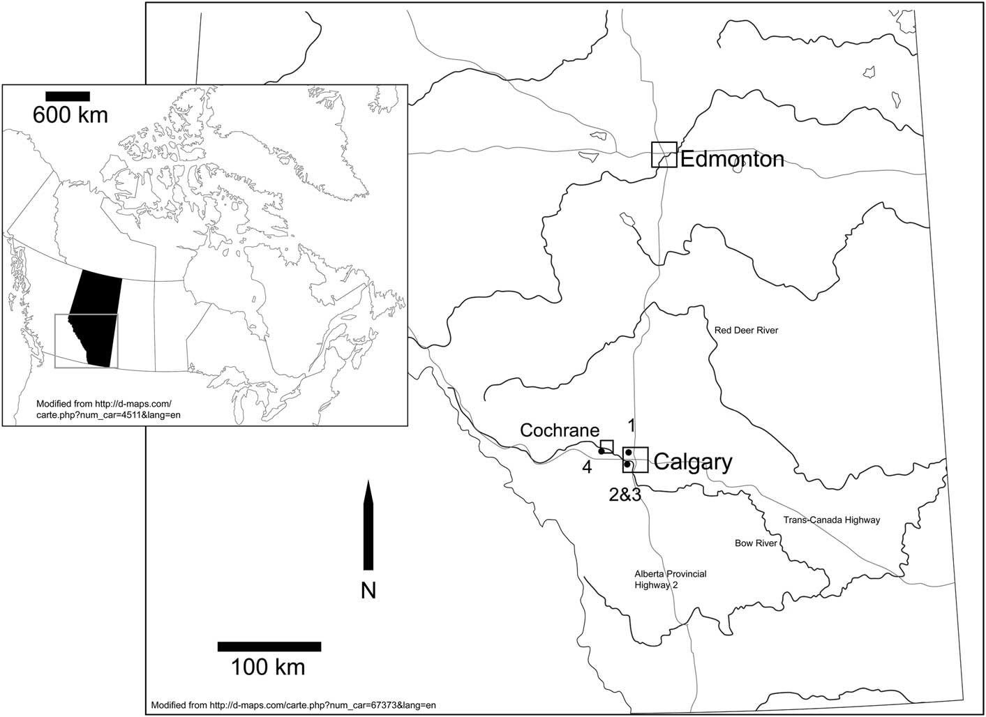
The fossils described in this paper occur in the Porcupine Hills Formation at relatively recently discovered localities in and near the City of Calgary, and near the town of Cochrane (Fig. 1). Legal land descriptions for each of the localities are provided here; more precise coordinates can be made available to qualified investigators through the Royal Tyrrell Museum of Palaeontology.
Map of southern Alberta showing the locations of the localities discussed. Gray rectangle in inset highlights the southern half of the province represented by the larger map. 1=Zagas Quarry; 2&3=Narcissus locality and Nordic Ski Quarry; 4=Jumpingpound Creek Site 1.

Zagas Quarry
Zagas Quarry (TMP locality L2391) was discovered in 2007 during construction of an interchange at Centre Street and Stoney Trail in the northeastern quadrant of Calgary, west of Alberta Highway No. 2 (Lsd5, S34, T25, R1, W5), at ~1110 m above sea level. The mammalian fossils occur in greenish to somber gray, fine-grained siltstone with abundant caliche, clay clasts, molluscan shells and steinkerns, and thin, laterally discontinuous lignite beds. The mammalian fauna from Zagas Quarry is largely unstudied, but the presence of several fossil mammals—including the multituberculates Ptilodus wyomingensis Jepsen, Reference Jepsen1940 and Baiotomeus cf. B. russelli Scott, Fox, and Youzwyshyn Reference Scott, Fox and Youzwyshyn2002, the hyopsodontid Promioclaenus thnetus Scott, Spivak, and Sweet, Reference Scott, Spivak and Sweet2013, and the eutherian Eudaemonema bohachae Scott, Spivak, and Sweet, Reference Scott, Spivak and Sweet2013—suggests correlation with the Bearspaw and Nordic Ski Quarry localities (see below), and hence a middle or late Torrejonian (To2 or To3) age for the local fauna.
Narcissus locality
The Narcissus locality (TMP locality L2395) occurs on the southern side of Bow River near the western limits of the city of Calgary (SE1/4 Lsd7, S1, T25, R3, W5), at ~1113 m above sea level. The fossils occur in channel lags containing caliche debris, clay rip ups, and fragmentary molluscan shells. The occurrence of several fossil mammals (e.g., the multituberculate Ptilodus wyomingensis and the eutherian Elpidophorus minor Simpson, Reference Simpson1937) suggest a middle Torrejonian (To2) age for the local fauna; a projection of the locality onto a composite regional stratigraphic section suggests that the fossils are preserved in sediments occurring in reversed polarity, which has been interpreted as representing chron 27r (Lerbekmo and Sweet, Reference Lerbekmo and Sweet2000; Scott et al., Reference Scott, Spivak and Sweet2013).
Nordic Ski Quarry
Nordic Ski Quarry (TMP locality L2383) occurs in Canada Olympic Park (COP), a high-performance athletic training facility in the southwestern quadrant of Calgary that was built for the 1988 Winter Olympic Games. Fossiliferous strata were exposed in COP during construction of ski jumps and a spectator bowl. The locality is south of Bow River and the Trans-Canada (No. 1) Highway (SE1/4 Lsd13, S27, T24, R2, W5), at ~1113 m above sea level. Two superposed fossil-bearing horizons have been discovered at Nordic Ski Quarry, the lower of which has produced the fossils discussed in this paper. The fossils at Nordic Ski Quarry occur in yellow to rusty orange, fine-grained siltstone in association with abundant gastropod and bivalve shells and steinkerns, clay clasts, and caliche debris. Nonmammalian vertebrate fossils are abundant and include the teeth and bones of actinopterygians (primarily amiids and gar), lissamphibians, crocodylians, choristoderes, and lizards. Age-diagnostic mammalian taxa have yet to be identified at this locality, but the presence of the multituberculate Baiotomeus sp., and the eutherians Elpidophorus cf. E. elegans Simpson, Reference Simpson1927 and Pararyctes cf. P. pattersoni Van Valen, Reference Van Valen1966 argue for a late Torrejonian age (To3) for the local fauna. A late Torrejonian age is further suggested by the work of Lerbekmo and Sweet (Reference Lerbekmo and Sweet2000), who determined that Nordic Ski Quarry occurs in an interval of mixed polarity interpreted by them as magnetochron 27n, correlative with strata containing fossil mammal localities of late Torrejonian age in the Crazy Mountains Basin of Montana, and the San Juan Basin of New Mexico (Butler et al., Reference Butler, Krause and Gingerich1987; Williamson, Reference Williamson1996; Lofgren et al., Reference Lofgren, Lillegraven, Clemens, Gingerich and Williamson2004).
Jumpingpound Creek Site 1
The Jumpingpound Creek locality (TMP locality L2404) was discovered in 2013. The locality occurs at outcrops along the southern bank of Jumpingpound Creek south of Cochrane, ~1 km upstream of its confluence with Bow River (SE1/4 Lsd11, S33, T25, R4, W5). The rock exposed on Jumpingpound Creek is referable to the Porcupine Hills Formation, demonstrating that the formation extends a significant distance west of Calgary. Fossils occur in extensive channel lags containing caliche debris, clay rip up clasts, and fragmentary molluscan shells. The mammalian fauna from Jumpingpound Creek Site 1 is largely unstudied, but the presence of several taxa, including the multituberculate Ptilodus wyomingensis and the eutherian Eudaemonema bohachae suggests correlation with the Bearspaw localities some 20 km to the east, and hence a middle Torrejonian (To2) age for the local fauna (Scott et al., Reference Scott, Spivak and Sweet2013).
Materials and methods
Dental terminology
Dental terminology follows that of Krause (1977, 1982, Reference Krause1987); dental measurements follow Krause (Reference Krause1987) and Scott (Reference Scott2003). Cusp number and their expression in formulae follow the methods and rationale of Krause (Reference Krause1977). Upper molar cusp formulae of the form x:y:z designate cusps of the labial (x), middle (y), and lingual (z) rows, respectively. Lower molar cusp formulae of the form x:y designate cusps of the labial (x) and lingual (y) rows, respectively.
Abbreviations for dentition
L/l, left; R/r, right; I/i, upper and lower incisor; M/m, upper and lower molar; P/p, upper and lower premolar.
Abbreviations for North American Land Mammal Ages
Pu, Puercan; Ti, Tiffanian; To, Torrejonian.
Repositories and institutional abbreviations
Specimens included herein are housed in the Royal Ontario Museum (ROM), Toronto, Ontario; the Royal Tyrrell Museum of Palaeontology (TMP), Drumheller, Alberta; the University of Alberta Laboratory for Vertebrate Paleontology (UALVP), Edmonton, Alberta; and the University of Wyoming Collection of Fossil Vertebrates (UW), University of Wyoming, Laramie, Wyoming.
Systematic paleontology
Class Mammalia Linnaeus, Reference Linnaeus1758
Subclass Allotheria Marsh, Reference Marsh1880
Order Multituberculata Cope, Reference Cope1884
Suborder Taeniolabidoidea Sloan and Van Valen, Reference Sloan and Van Valen1965
Family Uncertain
Genus Catopsalis Cope, Reference Cope1882
Type species
Catopsalis foliatus Cope, Reference Cope1882.
Other species
Catopsalis fissidens Cope, Reference Cope1884; C. calgariensis Russell, Reference Russell1926; C. alexanderi Middleton, Reference Middleton1982; C. johnstoni Fox, Reference Fox1989; C. waddleae Buckley, Reference Buckley1995; C. kakwa n. sp.
Catopsalis kakwa new species
urn:lsid:zoobank.org:act:3D891D17-76D8-4060-B5B3-510FC249DB59
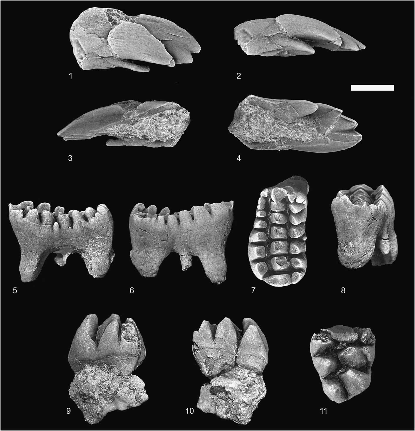
Catopsalis kakwa n. sp. from the Porcupine Hills Formation, early Paleocene, southwestern Alberta: (1–4) TMP 2011.110.0073, RI2 (ZQ) in (1) anterolateral, (2) lateral, (3) medial, and (4) occlusal views; (5–8) UALVP 57538, LM1 (NSQ) in (5) labial, (6) lingual, (7) occlusal, and (8) anterior views; (9–11) TMP 2012.024.0207, LM2 (ZQ) in (9) labial, (10) lingual, and (11) occlusal views. NSQ=Nordic Ski Quarry; ZQ=Zagas Quarry. Scale=2 mm.

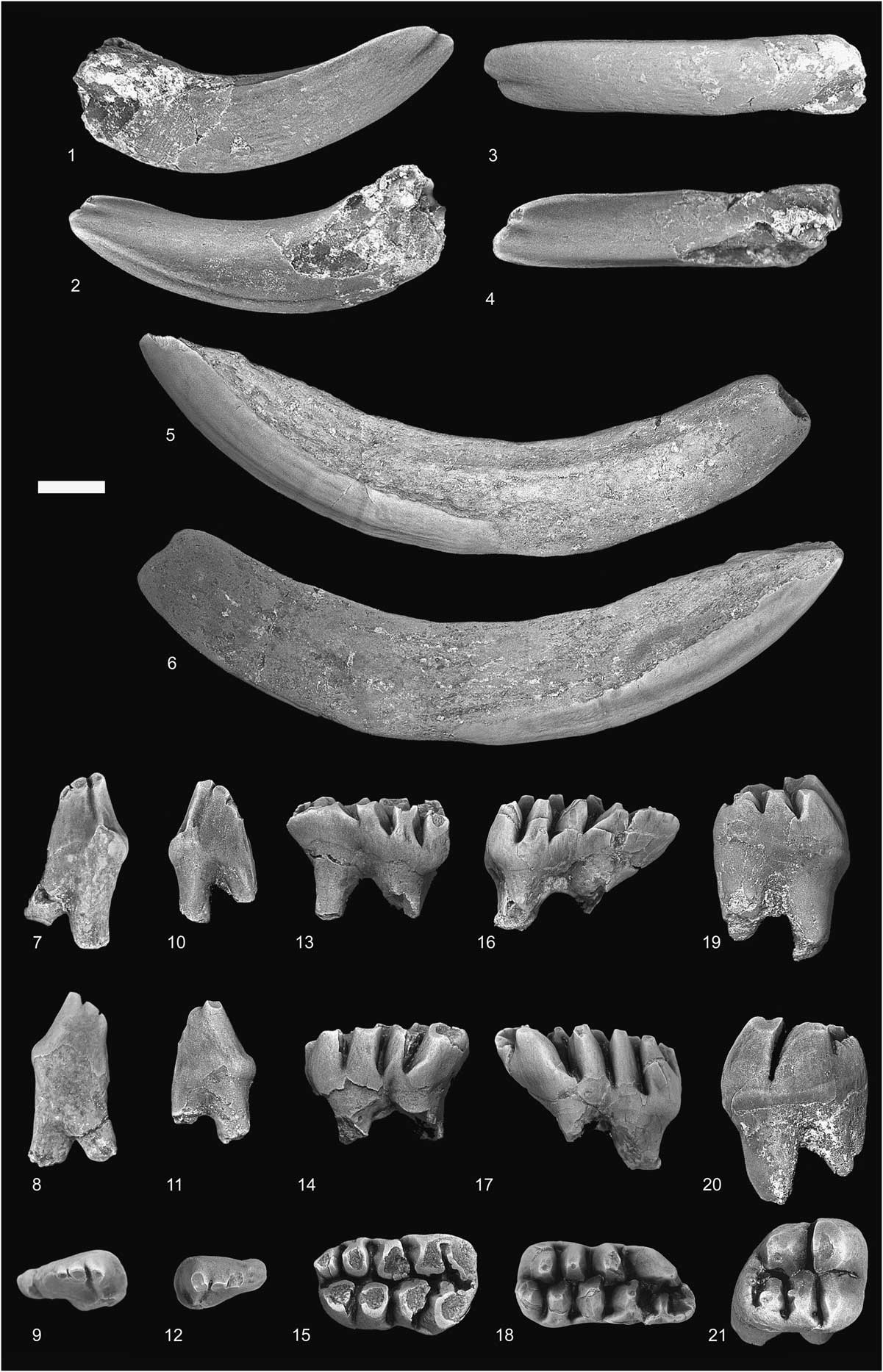
Catopsalis kakwa n. sp. from localities in the Porcupine Hills Formation, early Paleocene, southwestern Alberta: (1–4) UALVP 57537, Ri1 (NSQ) in (1) lateral, (2) medial, (3) anterior, and (4) occlusal views; (5–6) TMP 2009.133.0129, Li1 (ZQ) in (5) lateral and (6) medial views; (7–9) TMP 2015.071.0141, Lp4 (JPC) in (7) labial, (8) lingual, and (9) occlusal views; (10–12) UALVP 57541, Rp4 (NSQ) in (10) labial, (11) lingual, and (12) occlusal views; (13–15) TMP 2009.133.0114, Rm1 (ZQ) in (13) labial, (14) lingual, and (15) occlusal views; (16–18) TMP 2012.024.0063, holotype, Lm1 (ZQ) in (16) labial, (17) lingual, and (18) occlusal views; (19–21) TMP 2009.133.0041, Rm2 (ZQ) in (19) labial, (20) lingual, and (21) occlusal views. JPC=Jumpingpound Creek Site 1; NSQ=Nordic Ski Quarry; ZQ=Zagas Quarry. Scale=2 mm.

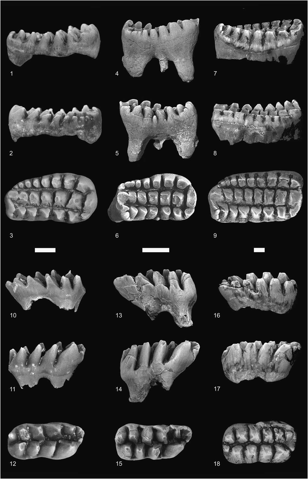
Comparison of upper and lower first molars of Valenopsalis joyneri (Bug Creek Anthills locality, Hell Creek Formation, northeastern Montana, earliest Paleocene), Catopsalis kakwa n. sp., and C. calgariensis (Shotgun locality, Fort Union Formation, Wyoming, late Paleocene) (for comparison, molars of V. joyneri and C. kakwa n. sp. have been enlarged to approximately the same size as those of C. calgariensis): (1–3) UALVP 28186, V. joyneri, RM1 in (1) labial, (2) lingual, and (3) occlusal views; (4–6) UALVP 57538, C. kakwa n. sp., LM1 (reversed from original), in (4) labial, (5) lingual, and (6) occlusal views; (7–9) UW 6407, C. calgariensis, RM1 in (7) labial, (8) lingual, and (9) occlusal views; (10–12) UALVP 21871, V. joyneri, Rm1 in (10) labial, (11) lingual, and (12) occlusal views; (13–15) TMP 2012.024.0063, C. kakwa n. sp., Lm1 (reversed from original), in (13) labial, (14) lingual, and (15) occlusal views; (16–18) UW 6388, C. calgariensis, Rm1 in (16) labial, (17) lingual, and (18) occlusal views. Scales=2 mm.

Comparison of molars of various multituberculates demonstrating cusp interlock (specimens have been scaled to approximately the same size): (1) UALVP 29240, Cimexomys antiquus (MR-6 locality, Milk River Formation of southern Alberta, Late Cretaceous), LM1 (reversed from original) in occlusal view (cusps of the middle row are subquadrate and show no interlock between successive cusps); (2) UALVP 7431, Mesodma sp. (Bug Creek Anthills, Hell Creek Formation, Montana, latest Cretaceous/earliest Paleocene), RM1 in occlusal view (cusps of the middle row are weakly crescentic and show slight interlock, with the convex posterior side fitting into the concave anterior side of the next cusp); (3) ROM 61836, Cimolomys sp. (Bug Creek Anthills, Hell Creek Formation, Montana, latest Cretaceous/earliest Paleocene), RM1 in occlusal view (cusps of the middle row are crescentic and show well-developed interlock between successive cusps); (4) ROM 61837, Meniscoessus robustus (Bug Creek Anthills, Hell Creek Formation, Montana, latest Cretaceous/earliest Paleocene), Lm2 in occlusal view (cusps in both rows are strongly crescentic and show strong interlock between successive cusps). Scale=1 mm.

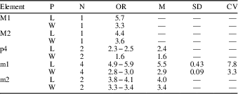
Combined measurements and descriptive statistics for the dentition of Catopsalis kakwa n. sp. from the early Paleocene (middle to late Torrejonian, To2−To3), southwestern Alberta, Canada. P=parameter; N=sample size; OR=observed range; M=mean; SD=standard deviation; CV=coefficient of variation; L=length; W=width.

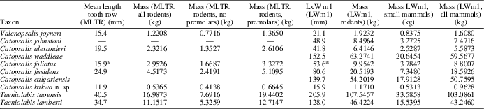
Body mass estimates for North American taeniolabidoid multituberculates. Mass estimates based on mean length of lower tooth row (MLTR) were calculated using the regressions of Freudenthal and Martin-Suarez (Reference Freudenthal and Martin-Suarez2013): ln (mass)=−0.447+2.723 (ln (MLTR)) (all rodents); ln (mass)=0.039+2.378 (ln (MLTR)) (rodents without premolars); ln (mass)=−0.381+2.747 (ln (MLTR)) (rodents with premolars). Correction factors (CF) were applied to each of the calculated body masses, where CF=antilog ((standard error of estimate2)/2). Mass estimates based on mean m1 occlusal area (LWm1) were calculated using the regressions of Legendre (Reference Legendre1986): ln (mass)=2.172+(1.767 (ln (LWm1))) (rodents); ln (mass)=1.786+(1.621 (ln (LWm1))) (small mammals); ln (mass)=1.810+(1.827 (ln (LWm1))) (all mammals). Resultant values in grams were converted to kilograms. For references for measurement data, see Williamson et al. (Reference Williamson, Brusatte, Secord and Shelley2015); for measurements for Catopsalis johnstoni, see Fox (Reference Fox1989). *Values for p4 length, m1 width, and m2 length for C. foliatus follow the estimates of Williamson et al. (Reference Williamson, Brusatte, Secord and Shelley2015).

Holotype
TMP 2012.024.0063, Lm1. Zagas Quarry (TMP locality L2391), Porcupine Hills Formation, early Paleocene (To2 or To3), southwestern Alberta, Canada.
Diagnosis
Differs from all other species of Catopsalis in being smaller (m1, C. kakwa n. sp.; ~65% shorter than m1, C. calgariensis; ~57% shorter than m1, C. fissidens; ~48% shorter than m1, C. foliatus; ~47% shorter than m1, C. alexanderi; ~66% shorter than m1, C. waddleae; and ~47% shorter than m1, C. johnstoni) and having lower M1:M2 and m1:m2 ratios. Differs from Valenopsalis in being smaller (m1, C. kakwa n. sp.; ~15% shorter than m1, V. joyneri) and having lower M1:M2 and m1:m2 ratios. Differs further from Valenopsalis and C. alexanderi in having p4 taller relative to length, with weaker posterolabial shelf. Differs further from C. fissidens in having fewer molar cusps, smoother molar enamel with infrequent infolding, and in having a weaker lingual cusp row on M1. Differs further from C. calgariensis in having smoother molar enamel with infrequent infolding, and in having a weaker lingual cusp row on M1.
Occurrence
Early Paleocene (middle to late Torrejonian, To2−To3) of southwestern Alberta, Canada.
Description
Catopsalis kakwa n. sp. is known from isolated teeth representing nearly all tooth positions, with only I3 and P4 yet to be discovered.
I2
The I2 of Catopsalis kakwa n. sp. is represented by one specimen (TMP 2011.110.0073) that preserves a small part of the crown, including the unworn tip. The tooth is extensively damaged, with most of the posterior surface having been destroyed prior to preservation. The crown is anteroposteriorly compressed and curved, although not to nearly the same extent as the i1 (see below), with convex anterior and concave posterior surfaces. The tip is bifid, with larger medial and smaller lateral cusps; a weak sulcus extends a short distance dorsally from the notch between the cusps on the posterior surface. A large, thumb-like accessory cusp is developed posterior and slightly lateral to the lateral cusp; the accessory cusp is approximately the same size as the lateral cusp, and its tip projects ventrally and slightly posteriorly. A deep sulcus extends dorsally and slightly posteriorly from the union of the lateral and accessory cusps along the lateral surface of the crown. The medial surface is somewhat flattened, and a distinct interdental facet is not evident. The tip is entirely covered in enamel but is thicker on the anterior and lateral surfaces of the crown, where it forms a conspicuous band; the extent of the enamel band cannot be determined given the state of preservation of TMP 2011.110.0073.
M1
The M1 is known from one complete (UALVP 57538) and two incomplete (UALVP 57539, TMP 2012.024.0225) specimens. The crown is wide relative to length (L:W=1.79; compare with L:W=1.84 in Valenopsalis, Lofgren, Reference Lofgren1995; 1.84 in Catopsalis alexanderi, Middleton, Reference Middleton1982; 1.84 in C. calgariensis, Middleton, Reference Middleton1982; 2.12 in C. fissidens, Lucas et al., Reference Lucas, Williamson and Middleton1997) and supports three well-developed cusp rows. The cusp formula is 7:7:8. The occlusal surface of the crown is concave, both anteroposteriorly and labiolingually around a spiral axis, with the labiolingual curvature being enhanced by the strongly oblique wear facets on the cusps of the labial and lingual rows. The anterior margin of the crown is shallowly concave, and a weak interdental facet occurs on the anterior face of the crest connecting the first cusps of the middle and labial rows; the posterior margin is smoothly rounded, and a faint interdental facet occurs on the posterior surface of the last cusp in the middle row. The labial and middle cusp rows are roughly parallel from the ultimate through fourth cusp, at which point the labial row curves slightly labially and the two cusp rows diverge anteriorly. The occlusal surface of the middle and lingual rows is faintly concave in lateral view, whereas that of the labial row is more deeply concave. The cusps of the labial row are nearly flat lingually but are otherwise smoothly rounded and appear semicircular in horizontal section; the cusps become more nearly conical toward the anterior part of the crown and appear circular in horizontal section. The labial cusps are nearly equal in size and height, except for the first and last cusps, which are smaller. A small accessory cusp occurs labial to the space between the fourth and fifth cusp in the labial row, and two others are developed on a short cingulum labial to the first cusp. The cusps of the middle row are subquadrate, with flat sides, and appear columnar and nearly vertical in the parts preserved (i.e., the cusps are essentially vertical in lateral view, and the apices, although worn, do not show any tendency to bend anteriorly). When unworn, the cusps of the middle row were likely of subequal height, but they decrease in size anteriorly, with the first and second cusps smallest in the row. The ultimate cusp of the middle row is connected to that of the labial row by a well-developed crest. The cusps of the labial and middle row are directly opposite one another, rather than staggered, and the intercusp valley becomes increasingly narrow posteriorly as the two cusp rows converge. The enamel on the cusps of the labial and middle rows is not infolded, and the conspicuous ridges that extend from the cusps to the intercusp valley in other species of Catopsalis (e.g., C. foliatus; see Lucas et al., Reference Lucas, Williamson and Middleton1997) are not present on any of the M1s of C. kakwa n. sp. The lingual cusp row extends for the entire length of the crown, resembling in this regard other species of Catopsalis and other taeniolabidoids more generally. The first three or four anterior cusps are subconical, whereas the more posterior cusps are subquadrate, and the cusps increase sharply in size and probably height posteriorly, imparting a somewhat teardrop, rather than subrectangular occlusal outline to the crown. Unlike those of the labial row, the cusps of the lingual row are staggered with respect to those of the middle row. The enamel is smooth, exhibiting little in the way of the wrinkling seen on the molars of several other taeniolabidoids (e.g., Kimbetopsalis; Williamson et al., Reference Williamson, Brusatte, Secord and Shelley2015). The anterior root is somewhat mediolaterally compressed, whereas the posterior root is strongly anteroposteriorly compressed. A small lingual accessory root occurs between the two main roots on UALVP 57538.
M2
The crown of M2 is subquadrate in occlusal outline, with the lingual side slightly longer than the labial side, and supporting three rows of large, columnar, swollen cusps. The anterior surface of the crown is weakly concave, and a faint interdental facet is developed at the first cusp of the middle row, marking the point of contact with the posterior side of M1. The M2 has a cusp formula of 1:3:3. The cusps of the middle and lingual rows are transversely opposed to one another, rather than staggered. The labial row consists of a single large cusp, and although extensively damaged on TMP 2012.024.0207, is nonetheless distinguishable from the first cusp of the middle row. The middle row consists of three large and transversely expanded cusps that have nearly flat anterior and strongly convex posterior surfaces. The first cusp of the middle row is anteroposteriorly compressed. The second cusp of the middle row is the largest on the crown, and its labial side bears elongate sulci anteriorly and posteriorly that extend the height of the cusp, suggesting the incipient development of a second labial cusp. The last cusp of the middle row forms the posterolabial corner of the crown and is connected to the second cusp by a low crest. The cusps of the lingual row are slightly smaller and lower than those of the middle row but are otherwise similar in overall morphology. The valley between the middle and lingual cusp rows is deep but extremely narrow, being essentially slit-like in occlusal view; the valley between each successive cusp is similarly deep but is significantly wider.
i1
The lower incisor of Catopsalis kakwa n. sp. is represented by three specimens, of which UALVP 57537 is the least worn and most informative. As in other taeniolabidoids, the i1 of C. kakwa n. sp. is gliriform and robust, with a dorsoventrally deep and somewhat mediolaterally compressed crown; the crown and root gently curve dorsally. The crown of i1 has mildly convex lateral, dorsal, and anterior sides, whereas the medial surface is essentially flat. Enamel covers the entire crown but is thickened as a prominent band on the anterior and lateral surfaces. The band is weakly fluted anteroposteriorly, is sharply delineated both laterally and medially, and is mostly absent from the remainder of the crown, likely a combination of it being generally thinner in areas other than the thickened band, and from the results of diagenetic processes. The enamel band extends dorsally to the level of approximately half the depth of the crown near the tip on the lateral surface, but becomes progressively lower posteriorly, where it continues before ending abruptly just past mid-length of the crown. The enamel on the medial surface also extends dorsally but terminates lower on the crown; as on the lateral surface, the enamel band terminates at an increasingly more ventral position posteriorly before ending at approximately mid-length of the crown. A heavy ridge runs along the posteroventral part of the crown on the medial surface, forming a flattened and slightly raised strip; the ridge and its associated strip weaken posteriorly. The unworn tip is bifid, with medial and lateral cusps; a shallow sulcus extends a short distance ventrally along the anterior surface from the notch between the cusps. The medial cusp is directed dorsally and slightly laterally, and although its tip is damaged in UALVP 57537, the lateral cusp was evidently only slightly smaller than the medial cusp and was similarly directed. A crest originating at the lateral side of the lateral cusp extends posteroventrally, sharply delimiting the lateral border of the thickened enamel band and separating it from the more weakly enamelled posterior surface; the crest weakens posterodorsally. A short, flattened area near the tip of the crown on the medial surface represents the point of contact with the contralateral i1. The tip of the crown in TMP 2009.133.0129 is slightly worn, with the wear facet oriented obliquely dorsolaterally-ventromedially, and this orientation is maintained even in more heavily worn specimens (e.g., UALVP 57540). The root is widely open posteriorly in TMP 2009.133.0129, but the aperture is significantly narrower in UALVP 57540.
p4
The crown of p4 is anteroposteriorly short relative to its height, triangular in lateral view, and wedge-shaped in occlusal outline, with the posterior part of the crown significantly wider than the anterior part. The anterior edge of the crown is nearly vertical and faintly convex in profile, whereas the posterior margin is shallower and weakly concave. The anterior side of the crown lacks a depression or concavity that might suggest the presence of p3 in life; the enamel extends ventrally along the anterior side of the anterior root and might have formed a lobe-like structure (the root is broken ventrally on each of the specimens at hand, so that the full extent of the enamel is unknown). The labial side of the crown is virtually flat, with the exception of a slight bulge posteriorly; a small cuspule is often developed on a conspicuous posterolabial shelf in this position in Valenopsalis and other species of Catopsalis (e.g., C. alexanderi, C. fissidens; see Sloan and Van Valen, Reference Sloan and Van Valen1965; Middleton, Reference Middleton1982; Lucas et al., Reference Lucas, Williamson and Middleton1997), but neither shelf nor cusp is present on any of the referred p4s. The lingual side of the crown is weakly convex, and the enamel extends less ventrally than on the labial side. Three swollen, cusp-like dorsal serrations occur along the cutting edge of the crown, each separated from one another by sharp notches. The anteriormost serration is broken in UALVP 57541 and heavily worn in both TMP specimens, but the dimensions of the parts that remain suggest that it was slightly smaller than the remaining two serrations. A weak carina extends ventrally from the first serration a short distance along the anterior margin of the crown. The first and second serrations are closely appressed, whereas the third serration is set off farther posteriorly and separated from the second by a deep, ventrally directed sulcus on the labial side of the crown. Although the dorsal serrations are worn on each specimen at hand, the strong convexity of the lingual sides of the serrations and the labially facing wear facets suggest that their apices probably faced slightly labially. Wear occurs primarily on the labial side of the dorsal serrations: on TMP 2015.071.0141, the facets are angled at ~60° from horizontal, whereas on UALVP 57541 they are nearly vertical, and the labial face of the crown shows significant wear; a flat, dorsolingually facing wear facet occurs on the third serration in UALVP 57541. The posterior slope of the crown is steeper than the anterior slope, and the posterior wall is virtually flat. A thick crest extends ventrolabially from the third dorsal serration to the level of the posterolabial bulge where it then curls lingually, enclosing a shallow pit posteriorly and labially. A conspicuous interdental facet is developed along the posterolabial crest; the facet faces posteriorly and dorsally, indicating that the anterior parts of m1 overlapped the posterior parts of p4. The p4 is two-rooted, with the posterior root slightly the thicker in horizontal section, and the anterior root projecting slightly anteriorly. The two roots are partially coalesced dorsally in TMP 2015.071.0141. Neither accessory roots nor an interradicular crest are developed.
m1
The m1 of Catopsalis kakwa n. sp. is known from four complete specimens. The crown is subrectangular in occlusal view, with a slight constriction occurring approximately mid-length imparting a faintly bilobed appearance to the outline. The anterior and posterior margins are smoothly rounded, and the crown is weakly arched dorsally in lateral view. A flat interdental facet occurs on the anterior side of m1 and faces anteriorly and ventrally. The crown supports two rows of large and somewhat inflated cusps that are parallel throughout their anteroposterior extent, but whose individual cusps are staggered, rather than transversely opposed to those in the opposite row. The cusp formula is 5:4. Each of the m1s at hand is worn to certain degree, and accordingly little can be said of the structure of the cusp apices; it is likely, although not with certainty, that the two rows of cusps were originally subequal in height. The first cusp of the labial row is relatively short and subconical, whereas the remaining cusps are nearly equal in both size and height. The labial cusps become increasingly subquadrate in horizontal section farther posteriorly on the crown and appear columnar in lateral view. Although the first and second labial cusps are well separated from one another, with a deep U-shaped valley developed between them, subsequent cusps are more poorly separated, and the U-shaped valley becomes shallower between each subsequent pair of cusps. The anterior and posterior surfaces of the labial cusps are virtually flat, but weak infolding of the enamel can occur on the anterior, posterior, and lingual surfaces of the fourth and fifth cusps; subvertical grooves can occur on the labial side of the first three cusps. The labial cusps are connected to one another on their labial sides by weak crests that are developed near their bases. The ultimate labial cusp can be large and bulbous (e.g., TMP 2009.133.0114, Fig. 3.15), or narrower and crest-like (e.g., TMP 2012.024.0063, Fig. 3.18). The cusps of the lingual row are nearly vertical in lateral view; the cusps increase in size posteriorly, are subquadrate to subovate in horizontal section, and are separated from one another by deep but narrow U-shaped valleys. The lingual surfaces are strongly convex, whereas the remaining sides are flat to slightly convex, and weak infolding of the enamel occurs on the anterior, posterior, and labial sides of the third and fourth cusps. A sharp crest connects the ultimate cusps of the labial and lingual rows, closing off the posterior part of the slit-like intercusp valley; the crest can be relatively short (e.g., TMP 2009.133.0114, Fig. 3.15) or expanded posteriorly (e.g., TMP 2012.024.0063, Fig. 3.15). The posterior side of the crown angles anteroventrally-posterodorsally, and a prominent interdental facet marks the area of contact with the m2. The crown is supported by two stout roots that are both anteroposteriorly compressed; a heavy interradicular crest is present in one specimen (UALVP 57542).
m2
The crown of m2 is both shorter and wider than that of m1, with the labial cusp row slightly longer than the lingual row. The anterior side of the crown is long and slopes steeply posterodorsally-anteroventrally, and the presence of a wide, strip-like interdental facet indicates that the anterior part of the m2 crown abutted the m1 underneath its sloping posterior margin. As with m1, the two cusp rows on m2 are parallel, but the individual cusps are staggered with respect to those in the opposite row. The three labial cusps decrease in size, although not height, posteriorly; each of the cusps is anteroposteriorly compressed, but the first cusp becomes appreciably swollen toward its base. The enamel of the second and third labial cusps is infolded posteriorly and lingually, and weak crests connect each of the cusps on their labial sides toward the base. The two cusps in the lingual row are massive: the first cusp is semicircular in horizontal section, with convex anterior, labial, and lingual sides, whereas the second cusp is more quadrate. A high crest extends labially and slightly posteriorly from the second lingual cusp, then curls anteriorly toward the ultimate labial cusp, closing off the posterior part of the deep, slit-like intercusp valley. Two stout roots, with the posterior one being the larger, support the crown.
Etymology
Kakwa, Cree, porcupine; the name is in reference to the occurrence of Catopsalis kakwa n. sp. in the Porcupine Hills Formation.
Materials
Specimens of Catopsalis kakwa n. sp. were discovered at four localities in southwestern Alberta, as follows:
From Zagas Quarry
TMP 2011.110.0073, RI2; TMP 2012.024.0225, incomplete RM1; TMP 2012.024.0207, LM2; TMP 2009.133.0129, Li1; TMP 2014.043.0027, incomplete Ri1; TMP 2009.131.0001, incomplete Ri1; TMP 2009.133.0114, Rm1; TMP 2009.133.0041, Rm2.
From Narcissus
TMP 2010.097.0020, Rm1; TMP 2010.097.0015, Rm2; TMP 2010.097.0037, incomplete Rm2.
From Nordic Ski Quarry
UAVLP 57538, LM1; UALVP 57539, incomplete LM1; TMP 2009.131.0149, incomplete RM2; UALVP 57540, 57537, Ri1; TMP 2009.131.0001, incomplete Ri1; UALVP 57541, Rp4; UALVP 57542, Rm1; UALVP 57543, incomplete Lm1.
From Jumpingpound Creek Site 1
TMP 2015.071.0141, Lp4.
Remarks
Taeniolabidoids are most obviously distinct among Multituberculata in their large size, but also in their possession of a suite of unusual dental features, including gigantoprismatic enamel, an anteroinferiorly restricted band of enamel on the hypsodont lower incisors, upper and lower fourth premolars that are greatly reduced in size, and enlarged molars (Granger and Simpson, Reference Granger and Simpson1929; Kielan-Jaworowska and Hurum, Reference Kielan-Jaworowska and Hurum2001; Weil and Krause, Reference Weil and Krause2008). These and several characters of the cranium were at one time thought to be synapomorphies of a larger group of multituberculates, the Taeniolabidoidea, which included taeniolabidids and, variously, eucosmodontids, microcosmodontids, cimolomyids, djadochtatheriids, and several other poorly known taxa (Sloan and Van Valen, Reference Sloan and Van Valen1965; Clemens and Kielan-Jaworowska, Reference Clemens and Kielan-Jaworowska1979; Kielan-Jaworowska, Reference Kielan-Jaworowska1980; Hahn and Hahn, Reference Hahn and Hahn1983; Simmons, Reference Simmons1993). The monophyly of Taeniolabidoidea, however, was challenged by Fox (1999, Reference Fox2005), who demonstrated that many of the purported taeniolabidoid synapomorphies, particularly the restricted enamel on i1, likely originated several times in multituberculates and other mammals. As such, ‘Taeniolabidoidea,’ in any of its previous incarnations, were then considered nonmonophyletic, and the phylogenetic relationships of the various ‘taeniolabidoid’ taxa (e.g., Eucosmodontidae, Microcosmodontidae, Djadochtatherioidea), both to one another and to other multituberculates, remained uncertain. The evolutionary relationships of the various taeniolabidid species have been explored more recently, resulting in Taeniolabidoidea once again being resurrected, although the constituent taeniolabidoid taxa differ among the studies (compare, e.g., Xu et al., Reference Xu, Zhang, Pu, Jia, Zhang, Lü and Meng2015, Williamson et al., Reference Williamson, Brusatte, Secord and Shelley2015, and Mao et al., Reference Mao, Wang and Men2016). We follow Williamson et al. (Reference Williamson, Brusatte, Secord and Shelley2015) in recognizing a monophyletic Taeniolabidoidea that minimally includes the Taeniolabididae, a North American family that contains Taeniolabis and Kimbetopsalis, and the genera Valenopsalis and Catopsalis, both of which are classified as Taeniolabidoidea incertae sedis. We provisionally concur with Xu et al. (Reference Xu, Zhang, Pu, Jia, Zhang, Lü and Meng2015), Williamson et al. (Reference Williamson, Brusatte, Secord and Shelley2015), and Mao et al. (Reference Mao, Wang and Men2016) in regarding Sphenopsalis and Lambdopsalis, from the Mongolian Plateau of East Asia, as constituting a monophyletic family, the Lambdopsalidae, the sister group to Taeniolabididae. The enigmatic Late Cretaceous taxon Bubodens magnus Wilson, Reference Wilson1987, to date known only from an isolated m1 from the Fox Hills Formation of South Dakota, has been previously considered a taeniolabidoid (e.g., Kielan-Jaworowska et al., Reference Kielan-Jaworowska, Cifelli and Luo2004; Weil and Krause, Reference Weil and Krause2008; Clemens, Reference Clemens2010). The species was included in the analyses of Williamson et al. (Reference Williamson, Brusatte, Secord and Shelley2015), and we agree with these authors in tentatively including Bubodens Wilson, Reference Wilson1987 as a Late Cretaceous taeniolabidoid. Prionessus, a diminutive multituberculate from the Tibetan Plateau, has been traditionally regarded as a lambdopsalid, but we consider this referral dubious.
Of the four North American taeniolabidoids, Catopsalis is the most speciose, but is poorly known, being represented largely by incomplete jaws with teeth, isolated teeth, and rare postcranial bones (e.g., Granger and Simpson, Reference Granger and Simpson1929; Middleton, Reference Middleton1982; Williamson and Lucas, Reference Williamson and Lucas1993; Buckley, Reference Buckley1995; Lucas et al., Reference Lucas, Williamson and Middleton1997); as a result, the relationships of the various species referred to Catopsalis continue to be uncertain (e.g., Simmons and Miao, Reference Simmons and Miao1986; Williamson et al., Reference Williamson, Brusatte, Secord and Shelley2015). The molars of Catopsalis are generally smaller than those of other taeniolabidids (excepting those of Valenopsalis, a genus that was until recently referred to Catopsalis; Williamson et al., Reference Williamson, Brusatte, Secord and Shelley2015) and have fewer cusps, although one of the largest species of Catopsalis, C. calgariensis, is larger than Taeniolabis lamberti Simmons, Reference Simmons1987, and exhibits significant overlap with T. taoensis in both size and cusp number (Middleton, Reference Middleton1982; Simmons, Reference Simmons1987). Catopsalis is first known from Late Cretaceous deposits in southwestern Saskatchewan, at the Medicine Hat Brick and Tile Quarry Long Fall horizon in the Frenchman Formation (Johnston and Fox, Reference Johnston and Fox1984; Fox, Reference Fox1989), although the age of this locality has been challenged and debate continues (Fox, Reference Fox1990, and see Lofgren et al., Reference Lofgren, Lillegraven, Clemens, Gingerich and Williamson2004; Redman et al., Reference Redman, Gardner, Scott and Braman2015). The earliest uncontested record of Catopsalis is from the earliest part of the Paleocene at several localities in the Western Interior of North America, during the Puercan North American Land Mammal Age (NALMA), where three relatively poorly represented species have been identified (C. alexanderi, C. waddleae, and the genotypic species C. foliatus; and see Archibald, Reference Archibald1982; Lofgren, Reference Lofgren1995; Lucas et al., Reference Lucas, Williamson and Middleton1997). A denser record is known from slightly younger, Torrejonian age rocks in New Mexico, where a single species, C. fissidens, has been documented (Wilson, Reference Wilson1956; Williamson and Lucas, Reference Williamson and Lucas1993; Williamson, Reference Williamson1996; Lucas et al., Reference Lucas, Williamson and Middleton1997). A final species, C. calgariensis, is known from the type locality at Calgary 2E in the upper part of the Scollard Formation in Alberta, and at the Shotgun locality in the Shotgun member of the Fort Union Formation in Wyoming, where its presence in the earliest Tiffanian represents the stratigraphically youngest occurrence of the genus (Russell, Reference Russell1926; Middleton, Reference Middleton1982).
The enlarged i1 having an anteriorly and laterally thickened enamel band, the reduced p4, and the large upper and lower molars having stout, columnar cusps are features that in combination are otherwise restricted among multituberculates to the Taeniolabidoidea (as currently conceived; Weil and Krause, Reference Weil and Krause2008; Williamson et al., Reference Williamson, Brusatte, Secord and Shelley2015), leaving little question of the higher-level affinities of C. kakwa n. sp. Williamson et al. (Reference Williamson, Brusatte, Secord and Shelley2015) restricted the Taeniolabididae to Taeniolabis and Kimbetopsalis, and considered Catopsalis likely to be nonmonophyletic (and see Simmons and Miao, Reference Simmons and Miao1986); given these results, we provisionally refer C. kakwa n. sp. only to the Taeniolabidoidea.
Comparisons
Among the taeniolabidoids so far discovered, Catopsalis kakwa n. sp. most closely resembles species of Catopsalis in comparable parts of the dentition; referral to this genus rests principally on smaller size and the lower number of molar cusps when compared to species of Taeniolabis and Kimbetopsalis (see Granger and Simpson, Reference Granger and Simpson1929; Weil and Krause, Reference Weil and Krause2008; Williamson et al., Reference Williamson, Brusatte, Secord and Shelley2015). The dentition of C. kakwa n. sp. differs from that of Valenopsalis and other species of Catopsalis most obviously in size, with upper and lower first molar lengths of C. kakwa n. sp. being significantly less than those of all other species of Catopsalis and being slightly smaller than those of V. joyneri. In addition to the difference in size, the teeth of C. kakwa n. sp. differ further from Valenopsalis and other species of Catopsalis: these differences are discussed most economically through comparisons first with Valenopsalis and the Puercan species of Catopsalis—C. alexanderi, C. waddleae, and C. foliatus—and then with the younger species, C. fissidens and C. calgariensis.
Despite being closest in size to Valenopsalis and Catopsalis alexanderi, the dentition of C. kakwa n. sp. differs from those of the smaller Puercan taxa in significant ways, e.g., the ratio of M1 length to M2 length is higher in Valenopsalis (1.55, data from Sloan and Van Valen, Reference Sloan and Van Valen1965) and C. alexanderi (1.58, data from Middleton, Reference Middleton1982) when compared to that of C. kakwa n. sp. (1.29). The p4 in V. joyneri and C. alexanderi is anteroposteriorly long and relatively short, and the posterolabial shelf is robustly developed. In contrast, the p4 of C. kakwa n. sp. is tall relative to its length, and the posterolabial shelf is weakly developed, forming only a slight posterolabial bulge of enamel. Perhaps the most salient differences between C. kakwa n. sp. and Valenopsalis and C. alexanderi are in the upper and lower molars (Fig. 4). The M1 is broadly similar in all three species: the coronal outline is somewhat teardrop shaped, with the lingual cusp row extending to the anterior margin of the crown; the cusp formula is generally similar (7:7:8 in C. kakwa n. sp. and Valenopsalis, 7:7:9 in C. alexanderi), and the lingual cusp row is smoothly arcuate lingually, with cusp size decreasing relatively soon anterior to the anteroposterior midpoint. Despite these similarities, the M1 of C. kakwa n. sp. differs importantly from that in Valenopsalis and C. alexanderi in the shapes of the cusps. In M1 of Valenopsalis and C. alexanderi, the cusps of the middle row are moderately to strongly crescentic, particularly the more posterior cusps. The anterior surface of each cusp is concave, and the posterior surface convex, with the convexity extending around to the lingual face; accordingly, the cusps of the middle row exhibit a weak to moderate overlap, or interlock, at the bases of successive cusps (Fig. 5). In contrast, the cusps in the middle row in C. kakwa n. sp. are generally subquadrate with near-flat surfaces, and appear columnar in labial or lingual view, erect, and are fully separated from one another, with no evidence of overlap of the bases of successive cusps. Further, several of the cusps in the middle row on M1 in Valenopsalis and C. alexanderi exhibit strong labial and lingual accessory crests, in contrast to C. kakwa n. sp., in which accessory crests are undeveloped. Differences are also seen in the cusps of the labial row of M1. In Valenopsalis and C. alexanderi, the labial sides of the labial cusps are anteroposteriorly compressed, imparting a subtriangular horizontal section to the cusps, and the cusps are separated from one another by wide valleys. In contrast, the labial sides of the labial cusps in C. kakwa n. sp. are smoothly rounded and somewhat swollen, imparting a subcircular cross section to the cusps, and a narrow valley separates successive cusps. Differences in the structure of m1 are equally apparent, e.g., the anterior cusps of the labial row in Valenopsalis and C. alexanderi are subconical to subquadrate anteriorly but become longer at their bases and increasingly crescentic posteriorly, with the posterior sides strongly concave. The labial cusps become increasingly reclined (i.e., less erect) posteriorly, and there is significant overlap at the bases of successive cusps. In contrast, the labial cusps in C. kakwa n. sp. are subconical to subquadrate, columnar, and are vertically oriented; the posterior surfaces of successive cusps are flat, and there is no overlap at the bases of successive cusps.
The comparative material for Catopsalis waddleae and C. foliatus is limited, but in the parts that can be compared adequately, C. kakwa n. sp. resembles these species more so than either Valenopsalis and C. alexanderi, e.g., the labial cusps on m1 of C. waddleae and C. foliatus are strongly quadrate anteriorly, similar to those in C. kakwa n. sp., but differ in that they become weakly crescentic posteriorly. The more posterior labial cusps in both C. waddleae and C. foliatus exhibit a faint recline posteriorly, in contrast with the more upright, columnar cusps in C. kakwa n. sp., but the m1s of all three taxa are generally similar in having the posterior surfaces of the labial cusps essentially flat, showing no evidence of cusp interlock.
The dentition of Catopsalis kakwa n. sp. differs from those of the younger species of Catopsalis, C. fissidens and C. calgariensis, primarily in their smaller size and in having fewer molar cusps, but despite these differences, the three species share a number of features, e.g., the p4 of C. kakwa n. sp. and C. fissidens are tall relative to length, and the posterolabial shelf and associated accessory cusps are weak or undeveloped. The molar cusps in each of the three species are essentially erect, columnar, and exhibit little development of the cusp interlocking that occurs with the more crescentic cusps in Valenopsalis and the older species of Catopsalis (Fig. 4). The cusps in the middle row on M1 are subquadrate in horizontal section, and the labial and lingual accessory crests, strongly developed in Valenopsalis and C. alexanderi, are only weakly developed in C. kakwa n. sp., C. fissidens, and C. calgariensis. The middle cusp row diverges lingually toward the anterior end of the crown in C. kakwa n. sp., C. fissidens, and C. calgariensis, as opposed to the subparallel labial and middle cusp rows in Valenopsalis and C. alexanderi. Additional similarities can be seen in the lower molars, e.g., the cusps of both rows are nearly vertical in their orientation, with little tendency to recline posteriorly; the spaces between successive labial cusps are narrow and slit-like; cusp interlock is undeveloped; and a prominent labial cingulid is developed on m1 in each of the three younger species. Although C. kakwa n. sp. shares several features with C. fissidens and C. calgariensis, it differs in significant ways, e.g., the ratio of M1 length to M2 length is higher in C. fissidens (1.66, data from Lucas et al., Reference Lucas, Williamson and Middleton1997) and C. calgariensis (1.81, data from Middleton, Reference Middleton1982) when compared to that of C. kakwa n. sp. (1.29), and the ratio of m1 length to m2 length is accordingly lower (1.7, 1.6, and 1.4, respectively). The lingual cusp row on M1 of C. kakwa n. sp. is smoothly arcuate, with the cusps rapidly decreasing in size anterior to the crown midpoint and imparting a roughly teardrop shaped outline to the crown, whereas the lingual row is essentially straight in both C. fissidens and C. calgariensis, with the cusps remaining relatively large and imparting a weakly subquadrate (C. fissidens) to quadrate (C. calgariensis) outline to the crown. Enamel infolding on the upper and lower first molars is weak or undeveloped in C. kakwa n. sp., as opposed to the strong infolding seen in the C. fissidens and C. calgariensis, and the enamel is smooth, rather than wrinkled.
Body mass
The body mass of North American taeniolabidoid species is of particular interest because the clade includes the largest multituberculates so far discovered. Exactly how large these mammals were, however, is a matter of some uncertainty, because different equations used in calculating the estimate give widely varying results. By way of example, Williamson et al. (Reference Williamson, Brusatte, Secord and Shelley2015, p. 20) presented three estimates of body mass for each taeniolabidoid species, based in whole or in part on calculations of occlusal surface area of m1. The use of m1 in body mass estimates became entrenched in paleontology following Gingerich et al. (Reference Gingerich, Smith and Rosenberg1982), who established that this tooth locus exhibited the least variation in relation to body mass. A considerably more recent body of work (e.g., Hopkins, Reference Hopkins2008; Millien, Reference Millien2008; Freudenthal and Martin-Suarez, Reference Freudenthal and Martin-Suarez2013) has confirmed that, particularly in the case of rodents, the total length of the lower cheek tooth row is a more reliable predictor of body mass. Body mass estimates of Multituberculata commonly make reference to equations derived from extant Rodentia (e.g., Wilson et al., Reference Wilson, Evans, Corfe, Smits, Fortelius and Jernvall2012; Williamson et al., Reference Williamson, Brusatte, Secord and Shelley2015): multituberculates are thought to have occupied a large part of the range of body size of extant rodents, and the cimolodontan tooth row is functionally similar to that of rodents when considered in its entirety, and particularly in light of the largely unidirectional power stroke in mastication, although the direction of the power stroke differs between the two (Gingerich, Reference Gingerich1973; Krause, Reference Krause1982). Despite these similarities, there are two principal differences in the cheek teeth between the two clades. First, most cimolodontans possess a distinctive blade-like p4 at the anterior extent of the lower cheek tooth row, although most of the surface of this tooth does not occlude with the upper teeth (e.g., Krause, Reference Krause1982). Although this is true of a majority of cimolodontans, the p4 of taeniolabidoids differs in being considerably smaller, lower, and somewhat triangular in lateral outline, and commonly exhibits occlusal wear on most of the crown, resembling in these respects the p4 of many extant rodents. The second difference, of greater importance, is that the identities of the teeth comprising the cheek tooth row differ between the two clades. The cimolodontan lower cheek tooth row consists of p4 and m1–2, with m1 being enlarged relative to m2 in many derived species, including taeniolabidoids (in many cimolodontan species, a small peg-like p3 occurs anterior to the enlarged p4, nestled in a concavity near the base of the latter; the p3 might have had a buttressing function [Gidley, Reference Gidley1909; Simpson, Reference Simpson1937], but it does not otherwise participate in occlusion with any of the upper teeth [Krause, Reference Krause1982]). In contrast, the lower cheek tooth row in most extant rodents consists of m1–3 (with the three molars generally being of nearly equal length) and variably zero, one, or two premolars. Because the m1 is elongated relative to the total length of the tooth row in cimolodontans, it does not have the same relationship to body mass as those established by Legendre (Reference Legendre1986) using the m1 occlusal area for either extant Rodentia or Mammalia more generally. However, because it has now been demonstrated that the length of the entire cheek tooth row is strongly correlated with body mass in extant rodents (Hopkins, Reference Hopkins2008), using this measure eliminates bias stemming from the increased relative length of m1 in taeniolabidoids specifically, and cimolodontans more generally.
Because there are no specimens of Catopsalis kakwa n. sp. at hand that preserve the lower tooth row in its entirety, we took a composite lower cheek tooth row measurement using the mean lengths of p4 (2.4 mm), m1 (5.5 mm), and m2 (4.0 mm). We then estimated the body mass of C. kakwa n. sp. using regressions for all rodents, rodents with premolars, and rodents without premolars from Freudenthal and Martin-Suarez (Reference Freudenthal and Martin-Suarez2013) and using the m1 occlusal area regressions for all mammals, small mammals, and rodents from Legendre (Reference Legendre1986). In addition to C. kakwa n. sp., we also estimated body mass for all but one of the North American taeniolabidoids included by Williamson et al. (Reference Williamson, Brusatte, Secord and Shelley2015; table 4) using both protocols (Table 2) (Kimbetopsalis was not included here, because the lower dentition remains unknown). Using the regressions of Freudenthal and Martin-Suarez (Reference Freudenthal and Martin-Suarez2013), C. kakwa n. sp. was estimated to have had a body mass between 0.4138 kg (all rodents regression) and 0.6645 kg (rodents with premolars regression), a range comparable to that of eastern gray squirrels (Nowak, Reference Nowak1999). Calculations using the regressions of Legendre (Reference Legendre1986) resulted in body mass estimates that were uniformly higher than those using the lower tooth row regressions of Freudenthal and Martin-Suarez (Reference Freudenthal and Martin-Suarez2013), with results ranging from 0.5313 kg (small mammal regression) to 1.1710 kg (rodent regression). Although the results using the lower tooth row regressions varied for C. kakwa n. sp., the estimated body mass range is comparable to that of single extant rodent species and should not be viewed as seriously discordant. Individuals of Ondatra zibethicus (Linnaeus, Reference Linnaeus1766) (muskrats), for instance, have a within-species body mass ranging from 0.681 kg to 1.820 kg, a mass that is seasonally variable, as well as encompassing sexual dimorphism (Nowak, Reference Nowak1999); discordant estimates are more of a conceptual problem in estimating the body mass of larger animals (Millien, Reference Millien2008; Wilson et al., Reference Wilson, Evans, Corfe, Smits, Fortelius and Jernvall2012). Our calculations confirm that C. kakwa n. sp. is the smallest taeniolabidoid so far discovered, with a body mass estimated to have been approximately one-half to two-thirds that of Valenopsalis joyneri, the next largest taeniolabidoid. Further, our body mass estimates for North American taeniolabidoids more generally using lower tooth row regressions are lower than those using m1 occlusal area (Williamson et al., Reference Williamson, Brusatte, Secord and Shelley2015). These are strikingly lower for larger species, e.g., body masses of 103.09 kg and 107.55 kg using m1 area were estimated for Taeniolabis taoensis using the all mammals and rodent regressions of Legendre (Reference Legendre1986), suggesting a body size that would have easily exceeded that of Hydrochoerus hydrochaeris (Linnaeus, Reference Linnaeus1766) (capybara, with maximum body mass of ~79 kg; Nowak, Reference Nowak1999), the largest extant rodent. Our body mass estimates for T. taoensis range from 7.6916 kg (rodents without p4 regression) to 19.4402 kg (rodents with p4 regression), suggesting a body size more comparable to that of the American beaver (Castor canadensis Kuhl, Reference Kuhl1820; typical body mass ~12–20 kg; Nowak, Reference Nowak1999). Although tooth row length could be better correlated with body size in extant rodents than m1 occlusal area, its utility in estimating body mass in Multituberculata should nonetheless be viewed with some caution, because there might be profound metabolic differences between rodents and multituberculates that likely affect the relationship between how much food can be processed and how nutrients are converted into body tissues. Given this caveat, our estimates should be viewed as preliminary.
Conclusion
Taeniolabidoidea is one of several groups of North American multituberculates that underwent a significant radiation in the first few hundred thousand years following the extinction of nonavian dinosaurs some 66 million years ago (Weil and Krause, Reference Weil and Krause2008; Wilson et al., Reference Wilson, Evans, Corfe, Smits, Fortelius and Jernvall2012). The discovery of a diminutive species of taeniolabidoid multituberculate in rocks of late early Paleocene age is unexpected, least of all because the general overall trend in taeniolabidoid evolution appears to have been one of increasing body size, but also increasing size in concert with larger molar size, greater cusp number, and increased dental complexity and morphological disparity (Middleton, Reference Middleton1982; Wilson et al., Reference Wilson, Evans, Corfe, Smits, Fortelius and Jernvall2012; Williamson et al., Reference Williamson, Brusatte, Secord and Shelley2015). Several recent phylogenetic analyses of multituberculates have included taeniolabidoids (Xu et al., Reference Xu, Zhang, Pu, Jia, Zhang, Lü and Meng2015; Williamson et al., Reference Williamson, Brusatte, Secord and Shelley2015; Mao et al., Reference Mao, Wang and Men2016), with the phylogeny of Williamson et al. (Reference Williamson, Brusatte, Secord and Shelley2015) being the most comprehensive with respect to Taeniolabidoidea. The results of Williamson et al. (Reference Williamson, Brusatte, Secord and Shelley2015) suggest that Valenopsalis and Catopsalis alexanderi, previously the two smallest North American taeniolabidoids, are the most basal, with larger body size generally being more characteristic of geologically younger, derived species, including C. calgariensis, the youngest and one of the largest known species of Catopsalis. The relatively late occurrence of C. kakwa n. sp., from rocks that are nearly contemporaneous with those that preserve C. calgariensis, implies either a significant ghost lineage during most of the early Paleocene, or reversal of several characters, including body size, during the latter part of the early Paleocene. Pending the results of our analysis of taeniolabidoid systematics, the more likely alternative must necessarily remain an open question.
Acknowledgments
We extend thanks to field parties from TMP and UALVP for their efforts in collecting microfossils from the localities included in this paper. We thank M. Tanner (WinSport Canada) for continued access to Canada Olympic Park, and the City of Calgary for continued access to Zagas Quarry. Thanks are extended to M. Caldwell and R. Fox (UALVP), M. Cassiliano (UW), J. Eberle and T. Culver (University of Colorado Museum of Natural History), S. Lucas and T. Williamson (New Mexico Museum of Natural History and Science), K. Seymour (ROM), and B. Strilisky (TMP) for access to specimens in their care. Comments by W. Clemens and an anonymous reviewer greatly improved the quality of the manuscript.