Introduction
Retinitis pigmentosa (RP) represents a large set of clinically and genetically heterogeneous retinopathies that are generally untreatable (for a review, see Daiger et al., Reference Daiger, Sullivan and Bowne2013). RP features an inherited photoreceptor cell (PRC) degeneration resulting in progressive visual loss and blindness in the end. It begins with the loss of night and peripheral vision due to the apoptotic death of the rod photoreceptors and generally continues with the irreversible loss of central visual acuity and visual perception of color due to secondary degeneration of cone photoreceptors. The current concept suggests that the death of rods in the first phase of the disease results in the loss of rod-specific trophic factors (Mohand-Said et al., Reference Mohand-Said, Hicks, Simonutti, Tran-Minh, Deudon-Combe, Dreyfus, Silverman, Ogilvie, Tenkova and Sahel1997, Reference Mohand-Said, Deudon-Combe, Hicks, Simonutti, Forster, Fintz, Léveillard, Dreyfus and Sahel1998, Reference Mohand-Said, Hicks, Dreyfus and Sahel2000)—the rod-derived cone viability factors RdCVF and RdCV2—which in turn leads to cone apoptosis (for reviews, see Cronin et al., Reference Cronin, Léveillard and Sahel2007; Sahel et al., Reference Sahel, Leveillard, Picaud, Dalkara, Marazova, Safran, Paques, Duebel, Roska and Mohand-Said2013). Alternatively, the loss of rods in early RP primarily may reduce oxygen consumption in the outer retina, and this may result in progressive oxidative damage of cones and gradual cone death. Considering that RdCVF may be important for the maintenance of photoreceptors during exposure to oxidative stress (see below), in this concept, cone degeneration in RP may result from an increase of oxidative stress in combination with a decrease of the defense against oxidative stress by RdCVF (Fridlich et al., Reference Fridlich, Delalande, Jaillard, Lu, Poidevin, Cronin, Perrocheau, Millet-Puel, Niepon, Poch, Holmgren, Van Dorsselaer, Sahel and Léveillard2009; Léveillard & Sahel, Reference Léveillard and Sahel2010).
The sequences of RdCVF and its paralogue RdCVF2 are homologous to the family of thioredoxins and correspond to unspliced protein products of nucleoredoxin-like genes Nxnl1 (also called thioredoxin-like 6; Txnl6) (Léveillard et al., Reference Léveillard, Mohand-Saïd, Lorentz, Hicks, Fintz, Clérin, Simonutti, Forster, Cavusoglu, Chalmel, Dollé, Poch, Lambrou and Sahel2004) and Nxnl2 (Chalmel et al., Reference Chalmel, Léveillard, Jaillard, Lardenois, Berdugo, Morel, Koehl, Lambrou, Holmgren, Sahel and Poch2007; Jaillard et al., Reference Jaillard, Mouret, Niepon, Clérin, Yang, Lee-Rivera, Aït-Ali, Millet-Puel, Cronin, Sedmak, Raffelsberger, Kinzel, Trembleau, Poch, Bennett, Wolfrum, Lledo, Sahel and Léveillard2012), respectively. Whereas they are enzymatically inactive—due to a truncation within the thioredoxin fold (Wang et al., Reference Wang, Tan, Sun, Ho and Ding2008)—the spliced products of nucleoredoxin-like genes, the longer isoforms RdCVFL and RdCVF2L encode putative thiol oxidoreductase enzymes and appear to convey resistance to photooxidative damage (Wang et al., Reference Wang, Tan, Sun, Ho and Ding2008; Fridlich et al., Reference Fridlich, Delalande, Jaillard, Lu, Poidevin, Cronin, Perrocheau, Millet-Puel, Niepon, Poch, Holmgren, Van Dorsselaer, Sahel and Léveillard2009; Cronin et al., Reference Cronin, Raffelsberger, Lee-Rivera, Jaillard, Niepon, Kinzel, Clérin, Petrosian, Picaud, Poch, Sahel and Léveillard2010).
A point of therapeutic intervention in RP could be the possibility of retarding cone death and thus the loss of central visual acuity. This leads to a therapeutic strategy by which protection of cones in RP could be achieved by increasing endogenous RdCVF expression in patients with RP, regardless of the nature of the mutation causing the primary rod degeneration. However, this requires knowledge of the transcriptional regulation of Nxnl genes.
Reportedly, the Nxnl1 promoter (Lambard et al., Reference Lambard, Reichman, Berlinicke, Niepon, Goureau, Sahel, Léveillard and Zack2010) can be activated by Pax4 (Reichman et al., Reference Reichman, Kalathur, Lambard, Aït-Ali, Yang, Lardenois, Ripp, Poch, Zack, Sahel and Léveillard2010), a homeobox gene that was observed to underlie daily regulation in the retina (Rath et al., Reference Rath, Bailey, Kim, Coon, Klein and Møller2009a), PRC (Kunst et al., Reference Kunst, Wolloscheck, Hölter, Wengert, Grether, Sticht, Weyer, Wolfrum and Spessert2013), and—outside the retina—photoreceptor-related pinealocytes (Rath et al., Reference Rath, Bailey, Kim, Ho, Gaildrat, Coon, Møller and Klein2009b). Therefore, daily regulation of Pax4 may result in a corresponding rhythm in Nxnl1, a hypothetical concept that has been tested in this study.
Materials and methods
Animals
Animal experimentation was carried out in accordance with the European Communities Council Directive (86/609/EEC). Adult male and female Sprague–Dawley rats (body weight: 150 to 180 g) were held under standard laboratory conditions under light/dark 12:12 (LD 12:12) for 3 weeks as described (Kunst et al., Reference Kunst, Wolloscheck, Hölter, Wengert, Grether, Sticht, Weyer, Wolfrum and Spessert2013). When indicated, the rats were then kept for one cycle under dim red light and killed during the next cycle. Rats were killed at the indicated time points by decapitation following anesthesia with carbon dioxide. All dissections during the dark phase were carried out under dim red light. Retinas were rapidly removed and immediately processed as follows.
Sample preparation
The preparation of the samples—including fixation by the Hepes-glutamic acid buffer-mediated organic solvent protection effect (HOPE) technique (DCS, Hamburg, Germany) (Goldmann et al., Reference Goldmann, Burgemeister, Sauer, Loeschke, Lang, Branscheid, Zabel and Vollmer2006)—was conducted as described (Kunst et al., Reference Kunst, Wolloscheck, Hölter, Wengert, Grether, Sticht, Weyer, Wolfrum and Spessert2013). Briefly, fresh retinas were incubated in an aqueous protection solution HOPE I (DCS), then dehydrated in a mixture of HOPE II solution (DCS) and acetone, followed by dehydration in pure acetone. Tissues were then embedded with low-melting point paraffin. Tissue sections (10 µm) from HOPE-fixed and paraffin-embedded retinas were prepared on membrane-mounted slides (DNase/RNase-free PALM MembraneSlides; P.A.L.M., Bernried, Germany). The sections were deparaffinized with isopropanol and stained with cresyl violet (1% w/v cresyl violet acetate in 100% ethanol), washed in 70% and 100% ethanol, and then air dried.
Laser microdissection and pressure catapulting
PRCs (rods and cones) were isolated from the stained sections in a contact- and contamination-free manner by using the laser microdissection and pressure catapulting (LMPC) technique as described (Schneider et al., Reference Schneider, Tippmann, Spiwoks-Becker, Holthues, Wolloscheck, Spatkowski, Engel, Frederiksen and Spessert2010). LMPC was conducted by using a PALM MicroBeam System (Zeiss MicroImaging, Munich, Germany). Under the 10× objective, PRCs were selected, cut, and catapulted into the caps of 0.5 ml microfuge tubes with an adhesive filling (PALM AdhesiveCaps; P.A.L.M.) by utilizing a pulsed UV-A nitrogen laser. Smaller areas of the sections were pooled to reach total average sample sizes of 4 million square microns per tube. To gain whole retina preparations, the entire retina was excised with a scalpel and collected in a 0.5-ml microfuge tube.
RNA extraction
RNA of the laser-microdissected tissue samples was isolated using the RNeasy Micro kit (Qiagen, Hilden, Germany) as described (Kunst et al., Reference Kunst, Wolloscheck, Hölter, Wengert, Grether, Sticht, Weyer, Wolfrum and Spessert2013). The amount of extracted RNA was determined by measuring the optical density at 260 and 280 nm.
Reverse transcription and quantitative polymerase chain reaction
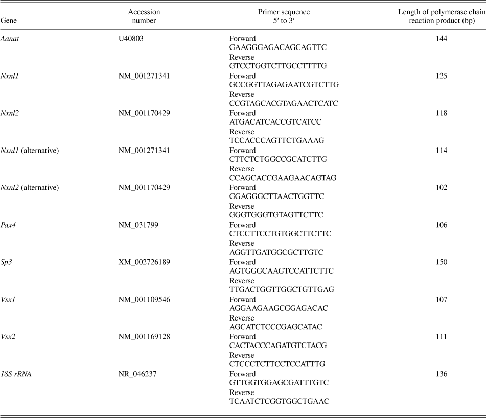
cDNA was synthesized using the Verso cDNA Kit (Abgene, Hamburg, Germany) as described (Kunst et al., Reference Kunst, Wolloscheck, Hölter, Wengert, Grether, Sticht, Weyer, Wolfrum and Spessert2013). Quantitative polymerase chain reaction (qPCR) was performed as described (Hölter et al., Reference Hölter, Kunst, Wolloscheck, Kelleher, Sticht, Wolfrum and Spessert2012). The amount of RNA was calculated from the measured threshold cycles (C t) using a standard curve. Values were then normalized with respect to the amount of 18S rRNA present. Primer sequences are listed in Table 1.
Primer sequences.

Statistical analysis
All PCR data are expressed as mean ± standard errors of the mean (SEM) of four independent experiments, including eight time points. Transcript levels were calculated relative to average expression of each data set throughout 24 h to plot temporal expression. Quantitative data were analyzed by one way analysis of variance to evaluate variations between the groups. Cosinor analysis was used to fit sine-wave curves to the circadian data to mathematically estimate the time of peaking gene expression (acrophase) and to assess the amplitude (Refinetti et al., Reference Refinetti, Lissen and Halberg2007; Cornelissen, Reference Cornelissen2014). The model can be expressed according to the equation: f(t) = A + B cos [2π (t + C)⁄T], with f(t) indicating relative expression levels of target genes, t specifying the time of sampling (h), A representing the mean value of the cosine curve (mesor; midline estimating statistic of rhythm), B indicating the amplitude of the curve (half of the sinusoid), and C indicating the acrophase [point of time, when the function f(t) is maximum]. T gives the time of the period, which is 24 h for this experimental setting. All statistical analyses should be regarded as being explorative, and p-values are given descriptively with no significance level being fixed.
Results
Nxnl1 and Nxnl2 transcript levels
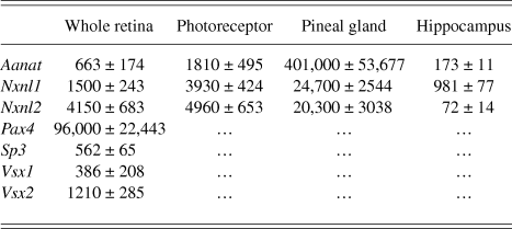
Using qPCR, Nxnl1 and Nxnl2 gene expression was seen in all tissues investigated, even though at very different levels (Table 2). Consistent with the formation of RdCVF and RdCVF2 in rods, both genes displayed a higher transcript amount in PRCs than in preparations of the whole retina—with a higher degree of enrichment for Nxnl1. Interestingly, the highest expression of both genes was seen in the pineal gland—an indirect light-sensitive part of the circadian system that harbors photoreceptor-related pinealocytes (for a review, see Rath et al., Reference Rath, Rohde, Klein and Møller2013). In this tissue, the expression of Nxnl1 and Nxnl2 was 16- and 5-fold, respectively, higher than in the retina. Among the genes, the mRNA levels were higher for Nxnl2 than they were for Nxnl1 in PRCs and the retina, whereas the situation was vice versa in the pineal gland.
Transcript levels of the genes in different tissues under LD 12:12. The transcript amount was calculated from respective peak expression during the 24-h period. It was determined by qPCR and represents the number of transcripts in relation to 18S rRNA × 105. Each value represents mean ± standard error of the mean (n = 4).
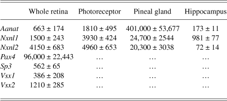
To test the validity of the experimental system used and to compare abundance and regulation of Nxnl genes with arylalkylamine N-acetyltransferase (Aanat)—a gene known to be under circadian regulation in PRCs and pinealocytes (for reviews see Iuvone et al., Reference Iuvone, Tosini, Pozdeyev, Haque, Klein and Chaurasia2005; Klein, Reference Klein2007)—in the same transcriptomes as those used for recording Nxnl genes, the amount of Aanat transcript was determined (Table 2). Consistent with previous reports (for reviews, see Klein, Reference Klein2007), among the tissues, Aanat mRNA level was found to be increased in the pineal gland and PRCs.
Daily profiling of Nxnl genes in the retina
To investigate daily regulation of Nxnl genes, the transcript levels of Nxnl1 and Nxnl2 were recorded throughout the 24-h period. The levels of both gene products were observed to undergo daily rhythms, irrespective of the primer pairs used (Fig. 1, left and middle columns). The daily profiles of both the Nxnl genes were similar, each with increasing values during the light phase and peak expression around the dark onset.
Daily profiling of Nxnl1, Nxnl2 and the reference gene Aanat in preparations of whole retina under LD 12:12 (left column), under LD with alternative primer pairs for Nxnl1, Nxnl2 (middle column) and under DD (right column) using qPCR. The solid bars indicate the dark period. The mRNA levels are plotted both as a function of ZT and Circadian time. The value of ZT0 was plotted twice at both ZT0 and ZT24. Each value represents mean ± SEM (n = 4). Data represent a percentage of the average value of transcript amount of each data set. The solid and dotted blue lines indicate the sinusoidal functions for genes under daily regulation (P ≤ 0.05) and those constitutively expressed (P > 0.05), respectively. LD: Nxnl1: P = 0.006, acrophase ZT10.6, amplitude 38.7%; Nxnl2: P = 0.01, acrophase ZT11.2, amplitude 30.7%; Nxnl1 (alternative primer): P = 0.014, acrophase ZT9.0, amplitude 34.8%; Nxnl2 (alternative primer): P = 0.016, acrophase ZT11.1, amplitude 48.6%; Aanat: P < 0.001, acrophase ZT19.3, amplitude 116.6%; DD: Nxnl1: P = 0.048, acrophase ZT11.5, amplitude 16.3%; Nxnl2: P = 0.43; Aanat: P = 0.04, acrophase ZT23.2, amplitude 56.8%.
In accordance with previous studies (for a review, see Iuvone et al., Reference Iuvone, Tosini, Pozdeyev, Haque, Klein and Chaurasia2005) and consistent with the validity of the experimental system used, the amount of Aanat transcript displayed a daily rhythm with peak expression at night in the retina (Fig. 1, left column).
Daily profiling of transcriptional regulators of the Nxnl1 gene
To investigate as to which extent daily regulation of the Nxnl1 gene may depend on 24-h changes of homeobox genes shown to regulate the Nxnl1 promotor (Reichman et al., Reference Reichman, Kalathur, Lambard, Aït-Ali, Yang, Lardenois, Ripp, Poch, Zack, Sahel and Léveillard2010), their mRNA levels were recorded as a function of daytime Zeitgeber time (ZT) (Fig. 2). In accordance to previous reports (Rath et al., Reference Rath, Bailey, Kim, Coon, Klein and Møller2009a; Kunst et al., Reference Kunst, Wolloscheck, Hölter, Wengert, Grether, Sticht, Weyer, Wolfrum and Spessert2013), the level of Pax4 mRNA was observed to undergo daily rhythms. On the contrary, the transcript levels of Sp3, Vsx1 and Vsx2 failed to show daily cyclicity. As a consequence, only Pax4 is a candidate for daily regulation of Nxnl1 transcription.
Daily profiling of homeobox genes Pax4, Sp3, Vsx1, and Vsx2 in preparations of whole retina under LD 12:12 using qPCR. For the relevant reference gene Aanat, see Fig. 1, left column. The mRNA levels are both plotted as a function of ZT. The value of ZT0 was plotted twice at both ZT0 and ZT24. The solid bars indicate the dark period. Each value represents mean ± SEM (n = 4). Data represent a percentage of the average value of transcript amount of each data set. The solid and dotted blue lines indicate the sinusoidal functions for genes under daily regulation (P ≤ 0.05) and those constitutively expressed (P > 0.05), respectively. Pax4: P = 0.014, acrophase ZT8.7, amplitude 41.8%; Sp3: P = 0.54; Vsx1: P = 0.83 Vsx2: P = 0.75.
Daily profiling of Nxnl genes under constant darkness
The daily regulation of the Nxnl genes may be promoted by light input and/or a circadian clock. To systematically investigate and compare the influence of both parameters between the genes, rats adapted to LD 12:12 were kept in constant darkness (DD) for one cycle and monitored during the subsequent cycle (Fig. 1, right column). Under these conditions, the cycling of Nxnl1 but not that of Nxnl2 continued. This suggests that daily regulation of Nxnl1 depends primarily on a circadian clock, whereas that of Nxnl2 is primarily promoted by environmental light. As expected for a gene under circadian regulation (for a review, see Iuvone et al., Reference Iuvone, Tosini, Pozdeyev, Haque, Klein and Chaurasia2005), the reference gene Aanat continued to cycle under DD (Fig. 1, right column).
Daily regulation of Nxnl genes in photoreceptor cells
To examine the contribution of PRCs to daily changes in the expression of Nxnl genes, the 24-h course of the transcript levels of Nxnl1 and Nxnl2 (Fig. 3, left column) were monitored in PRCs collected from retina using the LMPC technique. The mRNA levels of both genes exhibited daily rhythms in PRCs, with profiles resembling those obtained from preparations of the whole retina (Fig. 1, left column). Consistent with previous observations (Niki et al., Reference Niki, Hamada, Ohtomi, Sakamoto, Suzuki, Kako, Hosoya, Horikawa and Ishida1998; Schneider et al., Reference Schneider, Tippmann, Spiwoks-Becker, Holthues, Wolloscheck, Spatkowski, Engel, Frederiksen and Spessert2010), the amount of Aanat transcript was seen to cycle in PRCs with a peak expression at night (Fig. 3, left column).
Daily profiling of Nxnl1, Nxnl2, and the reference gene Aanat in microdissected photoreceptor cells (PRCs, left column), the pineal gland (PD, middle column) and the hippocampus (HC, right column) under LD 12:12 using qPCR. The solid bars indicate the dark period. The mRNA levels are plotted both as a function of ZT. The value of ZT0 was plotted twice at both ZT0 and ZT24. The value of ZT0 was plotted twice at both ZT0 and ZT24. Each value represents mean ± SEM (n = 4). Data represent a percentage of the average value of the transcript amount of each data set. The solid and dotted blue lines indicate the sinusoidal functions for genes under daily regulation (P ≤ 0.05) and those constitutively expressed (P > 0.05), respectively. Photoreceptor cells: Nxnl1: P = 0.008, acrophase ZT12.6, amplitude 32.0%; Nxnl2: P = 0.016, acrophase ZT11.4, amplitude 37.6%; Aanat: P < 0.001, acrophase ZT19.3, amplitude 106.2%; pineal gland: Nxnl1: P = 0.037, acrophase ZT9.2, amplitude 33.6%; Nxnl2: P = 0.20; Aanat: P < 0.001, acrophase ZT21.4, amplitude 149.1%; hippocampus: Nxnl1: P = 0.276; Nxnl2: P = 0.976; Aanat: P = 0.087.
Daily profiling of Nxnl genes in the pineal gland
To investigate the extent to which daily changes in the expression of Nxnl gene products occur outside the retina, gene profiling was also performed in the pineal gland (Fig. 3, middle column) and the hippocampus (Fig. 3, right column)—brain areas phylogenetically related and unrelated, respectively, to the retina. Consistent with the phylogenetic relationship between PRCs and pinealocytes (for a review, see Rath et al., Reference Rath, Rohde, Klein and Møller2013), the mRNA levels of Nxnl1 were found to undergo a daily rhythm in the pineal gland. In contrast to Nxnl1, the Nxnl2 transcript amount failed to exhibit a statistically relevant rhythm in the pineal gland. As expected (for a review, see Klein, Reference Klein2007), the amount of Aanat transcript displayed marked daily changes with peak expression at night (Fig. 3, middle column). The mRNA levels of none of the Nxnl gene products were found to display daily rhythms in the hippocampus (Fig. 3, right column).
Discussion
In the present study, the expression of Nxnl1 and its paralogue Nxnl2 (for a review see Sahel et al., Reference Sahel, Leveillard, Picaud, Dalkara, Marazova, Safran, Paques, Duebel, Roska and Mohand-Said2013) was seen not only in the retina and PRCs but also—even at a higher level—in the pineal gland. This finding is consistent with the phylogenetic relationship of PRCs and pinealocytes (for a review, see Rath et al., Reference Rath, Rohde, Klein and Møller2013) and suggests that Nxnl expression is a feature of not only direct (PRCs) but also of indirect (pinealocytes) photosensitive cells. Until now, Nxnl1 expression was believed to be restricted to the retina, whereas Nxnl2 expression also encompasses other areas of the brain, including the olfactory system (Chalmel et al., Reference Chalmel, Léveillard, Jaillard, Lardenois, Berdugo, Morel, Koehl, Lambrou, Holmgren, Sahel and Poch2007). Therefore, the present study for the first time demonstrates Nxnl1 expression in a tissue outside the retina.
Nxnl1 and Nxnl2 display 24-h rhythms in the retina, which may produce corresponding changes in the amount of RdCVF and RdCV2. However, a limitation of this study is that the postulated daily changes in the amount of RdCVF and RdCV2 have not been demonstrated. This is due to the malfunction of commercially available antibodies and the unavailability of antibodies from other groups. Daily cyclicity of Nxnl1 and Nxnl2 in the retina may evoke from PRCs. This becomes evident from the observation that the mRNA levels of both genes cycled in preparations of the whole retina and microdissected PRCs with similar profiles.
Homeobox genes Pax4, Sp3, Vsx1, and Vsx2 are reported to regulate Nxnl1 promotor activity (Reichman et al., Reference Reichman, Kalathur, Lambard, Aït-Ali, Yang, Lardenois, Ripp, Poch, Zack, Sahel and Léveillard2010). As a consequence, a putative rhythmicity of these genes may participate in daily regulation of Nxnl1 transcription. In accordance with previous findings (Rath et al., Reference Rath, Bailey, Kim, Coon, Klein and Møller2009a,Reference Rath, Bailey, Kim, Ho, Gaildrat, Coon, Møller and Kleinb; Kunst et al., Reference Kunst, Wolloscheck, Hölter, Wengert, Grether, Sticht, Weyer, Wolfrum and Spessert2013), Pax4 was seen to display a daily rhythm with a peak expression at ZT9. On the contrary, homeobox genes Sp3, Vsx1, and Vsx2 fail to cycle. These observations suggest that amongst the transcriptional regulators of Nxnl1, only Pax4 is a candidate gene for mediating the daily control of Nxnl genes. Consistent with a role of Pax4 in that matter, the cyclicity of Pax4 is approximately 2 h phase advanced to that of the Nxnl1 gene (Rath et al., Reference Rath, Bailey, Kim, Coon, Klein and Møller2009a; Kunst et al., Reference Kunst, Wolloscheck, Hölter, Wengert, Grether, Sticht, Weyer, Wolfrum and Spessert2013, Fig. 1vs. Fig. 2).
Daily cyclicity of Nxnl1 but not that of Nxnl2 persists in DD. As a result, transcriptional regulation of Nxnl genes as a whole is entrained by both the retinal clock system and light input (for reviews, see Tosini et al., Reference Tosini, Pozdeyev, Sakamoto and Iuvone2008; McMahon et al., Reference McMahon, Iuvone and Tosini2014). Nxnl genes increase the resistance of PRCs to photooxidative damage (Fridlich et al., Reference Fridlich, Delalande, Jaillard, Lu, Poidevin, Cronin, Perrocheau, Millet-Puel, Niepon, Poch, Holmgren, Van Dorsselaer, Sahel and Léveillard2009; Léveillard & Sahel, Reference Léveillard and Sahel2010; for a review see; Sahel et al., Reference Sahel, Leveillard, Picaud, Dalkara, Marazova, Safran, Paques, Duebel, Roska and Mohand-Said2013). Therefore, dual entrainment of Nxnl genes may facilitate PRCs to strengthen the protection from photooxidative damage in anticipation of and as a direct response to an increase in ambient light intensity.
The risk of PRCs undergoing photooxidative damage increases as a function of the duration of exposure (Cingolani et al., Reference Cingolani, Rogers, Lu, Kachi, Shen and Campochiaro2006). As a consequence, the risk of PRCs suffering photooxidative damage should vary throughout a 24-h light/dark cycle with an increase between light onset in the morning and dark onset in the evening. Strikingly, the presently observed daily profile in photoreceptor Nxnl transcription parallels that of damage risk. Taken into account that products of Nxnl genes increase the resistance to photooxidative damage (Fridlich et al., Reference Fridlich, Delalande, Jaillard, Lu, Poidevin, Cronin, Perrocheau, Millet-Puel, Niepon, Poch, Holmgren, Van Dorsselaer, Sahel and Léveillard2009; Léveillard & Sahel, Reference Léveillard and Sahel2010; for a review see; Sahel et al., Reference Sahel, Leveillard, Picaud, Dalkara, Marazova, Safran, Paques, Duebel, Roska and Mohand-Said2013), this observation suggests a hypothetical concept in which the up-regulation of Nxnl expression during daytime is a defense of the PRC against the steadily increasing risk of photooxidative damage. Accordingly, daily regulation of Nxnl genes may be a homeostatic factor in the maintenance of PRCs in retinal healthiness and—because oxidative damage appears to contribute to the progressive death of cones in RP (Cingolani et al., Reference Cingolani, Rogers, Lu, Kachi, Shen and Campochiaro2006; Komeima et al., Reference Komeima, Rogers, Lu and Campochiaro2006)—retinal disease.
RP is the most common form of inherited retinal degeneration, affecting more than one million human beings worldwide. To date, an effective treatment that can prevent or reverse the loss of vision in RP is not available. Although gene therapy is a promising treatment for RP, the genetic heterogeneity of this disease is a great challenge for its establishment. Thus, RP is associated with mutations in more than 50 gene loci and, in many cases of RP, the loci of mutation remain unidentified (for a review, see Daiger et al., Reference Daiger, Sullivan and Bowne2013). To treat RP in a mutation-independent manner is to take the genetically caused loss of rods, but to prevent the secondary death of cones and thus the irreversible loss of central visual acuity (for a review, see Sahel et al., Reference Sahel, Leveillard, Picaud, Dalkara, Marazova, Safran, Paques, Duebel, Roska and Mohand-Said2013). This could be achieved by compensating the decrease of RdCVF and/or RdCVF2 expression in patients with RP. One way to accomplish this would be through the delivery of exogenous RdCVF, for example, by injection of protein into the eye (Yang et al., Reference Yang, Mohand-Said, Danan, Simonutti, Fontaine, Clerin, Picaud, Léveillard and Sahel2009) or secretion from RdCVF-forming cells (for a review, see Sahel et al., Reference Sahel, Leveillard, Picaud, Dalkara, Marazova, Safran, Paques, Duebel, Roska and Mohand-Said2013). Another approach would be to enhance retinal levels of RdCVF by modulating expression of the endogenous gene. The present observation that transcription of Nxnl genes is clock and light dependent may provide a starting point for targeting endogenous RdCVF expression. Since the neurohormones melatonin and dopamine play central roles in mediating clock- and light-dependent regulation of the retina (for a review, see McMahon et al., Reference McMahon, Iuvone and Tosini2014), prospective investigations should reveal whether they also stimulate Nxnl transcription, and if yes, their application can retard cone death in rodent models of RP.
Nxnl1 was also seen to be under daily regulation in the pineal gland (for a review, see Maronde & Stehle, Reference Maronde and Stehle2007), a neuroendocrine transducer of the circadian system (for a review, see Hastings et al., Reference Hastings, Maywood and Reddy2008). The pineal gland in general and pineal gene expression in particular are under the control of the master clock in the suprachiasmatic nucleus (for reviews, see Møller & Baeres, Reference Møller and Baeres2002; Hastings et al., Reference Hastings, Maywood and Reddy2008; Bailey et al., Reference Bailey, Coon, Carter, Humphries, Kim, Shi, Gaildrat, Morin, Ganguly, Hogenesch, Weller, Rath, Møller, Baler, Sugden, Rangel, Munson and Klein2009). Therefore, Nxnl1 transcription appears to be clock dependent not only in the retina but also in the pineal gland. Mammalian PRCs and pinealocytes phylogenetically and ontogenetically descend from a common ancestor cell type (for a review, see Rath et al., Reference Rath, Rohde, Klein and Møller2013). Thus, the coincidence of Nxnl1 cyclicity in mammalian PRCs and pinealocytes suggests that daily regulation of Nxnl1 is evolutionally conserved.
In conclusion, the data of the present investigation suggest a hypothetical concept in which the circadian and daily regulation of Nxnl genes contributes to PRC survival in retinal healthiness and disease. They further may provide a basis for a putative point of therapeutic intervention in which endogenous stimulation of Nxnl genes is beneficial in retarding cone death in RP.
Acknowledgments
We thank Mr. Klaus Wolloscheck for statistical advice, Ms. Ute Frederiksen and Ms. Kristina Schäfer for their excellent technical assistance, and Ms. Susanne Rometsch and Ms. Bettina Wiechers-Schmied for secretarial help. The data contained in this study are included in the thesis of Ms. Stefanie Kunst as a partial fulfillment of her doctorate degree at the Johannes Gutenberg University, Mainz.