Schizophrenia has been infamous as the graveyard of neuropathology research (Plum, 1972). Over the years it must have received a lot of competition for that pride of place, because no psychiatric illness readily surrenders its secrets. However, the severity and heterogeneity of symptoms in schizophrenia, coupled with an absence of any obvious or profound change in the physical structure of the brain, makes schizophrenia particularly deserving of this reputation. Schizophrenia is a serious mental illness that typically begins in late adolescence or young adulthood. The illness cripples the aspirations of three million Americans and 1% of the population worldwide, and it is characterized by terrifying symptoms such as hallucinations of criticizing voices, believing others are scheming, or being unable to express a coherent thought. Parents' hopes for their ill sons and daughters are revised to reflect the possibility of a lifetime of dependency, unemployment, social withdrawal, and a 100-fold increased risk of suicide. Our best estimates indicate that schizophrenia costs society approximately $60 billion annually in the United States alone. Perhaps because of this complexity, as well as its similarity to aspects of neurological disorders like dementia, schizophrenia has long been considered from a reductionist, biological perspective. It is only more recently, however, that the challenges of the illness have begun to yield to the translational methods of modern neurosciences' armamentarium.
No review can cover the full breadth of theories about the nature of schizophrenia, much less the implications of this great multiplicity across various levels of analysis. For the sake of a tractable illustration of translational approaches to the symptoms and development of schizophrenia, we will restrict ourselves to a chain of evidence linking genes to the manifestation of schizophrenia that we find particularly compelling given the available evidence. This model, referred to as the N-methyl-D-aspartate (NMDA) synaptic deficit model, proposes that schizophrenia is a disconnection syndrome affecting NMDA receptor function at the glutamate synapse. Our discussion will bring evidence to bear from multiple levels of analysis (Cicchetti & Rogosch, 2002).
First, of the genes that have jumped to the lead in early association studies, a majority appear to play a role in glutamate transmission such that they have primary or perhaps secondary effects on NMDA synapse function. Second, blocking the NMDA receptor in healthy subjects mimics a number of symptoms of schizophrenia and greatly exacerbates symptoms in schizophrenia patients. The NMDA receptor drives one form of synaptic plasticity in the cerebral cortex, suggesting that schizophrenia may be a disease of synaptic plasticity. Third, neuroanatomical evidence from postmortem brain tissue suggests that schizophrenia physically disconnects neurons by destroying glutamatergic synapses, particularly in the prefrontal cortex. Fourth, neuroimaging has shown schizophrenia patients have reductions in prefrontal cortical activity that cannot be attributed to performance-related confounds, and appear to be specific to the disorder. These functional abnormalities also appear in the healthy first-degree relatives of schizophrenia patients, suggesting their relationship to the genetic liability to the disorder. Fifth, such prefrontal cortical abnormalities have been related to a process-specific deficit in context processing. This deficit appears to be most closely related to the disorganization symptoms of schizophrenia, which some evidence suggests is the most heritable component of the disorder. Sixth, there is an important developmental component to the NMDA synaptic deficit model. The onset of the illness in late adolescence is concurrent with a late developmental event involving the dramatic pruning of synapses in the prefrontal cortex. Figure 1 provides a heuristic model of the relationship among the different levels of analysis in this translational account.

The translational model of the hypothesized causal chain linking genes to neural systems to symptoms.
Given the vast number of data points in schizophrenia research, we assure our readers, including our critics, that the NMDA synaptic deficit model is not the only way in which these dots could be connected. Indeed, this plethora of evidence can be seen as consistent with multiple pathway models of disease development (Cicchetti & Cannon, 1999). Our purpose is, instead, to demonstrate that schizophrenia research has reached a point where a coherent, integrative explanation of schizophrenia can be hazarded, with reasonable evidence for each link in the chain. That is to say, coherent priorities for schizophrenia research now exist, and progress has brought us to a new and hopefully dynamic phase of research in which a translational perspective is front and center.
NMDA Synaptic Deficit Model of Schizophrenia
The lines of evidence reviewed below place the NMDA synapse of the glutamate neuron as the central, functional target of the disease process leading to schizophrenia. Glutamate is the neurotransmitter of the pyramidal neurons that form the primary excitatory and connective circuits of cortex. Conceptually, any insult to pyramidal neurons that adversely affects the functional integrity of the NMDA synapse or impedes the intracellular events mediated by its activation could increase the risk of the disease. Thus, factors contributing to schizophrenia need not be restricted to a mutation of the NMDA receptor itself, or a decrease in NMDA receptor number, but could include any factor that adversely impacts the complex chain of events triggered by NMDA receptor activation. There may be myriad such factors, with the impact of each summing to determine whether the synaptic system is sufficiently compromised to trigger the disease state. Thus, the NMDA synaptic deficit hypothesis posits that each risk factor imposes a “hit” at the NMDA glutamate synapse whose cumulative effects sum to drive NDMA synaptic function below a critical threshold. As a falling tide reveals a sandbar, any factor that lowers the efficacy of the NMDA synaptic function could increase the probability that cortical and subcortical networks will enter the pathophysiological state associated with schizophrenia.
This pathological state is proposed to be sustained by a complex interplay between the environment, the compromised operation of NMDA synapses, and the patterns of electrical activity that propagate through neural systems as the brain interacts with the environment. In this model, the environment, the synapse, neural systems, and electrical activity in the brain are mutually interdependent because of the phenomenon of synaptic plasticity. Activation of the NMDA channel sets in motion a sequence of events that leads to a change in the strength of the activated synapse. That is the reason why patterns of electrical activity generated by neural systems, the synaptic connections between neurons, and the environment exist in a state of dynamic tension, by which changes in one factor induces changes in the other two. This convergence of multiple developmental paths toward a common behavioral outcome has been expressed as the principle of equifinality in the developmental psychopathology literature (Cicchetti & Rogosch, 2002).
Although the NMDA synapse deficit model marks a significant departure from dopamine-derived accounts of the illness informed by pharmacological studies, the theory itself has been advanced in various forms by a number of investigators during the past decade or so. Friston and colleagues, for example, put forward a disconnection model of schizophrenia that was based on a defect in NMDA-mediated synaptic plasticity, supported by physiological evidence of functional disconnection between cortical areas (Friston & Frith, 1995; Stephan, Baldeweg, & Friston, 2006). NMDA synapse deficit models in many ways owe their existence to the identification of the ion channel associated with the NMDA receptor as the binding site of the psychotomimetic drug phencyclidine (Javitt, Jotkowitz, Sircar, & Zukin, 1987; Javitt & Zukin, 1991), one of the first and most important clues leading to the idea that neurotransmission at the NMDA synapse was altered in the disease. Hoffman and McGlashan (1993, 1997, 2001) developed neural network models to examine the effects of reducing excitatory synaptic connectivity, discovering that patterns of aberrant network activity emerged as a result of disconnection that bore resemblance in several respects to the symptoms of the disease. More recently, Phillips and Silverstein (2003) have posited NMDA hypofunction as underlying a cognitive coordination deficit. As reviewed below, many other researchers and studies have contributed essential information to the growing body of evidence supporting the NMDA synaptic deficit hypothesis. Thus, there is a strong consensus that a core event in schizophrenia is impairment of NMDA synapse function. Even so, the bridge we can establish from genetic mutation to the symptoms and ultimately the treatment of schizophrenia is marked by large gaps in knowledge in several crucial areas. This is inevitable, given the scope of the translational bridge that must be established and the current state of the relevant fields of research. However, the information reviewed here provides the skeleton of how the essential solution may someday appear.
Genes and Schizophrenia
Evidence for the key role of genes in schizophrenia dates back to pedigree studies early in the 20th century by German psychiatrist Ernst Rudin (Rudin, 1916). Contemporary interpretations of those data resulted in disastrous policies toward the mentally ill in prewar Germany, contributing to resistance to this view from psychoanalytic and behavioral camps in the following decades. This resistance was not seriously assailed until the work of Gottesman and Shields (1966) and Kety, Rosenthal, Wender, and Schulsinger (1971) clarified that liability to the disorder was highly heritable using twin and adoption study methods, respectively (see also Meehl, 1962). There is now wide convergence on the risk rates in the family members of schizophrenia patients, such that patients' first-degree relatives (whether raised together or adopted away) have an approximately 10-fold increased risk compared to the general population, whereas patients' monozygotic twins (whether raised together or apart) have an approximately 50-fold increased risk of developing the disorder. Considering the level of risk in the general population, this corresponds to about 10 and 50% risks, respectively. However, a 50% risk is far from genetically determined. That is to say, although this level of risk corresponds to a heritability of liability of .80, someone who inherits this liability is not “destined” to develop the disorder (these considerations have been discussed extensively in Gottesman, 1991).
When liability is heritable, but disease manifestation is less than 100%, the liability genes are said to be incompletely penetrant. The NMDA synaptic deficit model is consistent with the genetic epidemiology of the disorder if schizophrenia is manifest only when networks of pyramidal neurons settle into a suboptimal equilibrium. More specifically, because glutamate synapses are so exquisitely sensitive to the interaction between the brain and the environment (e.g., to behavior), manifestation of the network event associated with the disease state may be equally sensitive to the behavioral history of the brain. Given the close link between glutamatergic neurotransmission, NMDA synapse function, and synaptic plasticity, the functional integrity of excitatory synaptic transmission in schizophrenia is likely to be a function of at least two distinct parameters: first, the disease process itself attacking excitatory synaptic integrity, but also the functional history of the network. This latter consideration indicates that the manifestation of the disease state may be “activity dependent,” which is dependent on the functional state and history of the system. This interdependence between behavior, the environment, and the underlying genetic insult may ultimately explain why schizophrenia occurs in some individuals with a genetic predisposition and not others. One way to grapple with this dynamic interaction is to identify the first component of a gene–environment interaction; that is the genes.
Moving beyond genetic epidemiology and finding the actual genes associated with liability to the disorder has been the holy grail of psychiatry research for the past 20 years. Progress has followed in the wake of advances in genotyping technology, and there is now a cottage industry that reviews the strength of the latest genetic expression, linkage, and association studies (e.g., Owen, Williams, & O'Donovan, 2004, and others). For current purposes, it is sufficient to note that no gene alone or in combination has been able to completely distinguish schizophrenia patients from controls. Indeed, one would hardly expect this; the concordance rates between monozygotic twins, who share a nearly identical genetic liability to schizophrenia, suggest many would be classified as “controls” in such studies. However, even the candidate gene association studies, which show statistical relationships between DNA polymorphisms and the illness, still require moderately large sample sizes to detect the subtle differences in prevalence rates between schizophrenia patients and controls. Thus, there are unlikely to be any clear “schizophrenia genes,” which in and of themselves determine the presence or absence of schizophrenia. More humbling still, the genes (haplotypes) or single nucleotide polymorphisms (SNPs) that have been associated with schizophrenia are often found to be quite common in the general population, and are only slightly more prevalent in patients with schizophrenia. Nevertheless, a half dozen or so of these genes have amassed a number of independent replications in support of their association with schizophrenia. The largest subset of these genes share a thematic relationship: a role in glutamate neurotransmission.1
In addition to the glutamate modulating genes highlighted in the text, catechol-O-methyltransferase (COMT; Egan et al., 2001) and disrupted in schizophrenia-1 (DISC1; Millar et al., 2000) have shown association across a number of independent studies. COMT has a role in the methylation of dopamine and other catecholamines, whereas DISC1 contributes to the cytoskeletal elements that facilitate neuronal migration and axon growth during the development of cerebral cortex. It is important to consider that several systems may be compromised in psychosis and review like this one are unlikely to tie together all the loose ends in this field.
- RGS4 (regulator of G-protein signaling 4), located at 1q23, regulates G signaling proteins in glutamate neurons and thereby dampens the effects of neurotransmitter–receptor interaction at G-protein coupled receptors. It was originally found to be significantly underexpressed in dorsolateral prefrontal cortex in postmortem microarray studies (Mirnics, Middleton, Stanwood, Lewis, & Levitt, 2001). Several SNPs in this gene have been subsequently found to be associated with schizophrenia in at least four other samples (e.g., Chowdari et al., 2002).
- DTNBP1 (dystrobrevin binding protein 1, or dysbindin), located at 6p22, has recently been shown to modulate glutamate function through both the upregulation of presynaptic proteins and neurotrophic affects mediated by the Akt signaling pathway (Numakawa et al., 2004). Microarray studies of dorsolateral prefrontal cortex gene expression have also shown schizophrenia patients' brains have decreased dysbindin mRNA levels (Weickert et al., 2004). Association with schizophrenia has now been replicated in a number of samples (e.g., Straub et al., 2002).
- NRG1 (neuregulin-1), located at 8p21, is present in glutamatergic synaptic vesicles and also regulates NMDA-receptor expression. NRG1 hypomorph mice have fewer functional NMDA receptors than wild type (Stefansson et al., 2002), although this gene may also influence cytoskeletal elements such as glia (Moises, Zoega, & Gottesman, 2002). SNP and haplotype studies have consistently, albeit not universally, demonstrated association with schizophrenia in large samples (Moises et al., 2002; Stefansson et al., 2002).
- DAOA (formerly G72, d-amino acid oxidase activator), located at 13q24, activates NMDA receptors through a series of reactions and has been found to be abnormally expressed in schizophrenia patients' brains (Korostishevsky, Kaganovich, Cholostoy, Ashkenazi, Ratner, Dahary et al., 2004). The association between DAOA and schizophrenia has now been found in four cohorts of which we are aware (e.g., Chumakov et al., 2002).
In addition to the glutamate genes found within the hotspots of linkage analyses, a number of other candidate genes identified in various manners have been associated with schizophrenia. For example, GRM3 (glutamate receptor metabotropic 3), located at 7q21, is involved in the presynaptic inhibition of glutamate release through mGlu2 and mGlu3 receptors. GRM3 has recently been associated with schizophrenia in two samples (Egan et al., 2004; Fujii et al., 2003). Furthermore, variation in GRM3 SNPs has also been associated in differences in performance on cognitive markers of risk for schizophrenia (Egan et al., 2004). Other genes with a putative involvement in glutamatergic NMDA synapse function and preliminary support for a role in schizophrenia include GRIA2, located at 4q32–33 (Vawter et al., 2002); EPSIN 4, located at 5q33 (formerly SCZD1; Devlin et al., 2002); PPP3CC, located close to NRG1 at 8p21 (Gerber et al., 2003); GRIN, located at 9q34 (Rice, Niu, Berman, Heston, & Sobell, 2001); and DAAO, located at 12q24 (e.g., Chumakov et al., 2002). Two others, PRODH (Liu et al., 2002) and YWHAH (formerly 14-3-3 eta; e.g., Toyooka et al., 1999), are located near the 22q11.2 locus of COMT and may be related to successful linkage to this region of the genome.
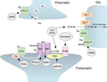
Although the work to establish linkage and association is necessarily clinical in nature, Figure 2 illustrates what basic studies have concluded about the putative mechanisms associated with these genes. They appear to play a number of different roles at the glutamate synapse, with the presynaptically active proteins like those associated with GRM3 being more distal and postsynaptic proteins associated with PPP3CC, GRIN 1, and dysbindin more proximal to NMDA synapse function. As described further below, both pre- and postsynaptic elements are of key importance at this site, contributing to the particular vulnerability of the mechanism. The interested reader is referred to Moghaddam (2003) for further discussion of the synaptic biochemistry.

Pre- and postsynaptic glutamate modulating genes. NR1 and NR2 are the primary subunits of the N-methyl-D-aspartate (NMDA) receptor. From “Bringing Order to the Glutamate Chaos in Schizophrenia,” by B. Moghaddam, 2003, Neuron, 40. Copyright 2003 by Elsevier. Reprinted with permission. [A color version of this figure can be viewed online at www.journals.cambridge.org]
Thus, were a scientist to hazard a theme for investigation from these early candidate schizophrenia genes, one might be well advised to consider NMDA dysregulation among the causes of the illness. Fortunately, this conclusion need not stand in isolation. We now step to the next rung on the translation ladder, in which pharmacological evidence for the NMDA synaptic deficit hypothesis is brought to bear.
NMDA Receptor Blockade and Schizophrenia Symptoms
The clinical link between glutamate receptor blockade and the behavioral manifestations of schizophrenia began accidentally with introduction of the drug phencyclidine (PCP) for use as a dissociative anesthetic in the 1950s, under the trade name Sernyl (for further review, see Domino & Luby, 1981). PCP and its derivative ketamine produce a catatonia-like state in which patients are expressionless, stare sightlessly, and are unresponsive to pain. Patients in this dissociative state are able to undergo minor surgical procedures, and exhibit the motor rigidity and “waxy flexibility” that is often characteristic of catatonia. During clinical tests, it was discovered that a substantial proportion of surgical patients recovering from PCP anesthesia suffered a psychotic reaction to the drug: patients became agitated and paranoid and experienced severe hallucinations. The symptoms were persistent. Episodes lasted for 12 to 72 hr typically, but in some cases for a week or more. These observations identified PCP as a viable drug model apparently reproducing some aspects of neuronal system malfunction in schizophrenia. Subsequent studies added support. In normal subjects, PCP induces negative symptoms such as apathy and flatness of affect, with concomitant thought disorganization and delusional ideation. Given to schizophrenia patients, PCP abruptly triggers a protracted intensification of psychosis. Likewise, PCP can induce a psychotic state in persons without a prior history of schizophrenia. One can only imagine the chaos this created as PCP became a drug of abuse: “In the fall of 1973, the admission rate for what appeared to be unusually long, severe, and treatment resistant initial schizophrenic psychosis suddenly tripled at the Area D Community Mental Health Center in Washington, D.C.” (Luisada, 1978). This apparent epidemic of new cases of schizophrenia was concomitant with a sudden increase in PCP availability and abuse in the area; the onset of psychosis in many patients admitted for schizophrenia could be traced to recent PCP abuse. Other reports confirm this drug-induced psychosis produces delusions of control, persecution, and religiosity, accompanied by the emergence of global paranoia, auditory hallucinations, and formal thought disorder (Luisada, 1978). Ketamine, a less potent analog of PCP, similarly exacerbates the symptoms of schizophrenia in patients (Krystal et al., 1994), and induces several aspects of the disease in normal subjects, including working memory dysfunction and disorganization symptoms (Krystal et al., 1994).
Basic studies utilizing animal models of disease and behavioral impairments have been useful in studying several links in the translational chain of evidence, including the pharmacology of this kind of glutamate antagonism. It is useful to consider the assumptions underlying cross-species generalization. In short, we believe there are strong reasons to think that essential aspects of the pathophysiology of schizophrenia will be replicable in animals, particularly nonhuman primates, which share many of the definitive attributes of human cortical organization and system function. It is conceivable that animal models of hallucination or thought disorder may be developed (and their neural underpinnings discovered), albeit in a much simplified form in comparison to their human counterparts. For example, neural representations of anticipated stimuli that do not appear, or disruptions in sequence processing are events that bear resemblance to these symptoms of schizophrenia but are well within reach of animal models. The feasibility of the translation from animal models to the human condition is supported by the overlap in cognitive functioning across species, especially nonhuman primates. Obviously, this area of overlap does not include all of the symptoms of schizophrenia (alterations in language perception or delusional ideation for example). However, the cognitive overlap between humans and nonhuman primates does include simpler cognitive abilities disrupted in human patients with schizophrenia (such as working memory and executive function), and these simpler functions are likely to establish the basis for a bridge between nonhuman primates and humans.
Such techniques have been essential for understanding the effects of PCP and ketamine on NMDA receptor functioning. Injecting monkeys with small doses of PCP for 2 weeks can induce neurocognitive deficits that outlast the injections by 4 months (Jentsch et al., 1997). This sustained change in behavior was consistent with impaired prefrontal cortical function, associated with a prolonged change in dopamine turnover in the prefrontal cortex, and it was reversed by the neuroleptic clozapine. Recently, it has been reported that acute ketamine administration in monkeys can induce a set shifting deficit analogous to that exhibited by schizophrenia patients in the Wisconsin Card Sorting task (Stoet & Snyder, 2005). Moghaddam and colleagues have studied the effects of NMDA receptor blockade in rodents and shown a physiological and functional impact on the operation of prefrontal cortical networks that model cognitive deficits in the human disease (e.g., Stefani & Moghaddam, 2005). Thus, these clinical and basic pharmacological considerations hang together with the evidence of genetic polymorphisms associated with schizophrenia affecting NMDA function. In the next section we will describe the role of NMDA in neurotransmission and the implications of an NMDA dysfunction.
NMDA-Dependent Synaptic Plasticity and Schizophrenia
The association between schizophrenia and altered NMDA receptor function implies that schizophrenia is a disease of reduced synaptic plasticity. Synaptic plasticity has been most thoroughly studied in basic studies of the hippocampus, but the biochemical principles of this synapse generalize to pyramidal neuronal networks throughout cortex. In the hippocampal slice preparation, it was discovered that high-frequency stimulation of afferent projections to the hippocampus lead to a long-lasting potentiation of the activated synapses (Bliss & Lomo, 1973). Long-term potentiation (LTP) of this type required at least two preconditions: (a) binding of glutamate to NMDA receptors (Herron, Lester, Coan, & Collingridge, 1986), and (b) depolarization of the postsynaptic neuron (Malinow & Miller, 1986). As illustrated in Figure 2, this latter condition is due to blockade of the ion channel associated with the NMDA receptor by Mg2+ ions at resting membrane potential (Mayer, Westbrook, & Guthrie, 1984). The NMDA channel is ligand gated but voltage dependent; both the neurotransmitter and depolarization of the postsynaptic neuron are required. This means that the NMDA synapse is operational only if the presynaptic and postsynaptic elements are simultaneously active, a gating condition that Hebb (1949) originally postulated could underlie learning in the nervous system. The ion channel associated with the NMDA receptor is permeant to Ca2+. The resulting influx of Ca2+ is sufficient to induce LTP by a process that can include the insertion of additional α-amino-3-hydroxy-5-methyl-4-isoxazolepropionic acid subtype glutamate receptors into the postsynaptic membrane, thereby increasing sensitivity to glutamate (Shi et al., 1999).
This basic scientific background, along with the genetic and pharmacological data reviewed above, provide the foundation for our clinical hypothesis: the aberrant patterns of electrical activity in the cortical systems that produce the symptoms of schizophrenia are the ultimate consequence of a failure to initiate and sustain behavior-dependent changes in synaptic strength through the NDMA receptor mechanism. This behavioral-dependent plasticity is the key process through which neurons are trained to work together to form complicated representations; a breakdown in synaptic plasticity could result in widespread miscoordination (Phillips & Silverstein, 2003) or functional disconnection (Friston & Frith, 1995). In the next section, we will evaluate the impact of these genetic, pharmacological, and biochemical findings on the next level of analysis: local cortical networks.
Anatomical Disconnection in Schizophrenia
One of the most useful ways of examining anatomical disconnection in the local cortical networks of schizophrenia patients has been the detailed examination of postmortem brain tissue. Broadly speaking, two measures of prefrontal synaptic connectivity, neuropil volume and dendritic spine density, are reduced. These two anatomical parameters reflect the extent to which the neuronal processes that provide the circuitry of cell to cell communication (axons, dendrites, and spines) are elaborated. Neuropil volume (the cortical compartment containing these processes, exclusive of cell bodies) and dendritic spine density (specializations associated with glutamate synapses) therefore provide at least indirect measures of interneuronal connectivity.
A reduction of neuropil in prefrontal cortex is supported by at least three observations. First, neuronal density in prefrontal cortex of schizophrenia patients is increased relative to controls (see Selemon & Goldman-Rakic, 1999). Second, prefrontal gray matter volume is reduced (e.g., Selemon, Kleinman, Herman, & Goldman-Rakic, 2002). Third, total neuron number is unchanged (Thune, Uylings, & Pakkenberg, 2001). Because neuronal density increases, neuronal number does not change but overall cortical volume is reduced; the above observations together suggest that the space between neurons, the neuropil, is reduced in schizophrenia. This loss is not uniform. Progressively greater increases in neuron density are found in primary visual cortex (10% increase), prefrontal Brodmann area 9 (BA 9; 17% increase), and prefrontal BA 46 (21% increase), but no increase in neuronal density is present in prefrontal BA 44 (Selemon, Rajkowska, & Goldman-Rakic, 1995). This suggests that the disease process that diminishes synaptic architecture in schizophrenia is not uniform, with subdivisions of prefrontal cortex being particularly hard hit.
In addition to a reduction in neuropil volume, there is evidence for reduced dendritic spine density in some cortical regions. For example, spine density is normal in primary visual cortex in schizophrenia, but reduced in lower layer III of prefrontal cortex (e.g., Glantz & Lewis, 2000), where cell size is also significantly reduced (e.g., Lewis, Glantz, Pierri, & Sweet, 2003). These findings are consistent with a loss of synapses and intercellular connectivity in schizophrenia that preferentially impacts prefrontal cortex.
There are some important caveats to this sweep of evidence. For example, not all neuropathological changes in schizophrenia are limited to glutamatergic pyramidal neurons in prefrontal cortex. A reduction in the density of GABAergic interneurons in the prefrontal and anterior cingulate cortex has also been reported (Benes & Berretta, 2001). Further support for a change in inhibitory circuits in schizophrenia has been provided by the finding that axon cartridges of GABAergic chandelier cells are significantly reduced in prefrontal cortex in the disease (Pierri, Chaudry, Woo, & Lewis, 1999). Other models that do not place the initial pathological insult in prefrontal cortex have also been advanced. For example, a well-developed rodent model involving an early postnatal insult to the ventral hippocampus has been successful in triggering the delayed emergence of cortical and behavioral abnormalities characteristic of schizophrenia in adolescence (O'Donnell, Lewis, Weinberger, & Lipska, 2002). This model has the important advantage of identifying a potentially causal chain of events starting in early neural development that can trigger changes in behavior and neural function that do not appear until adulthood. That, in turn, provides a potential account for how a neurodevelopmental insult could produce the delayed onset of schizophrenia. It has long been thought that changes in cortical structure in schizophrenia are most likely of developmental origin, as developmental insults are associated with increased incidence of the disease and the cytopathological signs of widespread neurodegeneration are essentially absent. It will be important to continue to seek bridges between glutamatergic and neurodevelopmental models of schizophrenia as these two parallel theoretical avenues advance.
In sum, the evidence of neuropathological changes in prefrontal cortex is consistent (if not fully constrained by) the kinds of neuronal abnormalities that reflect the genetic polymorphisms and NMDA synapse deficits discussed above. With these considerations in mind, we arrive at the next level of analysis to consider the impact of these cellular changes on the functional neuroanatomy of prefrontal cortex.
Prefrontal Cortical Dysfunction and Schizophrenia
A translation account of the development of schizophrenia must ultimately link evidence of neuronal pathophysiology with the manifestation of the disorder: that is, the symptoms and functional disabilities that are the hallmark of schizophrenia. One of the most powerful tools for building this link is cognitive neuroscience, and in particular, the marriage of clinical questions, experimental cognitive paradigms, and modern neuroimaging methods, including functional magnetic resonance imaging (fMRI). Thus, one may well ask whether findings from clinical imaging studies are consistent with the NMDA synaptic deficit hypothesis.
It had long been observed that patients with schizophrenia had difficulty on the same tasks that were difficult for patients with prefrontal cortex damage (see Davison, 1974, for a discussion of the difficulty in distinguishing brain damage from schizophrenia). The ground-breaking study in the imaging field is attributed to Ingvar and Franzen (1974), who found that the ratio of regional cerebral blood flow in the prefrontal cortex compared to the rest of the brain was lower in schizophrenia patients relative to controls performing various tasks with putative demands on executive functions. This relative “hypofrontality” became a common finding in the schizophrenia neuroimaging literature throughout the 1980s and 1990s, being replicated using a number of different methodologies including xenon inhalation (Weinberger, Berman, & Zec, 1986), positron emission tomography (PET; e.g., Jernigan, Sargent, Pfefferbaum, Kusubov, & Stahl, 1985), and single-photon emission computed tomography (e.g., Andreasen et al., 1992), although many other early studies also failed to observe hypofrontality particularly when looking at baseline levels of blood flow. The hypofrontality consensus was shaken in the mid-1990s by critiques of the methods employed until then. The most serious critiques came from investigators, who pointed out a performance confound, that being simply that activity in any part of the brain will be reduced whenever demands on the tissue are reduced (Ebmeier, Lawrie, Blackwood, Johnstone, & Goodwin, 1995; Gur & Gur, 1995). Thus, the prefrontal cortex will be less active in someone who is not performing a task that generally involves this region relative to someone who is engaged; should patients be less likely to perform the task, then they may systematically appear to have less prefrontal cortical activity.
Since that time, the popularity of fMRI and blood oxygenation level dependent (BOLD) fMRI in particular has made this neuroimaging modality open to many more investigators. The BOLD response describes the manner in which blood in the brain responds to local neural activity (Logothetis, 2002; Ogawa, Lee, Kay, & Tank, 1990). The popularity of this method has allowed for literally hundreds of fMRI studies of schizophrenia in the past decade, many of them demonstrating creative solutions to the performance confound.
Two recent meta-analyses of this burgeoning literature on prefrontal cortex confirm the basic finding of Ingvar and Franzen (1974). The first, by Hill et al. (2004), calculated a pooled effect size from 28 studies of about −.40 for reduced activations in prefrontal cortex. In the second review, Glahn et al. (2005) looked at the anatomical distribution of differences between controls and schizophrenia patients on a single task, the n-back continuous performance task, which allows a parametric manipulation of working memory load. They reported reliable bilateral hypofrontality in schizophrenia in dorsolateral prefrontal cortex, BA 9 (right Talairach center x = 33, y = 37, z = 28, volume = 1200 mm3; left Talairach center x = −33, y = 35, z = 23, volume = 1736 mm3). However, both of these reviews acknowledge that the performance confound has not been adequately addressed in this literature. To underline this point, Hill et al. (2004) report a correlation between the extent of hypofrontality and the extent to which patients were impaired on memory, attention, and executive functioning tasks, a finding perfectly consistent with this concern (albeit also consistent with the substantive conclusion that the prefrontal cortex is dysfunctional in schizophrenia). However, the performance confound need not hold back this methodology. The following two studies highlight one approach that has been used to address the performance confound, while at the same time addressing a cognitive construct of interest. Indeed, in considering functional neuroimaging studies, an inseparable component of the discussion is the cognitive construct and the corresponding behavioral task. We will therefore consider two studies of context processing to provide a window into this literature and illustrate how links between prefrontal cortical dysfunction, cognition, and the symptoms of schizophrenia can be built in a way that addresses the performance confound.
Context processing is conceptualized as a component of cognitive control that represents and actively maintains task-relevant information despite subsequent noise (Cohen & Servan-Schreiber, 1992; Miller, 2000). Task-relevant information includes the environmental stimuli, instructions, or goals that must be abstracted and integrated to guide behavior. Thus, context processing is more than merely storing recent experiences, as in the phonological loop or visuospatial scratchpad of working memory (Baddeley & Hitch, 1974). Instead, it can be conceptualized as overlapping with the central executive functions of this model. The use of context for guidance is particularly important for making novel or secondary responses. Thus, a change in a habitual routine requires the context representation to support an alternative response: if you usually have a relaxing cup of coffee in the morning, you may need to maintain the abstract representation of an early appointment to usher you out the door in a timely manner.
One task has been particularly useful for evaluating context processing deficits and their relationship to dorsolateral prefrontal cortical functioning in schizophrenia. This is an expectancy variant of the AX continuous performance test, originally introduced by Rosvold, Mirsky, Sarason, Bransome, and Beck (1956). In this task, a target was defined as an X if and only if it follows an A. Every other letter, including all As, required a nontarget response. Cohen and Servan-Schreiber (1992; Servan-Schreiber, Cohen, & Steingard, 1996) first recognized that increasing the frequency of the criterion trials (valid cues, or As, followed by valid probes, or Xs) would instill a prepotent pattern of alternating nontarget then target responses. Two critical trial-types would then emerge that had very different properties. First, context processing could be evaluated using invalidly cued, or BX trials (where B is any non-A stimulus). Under this circumstance, the appropriate representation and maintenance of the “B” makes it easy to prepare a nontarget response to the subsequent stimulus even should it be an “X” (which usually requires a target response). However, if this cue representation were often ablated or stored in an inefficient manner, then the “X” would appear in the absence of context; one would be tempted to make an incorrect target response. In contrast to BX trials, AY trials (where Y is any non-X stimulus) were introduced as a general difficulty control condition. That is, the occurrence of “Y” following an “A” was difficult if one had already prepared the prepotent target response. Alternatively, it was easier to respond accurately to the “Y” if no prepotent response was prepared. BY trials were included as a general manipulation check to determine whether participants understood the instructions. Thus, the expectancy AX task can produce a double-dissociation in performance whereby individuals with a context processing deficit show impaired performance on BX trials, whereas those with intact context processing perform worse on AY trials. As we will see below, patients with schizophrenia have shown selective deficits on this task and others like it, which makes it a good candidate task for functional neuroimaging.
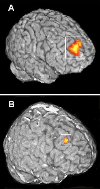
Although the expectancy AX task has been used in a number of imaging studies of both basic processes (Barch et al., 1997; Carter et al., 1998) and clinical conditions (Barch et al., 2001; Holmes et al., 2005; MacDonald & Carter, 2003; Perlstein, Dixit, Carter, Noll, & Cohen, 2003), we will describe just one with particularly straightforward results. In this study the AX task was administered during fMRI in 18 medication-naive first-episode schizophrenia patients, 12 medication-naive first-episode nonschizophrenic psychosis patients and 28 healthy controls (MacDonald, Carter, et al., 2005). On correct trials nonschizophrenic psychosis patients and controls activated bilateral dorsolateral prefrontal cortex following B cues. Schizophrenia patients did not activate this region even though they performed as accurately as controls on these trials. (Furthermore, average accuracy rates on B-cued trials were greater than 90% for all groups and therefore well above chance.) Direct contrasts of the neuroimaging data showed that in the region of dorsolateral prefrontal cortex illustrated in Figure 3A schizophrenia patients had significantly less activity than either of the other two groups. Thus, the centroid of this region was slightly anterior to the centroid of the region in the right dorsolateral prefrontal cortex found to be reliably less active in the meta-analysis of n-back group differences (Glahn et al., 2005).

The right dorsolateral prefrontal cortical dysfunction observed in (A) schizophrenia patients (BA 9, Talairach center x = 22, y = 51, z = 32, volume = 1388 mm3; MacDonald, Carter, et al., 2005) and (B) patients' healthy relatives (BA 9/8, Talairach center x = 37, y = 28, z = 43, volume = 1031 mm3; MacDonald et al., in press). Note: These figures have been modified from the originals only to highlight regions of interest. [A color version of this figure can be viewed online at www.journals.cambridge.org]
As we have seen, the finding of reduced functioning in the right dorsolateral prefrontal cortex in and of itself is not unusual in the schizophrenia literature. However, the sample in this study, unmedicated psychosis patients including both nonschizophrenia and schizophrenia patients, the behavioral results of which were suggestive of a specific deficit similar to that described below, and an analysis that controlled for the performance confound made these findings particularly useful. That is, they better reflect the nature of the functional neuroanatomy of schizophrenia and its link to the underlying neuropathology. One of the most useful findings was a relationship found within the schizophrenia patients. Here, we observed that there was a significant negative correlation among schizophrenia patients, such that those who showed the least activity in this right dorsolateral prefrontal cortex region had more prominent disorganization symptoms (r = −.53, p < .01). One could speculate that this was because these patients tended to have more symptoms overall. This was not the case. We were also able to show that the correlation between disorganization symptoms and hypofrontality was significantly stronger than the correlations between activity and the positive or negative symptom factors. This finding particularly supports a relationship between prefrontal cortical dysfunction and disorganization symptoms, and converges with the relationship between ketamine NMDA blockade, working memory problems, and disorganization symptoms reported by Adler, Goldberg, Malhotra, Pickar, and Breier (1998).
Whereas this study of unmedicated, first-episode patients connected prefrontal cortical dysfunction to symptoms and illness manifestation, a second study linked prefrontal cortical dysfunction to the genetic liability to schizophrenia. In this study, we used a variant of the continuous performance task called the Preparing to Overcome a Prepotent Response Task (MacDonald, Becker, & Carter, in press). This task took advantage of the effect described by Simon (1990) to both simplify the task and increase our efficiency in obtaining critical trials during fMRI. The Simon effect is the additional time required to make a response with the hand opposite where the stimulus occurs, relative to responding with the hand on the same side the stimulus occurs. To make the task similar to the AX task, the first part of the trial consisted of cues that indicated whether to respond on the same side (cue = green square) or overcome this prepotent response (cue = red square). The second part of the trial consisted of an arrow on either the left- or right-hand side of the screen. Green trials were therefore similar to AX trials, whereas red trials were similar to BX trials. To maintain the strength of the directional prepotency, 70% of trials were green cue trials. Using this task, we evaluated brain activity on correct trials in 21 healthy relatives of schizophrenia patients and 20 controls. Behaviorally, relatives were slower when overcoming the prepotent response, consistent with a slight context processing impairment. Analyses of the cue interval (before the response) on correct trials showed that both relatives and controls increased activity when preparing to overcome the prepotent response in inferior regions of bilateral dorsolateral prefrontal cortex. In the direct contrast illustrated in Figure 3B, however, patients' relatives showed significantly less activity than controls when preparing to overcome a prepotent response in the superior, right dorsolateral prefrontal cortex. The center was slightly posterior and superior along the right middle frontal gyrus to the regions identified in Glahn et al. (2005) and MacDonald, Carter, et al. (2005), with regions above the statistical threshold in the latter study close to overlapping. These findings suggest the similarity of regions affected by the genetic liability to schizophrenia and regions affected by the disorganization symptoms of schizophrenia as highlighted through the use of context processing tasks.
Thus, there does appear to be convergent evidence across different levels of analysis and different laboratories linking (a) the genetic liability to the illness to (b) NMDA synapse dysfunction and (c) a failure of neuronal networks that rely on coordinating firing, which, in turn, (d) appears to be associated a reduced ability to activate dorsolateral prefrontal cortex even for simple stimuli in a context processing task. In the next section we will consider the consequences of these failures at the next rung of the translational ladder, which is cognitive functioning.
Cognitive Functioning and Schizophrenia
“Cognitive differences between schizophrenia patients and healthy people have emerged systematically as the most powerful findings across hundreds of studies and 2 decades of neuroscience-based research. On average, cognitive measures yield [effect sizes] that are twice as large as those obtained with measures of regional brain volume (structural MRI), blood flow, metabolism, and receptor occupancy (PET/xenon)” (Heinrichs, 2005, p. 238). Cognitive differences also compare favorably, in terms of effect size measure, with postmortem tissue differences such as those described above. For all this, cognitive functioning should be at the forefront of research into the causes and cures of schizophrenia, but instead it risks becoming a backwater.
In some sense the cognitive level of analysis of schizophrenia is a victim of its own success. The primary findings that Heinrichs (2005) has drawn attention to are the large main effects for comparisons of patients and controls across a wide variety of behavioral tasks. Whether penciling pictures, declaiming digits, placing pegs, or memorizing mug shots, patients do it worse than controls. They do it about 1 SD worse, on average. This observation has come to be known as the “generalized deficit.” An NMDA synaptic dysfunction is consistent with a large generalized deficit. This synapse contributes to neurotransmission in many brain systems, after all. Such deficits should be accompanied by impairments in the sensory, cognitive, and motor functions mediated by these diverse cortical systems. However, if patients perform poorly on all tasks, then no task provides any particular guidance about where to look for neural signals associated with the causes of schizophrenia. Such guidance might be useful because the changes in neurotransmission mediated by the NMDA synapse in schizophrenia appear to impact some cortical systems more severely than others (as seen in the postmortem data described above, for example). Thus, an important task for this level of analysis is not merely to map the extent of the generalized deficit, but to identify specific cognitive deficits that map onto the anatomical distribution of the disease process in the brain.
To this end, experimental psychopathologists are interested in showing that novel cognitive impairments in schizophrenia pass a more stringent test. The more stringent test is based on the idea that an impairment only provides incremental information if it taps a cognitive domain that is demonstrably more impaired than other cognitive domains or the generalized deficit. However, the psychometric properties of the task must also be taken into account in demonstrating a specific or differential deficit like this, and there is the rub. Consider just one of the psychometric properties that is relevant to the comparison of two tasks: difficulty. A task of interest that is difficult will be more likely to show a group difference than a comparison task on which participants are close to ceiling even if both tasks are merely measuring the same generalized deficit. This is the essence of the generalized deficit confound highlighted by Chapman and Chapman (1973), bemoaned by others (e.g., Knight, 1984; MacDonald & Kang, in press; Strauss, 2001), and ignored by many. In contrast to the sanguine perspective on cognitive differences in schizophrenia lauded above, we propose that there is less here than meets the eye. Although a thorough review of differential deficits in schizophrenia has yet to be undertaken, to our knowledge only a small minority of studies rise to the level of a differential deficit and thereby provide incremental information about the neuropathology of schizophrenia (for further discussion of this state of affairs, see Joyce & Huddy, 2004; MacDonald & Carter, 2002).
As with the performance confound, struggling against the generalized deficit confound is likely to be worthwhile. Indeed, we have described the expectancy AX task that was designed in part with the generalized deficit confound in mind. Through the use of the expectancy AX and related tasks, a growing literature suggests that context processing is a specific deficit in schizophrenia patients (Barch, Carter, MacDonald, Braver, & Cohen, 2003; MacDonald, Goghari, et al., 2005; MacDonald, Pogue-Geile, Johnson, & Carter, 2003). On the one hand, this work illustrates the links between schizophrenia, disorganization symptoms, and context processing deficits. On the other hand, it demonstrates the relationship between context processing deficits and genetic liability to the disorder in a manner that complements the neuroimaging data presented above.
Context processing deficits distinguish schizophrenia patients from other psychosis patients. For example, among 42 medication-naive psychosis patients who were later diagnosed with schizophrenia, context processing deficits found during the acute phase of their illness were still present 1 month later despite the initiation of treatment (Barch et al., 2003). In contrast to this continuing deficit, we found that 21 psychosis patients who were later diagnosed with other conditions including psychotic affective disorders and psychosis not otherwise specified had largely recovered from initial context processing deficits at the 4-week follow-up. These data suggest that context processing deficits may be diagnostically specific and, among schizophrenia patients, trait related. However, another interpretation is possible. It may be that diagnosis is sensitive to an underlying dimension that is trait related, medication resistant, more characteristic of schizophrenia than other disorders, and tapped into by context processing measures. One such trait alluded to above is disorganization symptoms, which are both characteristic of schizophrenia and have shown a correlation of r = .41 with context processing measures in an earlier sample (Cohen, Barch, Carter, & Servan-Schreiber, 1999). To explore this possibility in the first-episode sample, we correlated error rates and signal detection rates from the AX task with index hospitalization indicators of disorganization, positive, and negative symptoms (Barch et al., 2003). The correlation between these measures and disorganization was consistently significant (r = .39–.41 in the expected directions) and significantly greater than the correlation with positive symptoms. However, negative symptoms were also correlated with context processing measures in this study (r = .28–.49 in expected directions).
In addition to their relationship to symptoms and diagnosis, context processing deficits also provide predictive outcome information. In an analysis of 38 first-episode schizophrenia patients, context processing scores at index hospitalization were measured as the difference between BX and AY errors on the AX expectancy task (Becker et al., 2003). This score showed significant correlations with both 1-year global assessment scores (r = −.34) and Strauss–Carpenter Outcome Scale scores (r = −.52), which broadly assess areas of functional outcome such as duration of nonhospitalization, social contacts, employment, and absence of symptoms. It may be, however, that context processing is simply measuring the severity of index hospitalization symptoms, and therefore, failing to provide incremental information about outcome. To examine this possibility, a hierarchical regression procedure was used in which manifest symptoms first accounted for outcome scores followed by the context processing difference score. In the analysis of the 1-year global assessment score, symptoms accounted for 10% of variance, whereas context processing contributed an additional and significant 22% of variance. In the analysis of the Strauss– Carpenter Outcome Scale score, symptoms accounted for 16% of variance, whereas context processing contributed an additional and significant 25% of variance. As these findings were derived from a small sample size, they will require extension and replication. Nevertheless, they provide intriguing preliminary evidence that the importance of context processing deficits extends beyond concurrent symptoms and into the domain of future functioning.
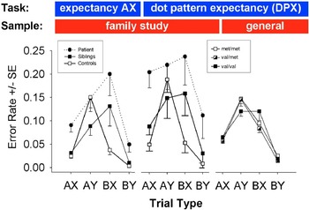
Thus far, we have described the relationship of context processing to higher levels of analysis, symptoms, diagnosis, and outcome. In the previous section we additionally showed that context processing has been related to prefrontal cortical dysfunction. It is not surprising, perhaps, that context processing paradigms have also been used to examine genetic liability to schizophrenia. Figure 4 illustrates the specific deficits in context processing that have been found in two genetically informative samples. In the first panel, the BX condition that measures context processing is one of the easiest conditions for control participants (n = 36, open squares; MacDonald, Pogue-Geile, et al., 2003). However, both patients with schizophrenia (n = 24, closed circles) and their healthy siblings (n = 24, open circles) find the BX context processing condition even more difficult than the AY condition. Although the schizophrenia literature shows many large behavioral main effects, the difference between patients and controls is an uncommonly large effect for an interaction (equivalent to Cohen's dBX-AY = .84). More importantly, for the purpose of demonstrating a genetically informative behavior, the siblings also showed a large interaction effect of nearly the same magnitude (dBX-AY = .80).

The results from two context processing tasks across two different samples (MacDonald, Goghari, et al., 2005; MacDonald, Pogue-Geile, et al., 2003). [A color version of this figure can be viewed online at www.journals.cambridge.org]
The expectancy AX paradigm can also be modified to use simple dot patterns that have the significance of valid and invalid cues and probes. Such a variant, called the dot pattern expectancy (DPX) task, allows the AY condition to be more difficult for healthy controls and the task to be conducted in a shorter amount of time. Results on the DPX task on a subset of patients and the healthy siblings from the above study are illustrated in Figure 4, panel 2 (MacDonald, Goghari, et al., 2005). As can be seen, again the AYDPX trials were harder for controls than the BXDPX trials, whereas siblings were significantly more impaired than controls on the BXDPX trials (dBX-AY = 0.72). This pattern of findings informs the third result illustrated in Figure 4. Panel 3 illustrates data from 464 healthy, unselected, European American individuals (50% male) from the general population that were genotyped on the COMT Val158Met polymorphism. Val158Met, located at 22q11.2, is an SNP that modifies the methylation of catecholamines such as dopamine. As alluded to above, several studies have reported the valine (Val) allele at this site increases susceptibility to schizophrenia (e.g., Egan et al., 2001; Shifman et al., 2002), although this finding has been challenged (Fan et al., 2005). In these data, individuals homozygous for the valine allele showed a pattern of context processing deficits on the DPX similar to that observed in the siblings of patients with schizophrenia. That is, they were impaired on BXDPX trials relative to methionine homozygotes and heterozygotes on BXDPX trials, but made fewer errors on AYDPX trials. A priori contrasts between homozygous groups on the BXDPX and AYDPX conditions of interest indicated a significant interaction (dBX-AY = .31). Note that magnitude of the effect size was less than half of that observed in patients' siblings on the DPX; however, it remained significant even when covarying for gender and removing low IQ individuals. Thus, we have preliminary evidence that individual differences in genotype can partially account for the differences in behavior found in the family members of schizophrenia patients.
Regardless of one's evaluation of the importance of context processing for understanding schizophrenia, these data present an illustration of how a cognitive construct can be used as a window to examine multiple levels of analysis, from genes to symptoms and functioning. Although the one gene so far linked with context processing, COMT, is likely to have only indirect effects on glutamate neurotransmission in cortex, this in no way contradicts the primary thrust of the NMDA synaptic dysfunction model. The relationship between the dopamine system, which is directly affected by COMT, and NMDA receptor functioning is explored more fully below. Furthermore, the particular aspect of schizophrenia illuminated by context processing deficits suggests it is related to prefrontal cortical dysfunction on a more elementary level of analysis, and disorganization symptoms and impaired functioning at higher levels of analysis. This is the final level of analysis to which we now turn.
Detangling the Symptoms of Schizophrenia
Schizophrenia is a nosologically complicated illness, and the final manifestation may be the result of a common mechanism acting in different cortical regions, or a number of independent causes with varying degrees of prominence. To address this heterogeneity, a great deal of effort has been spent to derive valid subtypes and symptom dimensions (e.g., Arndt, Alliger, & Andreasen, 1991; Crow, 1980; Peralta & Cuesta, 1999). As alluded to above, three symptoms dimensions are commonly extracted from patient samples, including positive, negative, and disorganization symptoms, with additional factors such as social dysfunction and mania emerging less consistently (Liddle, 1987; Peralta & Cuesta, 1999). Disorganization, or thought disorder, is characterized by derailment, illogicality, and inappropriate affect. This dimension is of particular interest to this version of the NMDA synaptic dysfunction model. This is because two studies using separate samples have found disorganization to be the most heritable symptom dimension, suggesting that it is the dimension most likely to be eventually linked to genes that influence glutamate neurotransmission.
The first study rated the standard three symptom dimensions as well as affect symptoms for a sample of 114 dually affected sibling pairs (Loftus, DeLisi, & Crow, 1998). Symptoms were scored as present if they had occurred in any episode, and a threshold was applied that assigned the dimension as either present or absent in that individual. Positive, negative, and affective symptoms did not reliably predict the symptoms that occurred in the cosibling (ps ≥ .10). However, 73 of the pairs were concordant for the presence or absence of disorganization symptoms (compared to 57 expected by chance), whereas 41 were discordant (compared to 57 expected by chance, p = .01). In a second study, twin pairs who had experienced any sort of psychosis during their lifetimes were ascertained from the Maudsley Twin Register (Cardno, Sham, Murray, & McGuffin, 2001). As with the previous study, there was no evidence of significant genetic contributions to positive symptoms across the 13 identical, or monoygotic, twins concordant for schizophrenia. Negative symptoms were correlated among monozygotic pairs (r = .42); however, this was not significant given the small sample. Disorganized symptoms were significantly and strongly correlated among monozygotic pairs with schizophrenia (r = .64). This pattern of findings was also found in the broader sample of 43 twins that included any pair concordant for psychosis, except that in this larger sample negative symptoms were also significantly correlated.
These studies suggest that among the dimensions of schizophrenia, disorganized symptoms have a privileged position. They may be somewhat closer to the genetic liability to the disorder than negative symptoms. Positive symptoms may not be very closely related to genes at all. This further strengthens the argument that more elementary mechanisms, such as context processing deficits and prefrontal cortical dysfunctions related to the disorganization dimension of schizophrenia may be optimally situated to provide insight into the relationship between the pathophysiology of schizophrenia and the illness manifestation. We will now turn our attention to the relationship between developmental changes in cortex and the onset of schizophrenia.
NMDA Synaptic Deficit Model and the Development of Schizophrenia
In addition to building a developmental account of schizophrenia across multiple levels of analysis (Cicchetti & Blender, 2004, in press), the NMDA synaptic deficit model also addresses the developmental nature of schizophrenia, and in particular, its occurrence generally in late adolescence or early adulthood. This model is compatible with, and indeed supported by, growing evidence demonstrating a dramatic change in the synaptic organization of the cerebral cortex during this time. Studies of synaptic organization in human (Huttenlocher, 1979; Huttenlocher & Dabholkar, 1997) and nonhuman primates (Bourgeois, Goldman-Rakic, & Rakic, 1994; Rakic, Bourgeois, Eckenhoff, Zecevic, & Goldman-Rakic, 1986) have indicated that the total number of synapses in prefrontal cortex peaks in early childhood (at 2 months of age in monkeys; 2 years in human prefrontal cortex), plateaus throughout later childhood, and then undergoes a marked reduction during adolescence. However, this pruning is not uniform. It appears to occur primarily in the pool of synapses that are located on dendritic spines. The density of dendritic spines visualized in Golgi-stained neurons follows a time course that is similar to that reported for synapses visualized at the electron microscope level (Lewis, 1997). Synapses on dendritic shafts, which comprise 25–40% of all synapses, do not decrease in number, whereas synapses on dendritic spines decrease by up to 50% during adolescence (Bourgeois et al., 1994; Lewis, 1997). The greatest drop in synaptic density during adolescence involves the dendritic spines of Layers II and III pyramidal neurons. It is just these layer III spines that are significantly reduced in schizophrenia. A central tenet of the NMDA synaptic deficit hypothesis is that the disease process driving schizophrenia and a normal developmental event by which synaptic density is reduced converge to produce catastrophic disconnection of pyramidal neurons in layer III of prefrontal cortex.
There is some evidence to indicate that this drop in synaptic connectivity involves local excitatory circuits within prefrontal cortex (Lewis & Gonzalez-Burgos, 2000). Axons of pyramidal neurons in prefrontal cortex give rise to a dense plexus of collaterals that travel horizontally in the cortex. The stripelike terminal zones of axon collateral projections undergo a reduction in of up to 40% in area during adolescence (Woo, Pucak, Kye, Matus, & Lewis, 1997), suggesting that recurrent local excitatory circuitry comprised of axon collaterals may be a target of synaptic pruning during late development. Thus, the development of cortex is consistent with a model in which ever decreasing connections in PFC lead to a systemwide failure in vulnerable circuits.
Conclusion: Issues for Translational Research in Schizophrenia
Translational schizophrenia research has reached a dynamic phase with convergent evidence aligning from a number of different domains, including genetic, pathophysiological, neuroanatomical, and cognitive. Modern genotyping, animal modeling, and neuroimaging technologies allow us to move more easily than ever across different levels of analysis to test novel hypotheses and modes of interaction. However, there are several outstanding issues that require addressing.
One first question to address is that if NMDA hypofunction underlies psychosis, or at least the symptoms of disorganization, then why is schizophrenia not treated using glutamate, or more specifically NMDA, agonists? The answer is that this very mechanism of action is currently being investigated. Even more so than other rational drug developments, such agents have required great care because direct antagonism of glutamate can have potential excitotoxic effects. One particularly promising approach has been to design agents, such as d-cycloserine, to activate the glycine B receptor, which influences the NMDA receptor (for review, see Coyle & Tsai, 2004). Initial reports from clinical trials that have used d-cycloserine as an adjunct to standard antipsychotic pharmacotherapy suggest a U-shaped dose–response curve with improvements in cognitive functioning and reductions in negative symptoms. Thus, the NMDA synaptic deficit model has already had an impact on drug development and may soon result in a positive impact on patients' lives.
A second question concerns the role of dopamine. The dopamine hypothesis was ascendant in the 1970s based on the effects of the first generation of antipsychotic medications, which were all dopamine antagonists. Furthermore, there was found to be a direct relationship between how much an agent reduced psychotic symptoms, which generally meant positive symptoms, and the agent's ability to bind with the dopamine D2-receptor subtype in striatum. There is now growing evidence that dopamine dysregulation may be downstream of glutamate, and the nature of glutamate–dopamine interactions is now just beginning to be uncovered (Laruelle, Kegeles, & Abi-Dargham, 2003). One relationship of particular interest from a neurodevelopmental and neuroplasticity perspective is one of the secondary effects of striatal dopamine blockade. Preliminary evidence from basic studies suggests that chronic treatment with a dopamine antagonist such as haloperidol increases the proliferation of neural stem cells in the adult forebrain, albeit in rat adult forebrain (Kippin, Kapur, & van der Kooy, 2005). Thus, one may speculate that a number of processes could be spawned by dopamine antagonists in such a way as to reduce, or in some cases eliminate, some of the downstream effects of NMDA synaptic deficits.
A third gap in the NMDA model we advance is the reason why a defect at the NMDA synapse, so ubiquitous in cortex, should impact the prefrontal cortex and its corticocortical networks more than other regions. A definitive answer awaits further work; however, available evidence from prior translational studies identifies some preliminary possibilities. Two aspects of NMDA glutamatergic synaptic capacity seem to be changed in schizophrenia; synaptic function (as implicated by genetic studies), and synaptic number (as implicated by neuroanatomical studies). Both aspects of NMDA neurotransmission may preferentially impact prefrontal function. With respect to NMDA synaptic function, one plausible reason for why prefrontal networks are particularly disrupted may be that the function of prefrontal cortex depends on the coordination of activity in particularly large and complex distributed cortical systems via corticocortical interactions. Perhaps because of the size and complexity of the corticocortical networks involving prefrontal cortex, they are in some sense less stable than sensorimotor networks, in such a way that they are more vulnerable to functional dysregulation of NMDA synapses. With respect to NMDA synaptic number, there is anatomical evidence suggesting prefrontal cortex is particularly impacted. Anatomical disconnection in schizophrenia, as evidenced by a loss of neuropil and dendritic spines, selectively targets prefrontal networks, leaving sensory and motor circuits in the cortex relatively spared (see the discussion of anatomical disconnection in schizophrenia, above).
A fourth unanswered question in this model likely to be addressed by future translational studies is whether all gene variations associated with schizophrenia produce a common effect on NMDA mediated neurotransmission (e.g., whether these mutations all lower the efficacy of transmission at this synapse, as suggested by the parallel between the behavioral effects of NMDA receptor blockade and schizophrenia). Knowledge of how SNPs in genes associated with schizophrenia impact the function of the proteins they encode, and therefore the function of the glutamate synapse, is incomplete.
One advantage to having a strong hypothesis that penetrates a number of levels of analysis is that it can spawn an enriched research agenda. Stepping back from the specifics of the NMDA synaptic deficit model provides us with an opportunity to consider a research agenda for psychosis much more broadly. For example, one development that would facilitate advances in translational research in schizophrenia would be a better mapping between behavioral assays and symptoms dimensions. One of the problems the field is struggling to overcome is the generality of the deficits associated with schizophrenia. In addition to minding psychometric concerns described above, a set of tasks that were demonstrably selective to a particular symptom type would allow us to build a more comprehensive story about the development of schizophrenia. In particular, there is a dearth of behavioral probes for positive symptoms.
The evidence reviewed here clearly demonstrates that translational research has been remarkably successful in recent years. For the first time since the initial description of the disease, it is possible to form initial if incomplete links between changes at multiple levels of cortical organization and analysis in schizophrenia. This work has begun to reveal the functional logic that may eventually unify alterations in gene structure, protein function, neuronal structure, connectional anatomy, physiological operation, cognitive function, and ultimately the clinical symptoms of schizophrenia. Changes at most of these levels of organization in schizophrenia have been reported and touched on in this review. Despite the gaps and outstanding questions noted above, current evidence is very strong that such a causal chain from molecules to thought drives schizophrenia, and gives good reason for confidence that this causal chain will prove intelligible and amenable to neuroscientific analysis, at least in its major features. As reviewed here, and perhaps most importantly, it is becoming increasingly evident that this causal chain will course through the glutamate synapse and synaptic plasticity mediated by the NMDA glutamate receptor, with changes in the function of this specific component of cortical systems driving the disease process that produces schizophrenia. Already this knowledge is producing new avenues of treatment, perhaps the ultimate test of the success of a translational approach.