Implications
The newly developed assay is accurate, sensitive and specific for FSH determination in bovine plasma. It has the advantage that it does not use radioactivity, unlike the previous FSH assays dedicated to the bovine species. This enzyme immunoassays (EIA) provides a useful tool for future research into FSH physiology and actions.
Introduction
Two gonadotropins, named FSH and LH, both secreted by the pituitary gland, control the function of male and female gonads. Follicle-stimulating hormone and LH are heterodimers, consisting of two non-covalently bound peptide sub-units. They share the same alpha sub-unit and the beta sub-unit provides specificity for receptor interactions (Combarnous, Reference Combarnous1992; Cooke et al., Reference Cooke, Crowe, Roche and Headon1996). In the ovary, the granulosa cells of all growing follicles have FSH membrane receptors, whereas LH receptors are present on the theca cells of these follicles, the granulosa cells of the pre-ovulatory follicles and the luteal cells of the corpora lutea (Richards, Reference Richards1994). Gonadotropins co-operate in controlling the development of ovarian follicles. In all mammalian species, FSH orchestrates the growth and differentiation of follicles, and LH supports their final maturation and triggers ovulation (McGee and Hsueh, Reference McGee and Hsueh2000; Hillier, Reference Hillier2001; Scaramuzzi et al., Reference Scaramuzzi, Baird, Campbell, Driancourt, Dupont, Fortune, Gilchrist, Martin, McNatty, McNeilly, Monget, Monniaux, Vinoles and Webb2011).
Our knowledge of the role and regulation of FSH in cattle has been established using radioimmunoassays (RIA), which are not commercially available. To meet the increasing societal demand for avoiding the use of radioelements in laboratories, RIA are now being replaced by EIA. Presently, there is no commercially available sensitive FSH EIA specific to the bovine species for determining blood FSH concentrations, which are generally lower than 1 ng/ml in cows, heifers and calves (Evans et al., Reference Evans, Adams and Rawlings1994; Burns et al., Reference Burns, Jimenez-Krassel, Ireland, Knight and Ireland2005; Ginther et al., Reference Ginther, Siddiqui, Baldrighi, Wolf and Castro2016). Moreover, despite high structural similarities between human, ovine and bovine FSH molecules (Haj Hassan et al., Reference Haj Hassan, Cahoreau, Jegot, Jouanny, Mariot, Lecompte, Klett and Combarnous2015), presently there is no human FSH EIA validated for the measurement of bovine FSH, and the existing ovine EIAs (Faure et al., Reference Faure, Nicol, Fabre, Fontaine, Mohoric, McNeilly and Taragnat2005; Torres-Rovira et al., Reference Torres-Rovira, Succu, Pasciu, Manca, Gonzalez-Bulnes, Leoni, Pennino, Spezzigu, Gallus, Dattena, Monniaux, Naitana and Berlinguer2016) are not sensitive and specific enough for the measurement of FSH in bovine plasma.
FSH is essential to drive the terminal waves of follicular development leading to the development of a pre-ovulatory follicle in cows (Fortune, Reference Fortune1994; Ireland et al., Reference Ireland, Mihm, Austin, Diskin and Roche2000; Adams et al., Reference Adams, Jaiswal, Singh and Malhi2008; Ginther, Reference Ginther2016). However, the role of FSH in ovarian function has not been fully established before puberty. Follicular growth is initiated between days 90 and 140 of gestation in the ovaries of fetal calves, but at birth the number of antral follicles remains low and their size rarely exceeds 1-mm diameter (Tanaka et al., Reference Tanaka, Nakada, Moriyoshi and Sawamukai2001; Yang and Fortune, Reference Yang and Fortune2008). From daily ultrasound examination of postnatal ovaries of calves as young as 2 weeks of age, the emergence of waves of development of antral follicles growing from 4 to 8 mm diameter was shown to occur every 6 to 10 days, each wave being preceded by a transient increase in plasma FSH concentrations (Evans et al., Reference Evans, Adams and Rawlings1994). However, the population of small growing follicles up to the stage when they enter a terminal wave of development remains difficult to assess using ultrasonography, and the role of FSH on this follicular population has not been established in young calves. The possibility should now exist to follow indirectly the changes in this follicular population in calves through the measurement of anti-Müllerian hormone (AMH) in plasma. Indeed, AMH has been established to be an endocrine marker of this follicular population making up a reserve pool for the recruitment of follicular waves in cows (Monniaux et al., Reference Monniaux, Drouilhet, Rico, Estienne, Jarrier, Touzé, Sapa, Phocas, Dupont, Dalbies-Tran and Fabre2013; Monniaux et al., Reference Monniaux, Clément, Dalbies-Tran, Estienne, Fabre, Mansanet and Monget2014).
In this study, we aimed to develop a bovine FSH EIA and validate it for the specific determination of FSH in bovine plasma. Owing to the high structural similarity existing between human and bovine FSH molecules, we assumed that bovine FSH could be detected by a sandwich-type EIA using anti-human FSH antibodies. After technical validation of the EIA, we carried out its biological evaluation on cows under different physiological situations. In a second objective, the newly developed EIA was used to study the role of FSH in regulating the postnatal development of ovarian follicles up to the small antral stage in calves. For this purpose, we established the concomitant FSH and AMH changes in the plasma of female calves during their first three months of life, with the assumption that AMH could be an endocrine marker of the population of small antral follicles in calves, as in adults.
Material and methods
Enzyme immunoassay of human/bovine FSH
The anti-human α-subunit antibody used for the detection of FSH bound on the plates was a mouse anti-hCG (α 4 epitope) monoclonal antibody, clone INN-hFSH-132 (MCA 1026; Bio-Rad, Marnes-la-Coquette, France). For the preparation of the biotinylated antibody, 0.5 mg anti-human α-subunit antibody was first diluted in 500 μl phosphate-buffered saline (PBS, containing 0.09% NaN3) and dialyzed with NaHCO3 (100 mM solution, pH = 8.2) during 24 h at 4°C with three changes of the NaHCO3 solution. A solution of Biotinamidohexaonic acid N-hydroxysuccinimide ester (B2643; Sigma, Saint-Quentin Fallavier, France) was dissolved and concentrated at 4 mM in dimethyl sulfoxide extemporaneously, then 25 μl of this solution was added to the dialyzed antibody and the mixture was vortexed and incubated for 2 h at room temperature under agitation. For the purification of the biotinylated antibody, the mixture was dialyzed during 36 h at 4°C in PBS (10 mM/NaCl 150 mM, pH = 7.4). After dialysis, the biotinylated antibody was diluted with an equal volume of glycerol and stored at −20°C.
The antibody used for the capture of FSH was a mouse anti-human FSH β-subunit monoclonal antibody (ref: LS-C6871; CliniSciences, Nanterre, France). For coating of the antibody on plate wells, 100 μl coating buffer (50 mM carbonate/bicarbonate buffer, pH = 9.6) containing 0.2 μg anti-human FSH β-subunit antibody were dispensed into each well of microtiter plates (Nunc-Immuno Maxisorp C96; Sigma). The plates were then incubated overnight at 4°C. The coated wells were washed with 400 μl washing solution (PBS 10 mM/NaCl 150 mM, pH = 7.4) per well using an automated microtiter plate washer (405LS; BioTek, Colmar, France). For surcoating, 300 μl washing solution containing 0.09% Tween 20 and 12.5% Sea-Block (UP40301A; Interchim, Montluçon, France) were added to the wells. After incubation of the plates for 1 h at 37°C, the surcoating buffer was removed and the wells were washed once.
Plasma samples to analyze by FSH EIA were thawed, mixed and centrifuged. For the preparation of the standard curve, samples of purified bovine FSH (bFSH-I-2, USDA) were diluted in assay buffer (PBS containing 1% BSA) to obtain concentrations ranging from 0.1 to 3.2 ng/ml. Duplicates of 200 μl plasma samples and FSH standard samples were then dispensed into their respective wells along with 50 μl of first incubation buffer (PBS containing 0.09% Tween 20, 12.5% Sea-Block and 2% normal rat serum). After overnight incubation at 4°C, the plates were washed three times with the washing solution. Then, 100 μl of second incubation buffer (PBS 10 mM/NaCl 1 M, pH = 7.2, with 0.1% Tween 20 and 1% rat serum) containing 30 ng biotinylated anti-human α-subunit antibody were added to each well and the plates were incubated for 1 h at 37°C. After three washings, 100 μl of second incubation buffer, without rat serum but containing 25 ng horseradish peroxidase-labeled neutravidin (31001; Thermo Fisher Scientific, Artenay, France), were added to each well and the plates were incubated for 25 min at room temperature in the dark. After three washings, the staining was revealed by the incubation of 100 μl of substrate solution (TMB SureBlue KPL; Eurobio, Courtaboeuf, France) per well for 1 h at room temperature in the dark. The reaction was stopped by the addition of 50 μl HCl/H2SO4 per H2O (1 V/1 V per 6 V) per well and the optical density was measured at 450 nm (OD450, specific staining) and 620 nm (OD620, non-specific background) using a microtiter plate reader (Sunrise, Tecan, Lyon, France). The OD450 to OD620 absorbance values were used for calculations. The concentrations of FSH in plasma samples were calculated from the standard curve plotted for known bovine FSH concentrations in the range of 0.1 to 3.2 ng/ml by using Magellan software (Tecan) with a four-parameter logistic regression.
The sensitivity of the FSH EIA was assessed for FSH standard samples by determining the lowest FSH concentration giving absorbance values significantly different from the absorbance values of the zero FSH concentration in six independent experiments (comparisons done using paired t tests). The accuracy of the FSH EIA was assessed by overloading bovine plasma samples with known amounts of purified bovine FSH (bFSH-I-2, USDA). For this purpose, the FSH concentrations of plasma samples recovered from 13 different cows were measured before and after the addition of bovine FSH to obtain expected increases in plasma FSH concentrations ranging from 0.5 to 2.5 ng/ml, and the experiment was performed twice. The specificity of the FSH EIA was studied by testing the cross-reaction of the immunoassay with different concentrations (ranging from 0.1 to 100 ng/ml) of purified bovine LH (bLH CY1042, 1.19xbLH LER1072.2), purified bovine TSH (thyroid-stimulating hormone, bTSH gifted by Dr F. Frankenne), and purified FSH of other animal species (porcine FSH: pFSH CY1890-II, ovine FSH: oFSH VAL253-II, equine FSH: eFSH CY1857-III, human FSH: hFSH SIAFP1) (Haj Hassan et al., Reference Haj Hassan, Cahoreau, Jegot, Jouanny, Mariot, Lecompte, Klett and Combarnous2015). The cross-reactivity of each hormone in the FSH EIA was estimated for the lowest concentration of the hormone which could be detected, after calculation of its found concentration in the assay. The intra- and inter-assay coefficients of variations of the FSH EIA were determined in different experiments, using six quality control plasmas, with FSH concentrations ranging from 0.3 to 2.2 ng/ml. A comparison between results obtained by the FSH EIA and an existing, previously validated, bovine FSH RIA (Crowe et al., Reference Crowe, Padmanabhan, Hynes, Sunderland, Enright, Beitins and Roche1997) was performed on 12 bovine plasma samples.
Animals and experimental protocols in cattle
Adult 4 to 7-year-old cycling cows and newborn female calves were allocated to different studies involving FSH measurement. Animals were housed in free stalls and provided feed and water ad libitum. All procedures were approved by the French Agricultural and Scientific Research Government Committee (Approval number E37-175-2) and the Val de Loire ethics committee for animal experimentation, in accordance with the guidelines for Care and Use of Agricultural Animals in Agricultural Research and Teaching.
For a first biological evaluation of the FSH EIA, FSH changes were studied in the plasma of three Prim’Holstein dairy cows during an estrous cycle. Blood samples were recovered daily from the time natural estrous behavior occurred to the next estrus. For one cow, blood sampling was performed twice a day during 48 h after estrus detection. Blood samples were collected by jugular venipuncture in heparinized tubes, and plasmas were stored at −20°C until FSH and progesterone assays were carried out. In addition, the two other cows underwent ovarian ultrasonography scanning with a transrectal probe, model LV 513 (6.0/8.0-MHz transducer; MyLab30; Esaote, Mauguio, France) three times weekly to follow the follicular waves, as previously described (Rico et al., Reference Rico, Médigue, Fabre, Jarrier, Bontoux, Clément and Monniaux2011). All follicles with an antrum larger than 3mm diameter were measured and counted, and individual follicular growth curves were constructed for follicles with an antrum larger than 5 mm in diameter.
A further biological evaluation of the FSH EIA was achieved by studying FSH and LH pulsatility during the peri-ovulatory period in four Rouge-des-Prés beef cows. Cows were first submitted to a cycle synchronization protocol. For this purpose, each cow received a subcutaneous progesterone implant (Crestar; Intervet, Angers, France) over 10 days and was treated with a Gonadotropin-releasing hormone analogue (0.01 mg busereline acetate, Receptal, Intervet) and prostaglandin PGF2α (15 mg Prosolvin, Intervet) when loading and removing the implant, respectively. Blood samples were collected via jugular cannulae at 10-min intervals for 5 h at 24 h and 42 h after implant removal, then the cannulae were removed and blood sampling was continued by jugular venipuncture every 2 h during an additional 24-h period to allow the detection of the LH/FSH pre-ovulatory peak. Plasmas were recovered and stored at −20°C until FSH and LH assays were carried out.
The newly developed FSH EIA was then used to study the role of FSH on ovarian follicular development during the postnatal period in six Prim’Holstein female calves. Blood samples were recovered weekly on calves between 1 week and 3 months of age by jugular venipuncture in heparinized tubes and plasmas were stored at −20°C until FSH and AMH assays were carried out. Three-month calves were slaughtered in a local slaughterhouse, their ovaries were immediately recovered and finely dissected to isolate all the antral follicles larger than 1 mm in diameter. For each animal, the follicles were classified according to three follicular size classes ((1 to 3 mm), (3 to 5 mm) and >5 mm) and counted. Follicular fluids were recovered by puncture using a syringe/needle. Follicular fluids from follicles larger than 5 mm in diameter were recovered individually. For smaller follicles, due to their small size, the follicular fluids recovered from each calf were pooled according to their size (1 to 3 mm or 3 to 5 mm). All follicular fluid samples were stored at −20°C until AMH assay was performed.
Progesterone assay
The concentration of progesterone in plasma was determined by EIA as described previously (Canepa et al., Reference Canepa, Lainé, Bluteau, Fagu, Flon and Monniaux2008). The limit of detection of the assay was 0.4 ng/ml and the intra-assay CV for quality control plasma samples containing 1.5 ng/ml progesterone was 6.6%.
LH assay
The concentration of LH in plasma was determined by EIA as described previously (Faure et al., Reference Faure, Nicol, Fabre, Fontaine, Mohoric, McNeilly and Taragnat2005). Luteinizing hormone was measured on 20 μl aliquots of undiluted plasma and all samples were analyzed in the same assay. The limit of detection of the assay was 0.1 ng/ml and the intra-assay CV for quality control plasma samples containing 0.5 ng/ml LH was 5.9%.
Anti-Müllerian hormone assay
Anti-Müllerian hormone concentrations in plasma and follicular fluids were determined using the AMH Gen II ELISA kit (Beckman Coulter, Villepinte, France), as previously described for the analysis of bovine samples (Rico et al., Reference Rico, Drouilhet, Salvetti, Dalbies-Tran, Jarrier, Touzé, Pillet, Ponsart, Fabre and Monniaux2012). Anti-Müllerian hormone concentrations were determined in 50 μl aliquots of undiluted plasma and of follicular fluids diluted 1/1000 for 1 to 3 mm and 3 to 5 mm follicles, and 1/100 for follicles larger than 5 mm in diameter. All samples were analyzed in the same assay. In our working conditions, the limit of detection of the assay was found to be 1 pg/well. The intra-assay coefficients of variation were 11.8% and 3.6%, for quality control plasma samples containing 0.033 and 0.125 ng/ml AMH, respectively.
Data analysis
The Dynpeak Software previously validated to analyze the pulsatile secretion of LH in jugular blood (Vidal et al., Reference Vidal, Zhang, Medigue, Fabre and Clement2012) was used for the detection of LH and FSH pulses on the eight time series of plasma samples collected on cows at 10-min intervals for 5 h. Pulse detection was used to calculate the time interval between two successive pulses and determine the percentage of co-occurrence of LH and FSH pulses. This percentage was estimated from the number of LH and FSH pulses occurring simultaneously, divided by the total number of detected pulses.
The amount of AMH present in the follicular fluid for each size class of follicles was estimated from the formula: Q = C × V × N, where C is the AMH average intra-follicular concentration of the class (expressed in ng/ml), V the average follicular volume (expressed in milliliters) and N the number of follicles of the class. For the class of follicles larger than 5 mm in diameter, the individual values of AMH concentrations and follicular volumes were used for the estimation AMH intra-follicular amount.
GraphPad Prism 6 Software (GraphPad Software, La Jolla, CA, USA) was used for the statistical analyses of data. Data are expressed as mean ± SEM. Data of the FSH standard curves were fitted to a four-parameter logistic regression, or to a linear regression after Log transformation. For analysis of hormonal pulsatility in the plasma of cows, means were compared by t test with Welch’s correction when variances were heterogeneous, and correlations were assessed using Pearson correlation coefficients (r). For the analysis of follicular and hormonal data in the six studied calves, non-parametric analyses were performed. Follicle numbers, AMH concentrations and AMH intra-follicular amounts were compared between follicular size classes using Friedman ANOVA followed by Dunn’s multiple comparisons tests, and correlations were assessed using Spearman correlation coefficients (r s).
Results
Characterization of the human/bovine FSH immunoassay
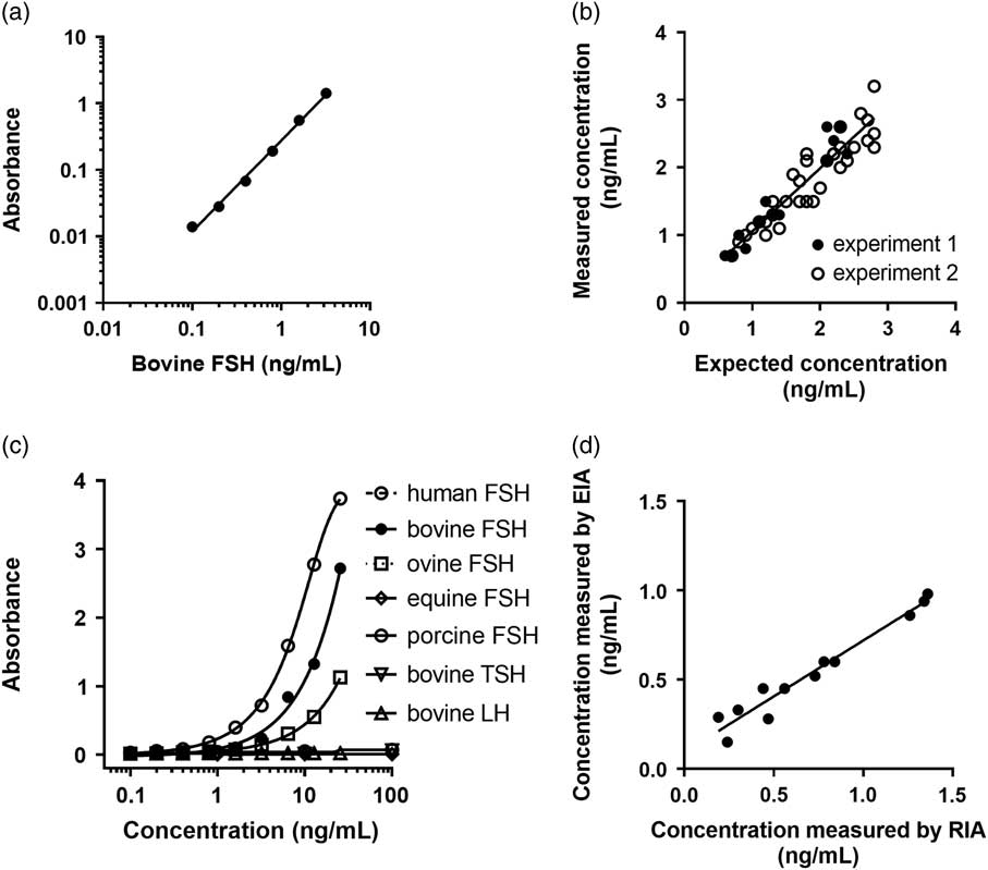
The absorbance found with the ‘zero’ of the standard curve was not different from the absorbance found with plasma containing no FSH (plasma ‘zero’, recovered from hypophysectomized sheep), both varying in the range of 0.037 to 0.039. The lowest amount of bovine FSH detected by the FSH EIA was 20 pg/well (absorbance values: 0.037 ± 0.006 v. 0.043 ± 0.007, 0.0 v. 20 pg/well, P < 0.01, n = 6), which corresponded to a sensitivity of 0.1 ng/ml. The absorbance increased with bovine concentrations, fitting to a four-parameter logistic regression. When plotted on logarithmic scales, the absorbance increased linearly with bovine FSH concentration in the range from 0.1 to 3.2 ng/ml (Figure 1a).
Characteristics and technical validation of the FSH enzyme immunoassay (EIA). (a) Example of a standard curve with bovine FSH, plotted on logarithmic scales and fitted to a linear regression (Y = 1.365 × X − 0.561, r 2 = 0.996, n = 6). (b) Accuracy test; plasma of 13 cows were overloaded by known amounts of bovine FSH in two independent experiments; the figure depicts the relationship between the expected FSH concentrations and the concentrations of FSH measured by the FSH EIA (Y = 0.931 × X + 0.128, r 2 = 0.88, n = 48). (c) Specificity test; data represent the cross-reactions of the FSH EIA with thyroid-stimulating hormone (TSH), LH and FSH from different animal species, all tested at different concentrations. (d) Relationship between concentrations obtained using the FSH EIA and a bovine FSH radioimmunoassay (RIA) (Crowe et al., Reference Crowe, Padmanabhan, Hynes, Sunderland, Enright, Beitins and Roche1997) for different bovine plasma samples (Y = 0.626 × X + 0.093, r 2 = 0.95, n = 12).

The accuracy of the FSH EIA was assessed by overloading bovine plasma samples with 0.5, 1.0, 1.5, 2.0 or 2.5 ng/ml of bovine FSH, giving respective recoveries of 0.55 ± 0.04 (n = 6), 1.13 ± 0.04 (n = 13), 1.63 ± 0.11 (n = 8), 2.08 ± 0.07 (n = 13) and 2.46 ± 0.10 ng/ml (n = 8). A linear regression with a slope of 0.931 ± 0.051 (n = 48) was found between the expected and the measured FSH concentrations in the studied plasma samples (Figure 1b).
The cross-reactivity of bovine TSH and bovine LH in the FSH EIA was low (0.4% and 0.01%, respectively), indicating that the immunoassay is highly specific to FSH measurement. The FSH EIA detected human FSH with a high (149%) cross-reactivity compared to bovine FSH (set at 100%), as expected due to the anti-human FSH antibodies used for the capture and detection of FSH. The cross-reactivity of ovine FSH in the FSH EIA was found to be 51%, and it was <0.1% for porcine FSH and <0.01% for equine FSH (Figure 1c).
FSH concentrations found in bovine plasma samples using the FSH EIA were highly correlated with those found using the previously validated FSH RIA developed by Crowe et al. (Reference Crowe, Padmanabhan, Hynes, Sunderland, Enright, Beitins and Roche1997) (r = 0.98, P < 0.001, Figure 1d). A linear regression with a slope of 0.626 ± 0.045 was found between results, indicating that the FSH EIA gives lower values than the FSH RIA in a systematic way.
The intra- and inter-assay coefficients of variation of the FSH EIA were found to be 13.9% (n = 8) and 10.0% (n = 5), respectively, when estimated using a quality control plasma containing 1 ng/ml FSH. For a quality control plasma containing a low FSH concentration (0.3 ng/ml), the inter-assay CV was found to be 18.9% (n = 6). For four quality control plasmas containing high FSH concentrations (1.8 to 2.2 ng/ml), the inter-assay coefficients of variation were found to vary in the range of 4% to 12% (n = 3 for each sample).
Biological evaluation of the FSH immunoassay for endocrine studies in cattle
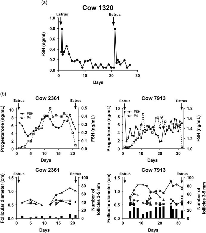
A short duration pre-ovulatory FSH peak was observed in the plasma of cycling Prim’Holstein cows about 8 h after the first detection of estrus behavior, as exemplified for Cow 1320 (Figure 2a). During the estrous cycle of cows submitted to daily blood sampling, progesterone concentrations increased after estrus, indicating that ovulation has normally occurred, then plateaued and finally sharply decreased shortly before the following estrus. In Cow 2361, periodic fluctuations in FSH plasma concentrations accompanying the follicular waves of terminal development were observed during the cycle. For instance, the high FSH levels found at days 1 and 11 of the cycle preceded the respective emergence of the first and the second follicular waves of the cycle, 1 day later (Figure 2b). In Cow 7913 with an abnormally long luteal phase, two follicular waves also occurred, preceded by increases in FSH concentrations, but the second follicular wave led to the development of a persistent follicle with an ovulary size from day 15 up to the next estrus at day 32.
Biological validation of the FSH enzyme immunoassay (EIA) for endocrine studies in the bovine species: plasma FSH during the estrous cycles of Prim’ Holstein dairy cows. (a) Example of an endocrine profile with a pre-ovulatory FSH peak detected 8 h after the beginning of each estrus. (b) Examples of daily endocrine changes (upper panels) and follicular growth in two cycling cows. The upper panels represent FSH (full circles) and progesterone (P4, open squares) endocrine profiles between two successive estrus. The lower panels represent the numbers of small 3 to 5 mm follicles (black bars) and the growth curves of individual follicles larger than 5 mm in diameter, established from ovarian ultrasonography scanning. Periodic fluctuations in FSH plasma concentrations accompany the follicular waves of terminal development.

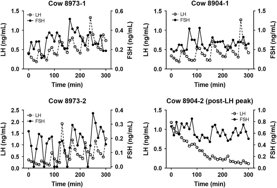
The pattern of LH and FSH secretion was studied during the peri-ovulatory period on plasma samples recovered at 10-min intervals for 5 h on Rouge-des-Prés cows. For seven among the eight time series of data which were analyzed, plasma samples were found to be recovered before the occurrence of the LH/FSH pre-ovulatory peak. For these time series, the average time interval between two successive pulses was not different for LH and FSH (50.00 ± 1.85 min, n = 32, v. 50.69 ± 3.78 min, n = 29, LH v. FSH, not significant). The time interval between each LH pulse and the LH/FSH pre-ovulatory peak was correlated with the time interval between successive LH pulses (r = 0.39, n = 32, P < 0.05), indicating that the frequency of LH pulses increased during the follicular phase of the cycle, whereas no such relationship was found for FSH pulses (r = 0.15, n = 29, not significant). Among the 75 detected pulses of LH or FSH, the percentage of co-occurrence of LH and FSH pulses was only 17.3%, suggesting that LH and FSH pulsatility was mainly asynchronous (Figure 3). For the time series of data corresponding to plasma recovered during the descending part of the LH pre-ovulatory peak, no synchrony between LH and FSH pulses was observed (Cow 8904-2, Figure 3).
Biological validation of the FSH enzyme immunoassay (EIA) for endocrine studies in the bovine species: plasma FSH and LH pulses during the peri-ovulatory period in Rouge-des-Prés beef cows. The figure depicts examples of FSH (full circles) and LH (open circles) profiles, determined on plasma samples recovered at 10-min intervals in two cows for 5 h at 24 h (time series 1) and 42 h (time series 2) after progesterone implant removal. The pulses of FSH (full arrows) and LH (open arrows) were detected using the Dynpeak Software (Vidal et al., Reference Vidal, Zhang, Medigue, Fabre and Clement2012).

Relationship between FSH and ovarian activity in female calves
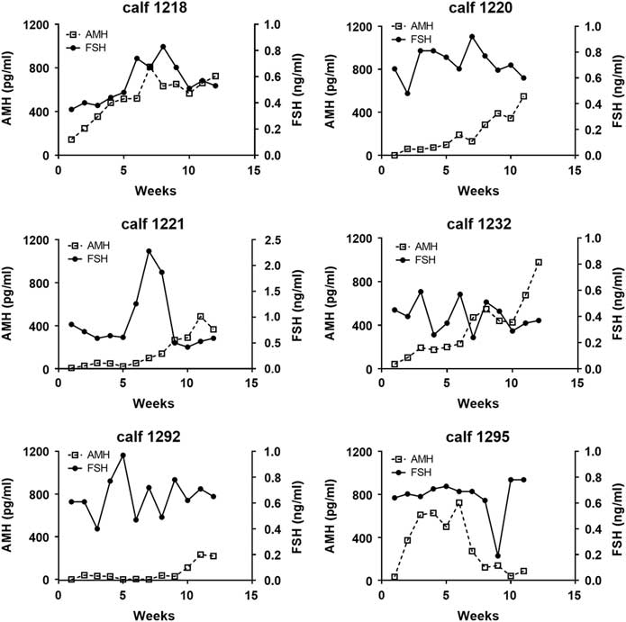
In Prim’Holstein calves submitted to weekly blood sampling, temporal changes in FSH plasma levels were studied concomitantly with those of AMH. Between 1 week after birth and 3 months of age, both FSH and AMH plasma concentrations followed very different patterns between the six studied calves (Figure 4). For instance, AMH plasma concentrations increased as soon as week 2 in calf 1295, but only after week 9 in calf 1293. When considering FSH plasma concentrations, only calves1221 and 1292 had a clear FSH peak at weeks 7 and 5, respectively. There were no correlations between FSH and AMH plasma concentrations of calves before three months of age (Figure 4).
Weekly changes in FSH (full circles) and anti-Müllerian hormone (AMH) (open squares) concentrations in the plasma of six Prim’ Holstein calves between 1 week and 3 months of age.

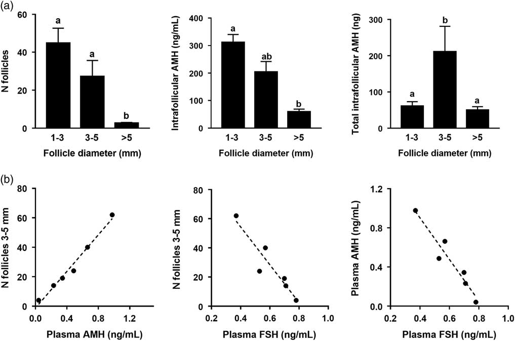
Most follicles recovered on the ovaries of the 3-month calves had diameters between 1 and 5 mm. Anti-Müllerian hormone concentrations in follicular fluid decreased with increasing follicle diameter (P < 0.05, Figure 5a). The population of 3 to 5 mm follicles contained the highest intra-follicular AMH amount (P < 0.05, Figure 5a) and the number of follicles of this size class was positively correlated with AMH concentration in the plasma of calves at the time of slaughtering (Spearman correlation coefficient: r s = 0.94, P < 0.05, n = 6, Figure 5b). Follicle-stimulating hormone concentration in the plasma of calves at the time of slaughtering was negatively correlated with the number of 3 to 5 mm follicles (r s = −0.94, P < 0.05, n = 6) and AMH plasma concentration (r s = −0.94, P < 0.05, n = 6, Figure 5b). No correlation was found between the numbers of 1 to 3 mm or >5 mm follicles, and AMH or FSH concentrations.
Ovarian and endocrine features of Prim’ Holstein calves at 3 months of age. (a) Numbers of antral follicles larger than 1 mm in diameter (left panel), anti-Müllerian hormone (AMH) concentrations in follicular fluid (middle panel) and total intra-follicular AMH amounts (right panel) in the ovaries of six calves; follicles were dissected on the ovaries of slaughtered calves and allocated to three size classes; AMH concentrations were determined on the recovered follicular fluids and the amount of AMH present in the follicular fluid was estimated for each size class of follicles, as described in The Materials and Methods section; (a) v. (b): P < 0.05 (n = 6). (b) Relationships between the numbers of 3 to 5 mm follicles on ovaries, plasma AMH and plasma FSH concentrations in the six calves at 3 months of age.

Discussion
The newly developed FSH assay is a sandwich EIA using antibodies raised against the α- and FSH β-subunits. It is specific for FSH determination but does not detect other heterodimeric proteins belonging to the same hormone family such as LH and TSH, which share with FSH the same α-subunit but have hormone-specific β-subunits. The use of antibodies raised against human hormonal sub-units explains why the sensitivity of the FSH EIA is the highest for the measurement of human FSH. From our cross-reaction tests, the assay detects bovine and ovine FSH, but not equine and porcine FSH. The insensitivity of the assay to porcine and equine FSH could be explained by the inability of the anti-human α-subunit antibody used, raised against the human α-4 epitope (Dirnhofer et al., Reference Dirnhofer, Lechner, Madersbacher, Klieber, de Leeuw, Wick and Berger1994), to detect the same region in equine and porcine FSH. Indeed, it is likely that the replacement of a proline residue, present in the human, bovine and ovine sequences, by a leucine residue in the equine and porcine sequences at this location, modifies the three-dimensional structure of the α-4 epitope and impairs its binding to the antibody (Haj Hassan et al., Reference Haj Hassan, Cahoreau, Jegot, Jouanny, Mariot, Lecompte, Klett and Combarnous2015). Rather, the lower sensitivity of the assay for the measurement of ovine, compared to bovine FSH, could result from differences between β-subunits, whose sequences differ by three amino acids located at the C-terminus after the last cystine residue, in a region known to be exposed in the heterodimer (Fox et al., Reference Fox, Dias and Van Roey2001) and potentially important for antibody binding.
We have shown that the present FSH EIA is highly accurate and sensitive for the measurement of bovine FSH in the plasma of cows. A biological validation of this assay was conducted on cows under different physiological situations. Using this assay, we could detect the FSH pre-ovulatory peak occurring at each estrus of the cycle. Moreover, through daily measurements in the plasma of cows, we have confirmed the existence of periodic FSH fluctuations known to give rhythm to the terminal waves of follicular development during the cycle (Ireland et al., Reference Ireland, Mihm, Austin, Diskin and Roche2000; Adams et al., Reference Adams, Jaiswal, Singh and Malhi2008). Furthermore, we have characterized the pulsatile pattern of FSH concentrations in the plasma of cows during the peri-ovulatory phase of the cycle, through measurements every 10 min on several time series of samples. From our results, among all the episodic FSH and LH surges identified as pulses, only about 17% occurred at the same time. In ovariectomized ewes submitted to highly intensive, central cavernous-sinus blood sampling, most FSH secretion, measured by both RIA and bioassay, is not associated with LH release, but only 13% of discrete FSH and LH pulses are concordant (Clarke et al., Reference Clarke, Moore and Veldhuis2002). From a physiological perspective, the pulsatile pattern of FSH secretion accompanying the LH pulses remains controversial (Walters and Schallenberger, Reference Walters and Schallenberger1984; Rhodes et al., Reference Rhodes, Fitzpatrick, Entwistle and Kinder1995; Dhali et al., Reference Dhali, Mishra, Karunakaran, Mech and Rajkhowa2006) and the mechanisms controlling FSH episodic secretion are not fully understood in the bovine and other species (Padmanabhan and Sharma, Reference Padmanabhan and Sharma2001; McNeilly et al., Reference McNeilly, Crawford, Taragnat, Nicol and McNeilly2003; Thompson and Kaiser, Reference Thompson and Kaiser2014). From a technical viewpoint, the presence of important differences between the pulsatile patterns of FSH and LH found in the present study on cows has confirmed the high specificity of each assay.
When using EIA for FSH determination, FSH values varied between cows in a broader extent (about 10-fold variations) than found in previous studies using RIA (fourfold variations), as recently exemplified (Boer et al., Reference Boer, Butler, Stotzel, Te Pas, Veerkamp and Woelders2017). It is currently accepted that FSH is synthesized and released as a large number of circulating isoforms varying in their glycan contents (Ulloa-Aguirre et al., Reference Ulloa-Aguirre, Timossi, Barrios-de-Tomasi, Maldonado and Nayudu2003). In cows, the distribution of FSH isoforms varies between animals and with their reproductive status (Butler et al., Reference Butler, Pelton, Knight and Butler2008). We can speculate that, in a sandwich-type EIA, the molecular conformation of the heterodimeric molecule and its glycosylation level could affect the molecular recognition of FSH to a larger extent than in a RIA. Further studies are needed to assess the detection of the different FSH isoforms in the bovine FSH EIA.
We have used this FSH EIA as a tool to study FSH endocrine changes in female calves during their first 3 months of life, based on weekly hormonal measurement. This interval between blood samples is not adequate to determine the transient FSH increase which triggers a wave of terminal follicular development every 6 to 10 days on the calf ovaries (Evans et al., Reference Evans, Adams and Rawlings1994). Rather, weekly values of FSH can provide some information on the presence of FSH long-term fluctuations, which might be involved in the growth of the small antral follicles on calf ovaries. These small follicles, which constitute a reserve pool for the recruitment of follicular waves, are known to grow slowly since more than 30 days are needed for an early antral follicle to reach the size of 3 mm in diameter in bovine ovaries (Lussier et al., Reference Lussier, Matton and Dufour1987). Even if they can develop in the absence of gonadotropins, their granulosa cells express FSH receptors (Wandji et al., Reference Wandji, Pelletier and Sirard1992), and FSH is probably important for their optimal development.
The pattern of FSH secretion remains a subject of controversy in pre-pubertal heifers. Some studies have shown that serum concentrations of FSH are low during the 1st month after birth, increase to a peak value between 2 and 5 months of age, and then decrease to 6 months of age (Evans et al., Reference Evans, Currie and Rawlings1992; Evans et al., Reference Evans, Adams and Rawlings1994). In a recent study, FSH concentrations were found to become detectable from 4 weeks of age and show little variation thereafter until puberty (El-Sheikh Ali et al., Reference El-Sheikh Ali, Kitahara, Takahashi, Mido, Sadawy, Kobayashi, Hemmi and Osawa2017). However, others have noted that serum FSH concentrations are high at birth and decrease to a nadir at about 23 weeks of age, then increase to 39 weeks of age (Dodson et al., Reference Dodson, McLeod, Haresign, Peters and Lamming1988). The discrepancies existing between all these previous observations could be due to timing differences between breeds. From our results, we propose that the large variability of FSH patterns observed between animals of the same breed indicate that young female calves undergo different patterns of postnatal pituitary activation, as recently observed in ewe lambs (Torres-Rovira et al., Reference Torres-Rovira, Succu, Pasciu, Manca, Gonzalez-Bulnes, Leoni, Pennino, Spezzigu, Gallus, Dattena, Monniaux, Naitana and Berlinguer2016).
This study is the first report of individual FSH and AMH endocrine changes with time in ruminants after birth. Anti-Müllerian hormone is an ovarian hormone secreted by the granulosa cells of the growing follicles, known to be an endocrine marker of the population of small antral follicles in cows (Monniaux et al., Reference Monniaux, Drouilhet, Rico, Estienne, Jarrier, Touzé, Sapa, Phocas, Dupont, Dalbies-Tran and Fabre2013; Monniaux et al., Reference Monniaux, Clément, Dalbies-Tran, Estienne, Fabre, Mansanet and Monget2014). In this study, AMH concentrations in the plasma of 3-month calves were closely correlated with the number of 3 to 5 mm follicles on ovaries at this age, highlighting that, also before puberty, AMH is an endocrine marker of this population of follicles producing the highest AMH amounts. The relationship of FSH concentrations with ovarian follicle dynamics is not yet understood in young calves. It has been speculated that the early transient postnatal increase in gonadotrophin secretion observed after birth in some studies certainly stimulates the development of the ovarian antral follicle pool (Rawlings et al., Reference Rawlings, Evans, Honaramooz and Bartlewski2003). Our data do not support this hypothesis, since from our results no relationship was found between the patterns of FSH and AMH concentrations in the plasma of calves between birth and 3 months of age.
In the studied 3 month-calves, a negative relationship was found between plasma FSH concentrations and the number of 3 to 5 mm follicles or plasma AMH concentrations. The findings that animals with high numbers of small antral follicles, or high AMH endocrine levels, have low FSH endocrine levels, might indicate the establishment of a negative feedback loop mediated by inhibin secreted by these small antral follicles on the pituitary gland. In bull calves, inhibin produced by the Sertoli cells of testis participates in the regulation of FSH secretion from infancy to early prepubertal stage, but before 13 weeks of age, no inverse relationship has been observed between plasma concentrations of inhibin and FSH (Kaneko et al., Reference Kaneko, Noguchi, Kikuchi, Akagi, Shimada, Taya, Watanabe and Hasegawa2001). It is suggested that the endocrine importance of inhibin becomes significant from about three months of age, in female as in male calves.
In conclusion, we have developed a FSH EIA, which detects bovine FSH with a high sensitivity and specificity, and validated it for FSH determination in the plasma of bovine females from different breeds and in different physiological situations. The concomitant study of FSH and AMH endocrine changes in calves between birth and 3 months of age has led to the assumption that the postnatal activation of follicular growth up to the small antral stage is not directly related to circulating FSH levels.
Acknowledgements
This work was supported by the INRA/DGER PREDICTOV development project. The authors acknowledge the staff of the UEPAO experimental unit in Nouzilly who took care of all experimental animals during the experiment, and participated in blood sampling and ovarian ultrasonography scanning, and Dominique Gennetay for her technical participation in hormonal assays. The authors thank also Dr Yves Combarnous who provided the hormones for cross-reaction studies, Dr AF Parlow of the National Hormone and Pituitary Program of the NIDDK who supplied FSH standard preparations for assays and FSH antibodies for FSH RIA, Dr Stéphane Fabre who participated in the design and management of the experiment dedicated to the study of FSH and LH pulses on cows, and Dr Frédérique Clément who helped to analyze hormonal pulses with Dynpeak Software.
Declaration of interest
The authors declare they have no competing interests.
Ethics statement
All procedures were approved by the French Agricultural and Scientific Research Government Committee (Approval number E37-175-2) and the Val de Loire ethics committee for animal experimentation, in accordance with the guidelines for Care and Use of Agricultural Animals in Agricultural Research and Teaching.
Software and data repository resources
None of the data were deposited in an official repository.