Introduction
The term ‘cervicogenic dizziness’ is used in clinical practice to describe dizziness originating from the cervical spine.Reference Reid and Rivett 1 , Reference Han, Song and Kim 2 Other names include cervical vertigo, proprioceptive vertigo, cervicogenic vertigo and cervical dizziness.Reference Lystad, Bell, Bonnevie-Svendsen and Carter 3 It has been found that the neck plays a key role in balance.Reference Furman and Whitney 4 Physiological mechanism related reasons for cervicogenic dizziness include vasomotor changes secondary to irritation of the cervical sympathetic chain, vertebrobasilar insufficiency, and vascular compression or upper cervical vertebra spine compression.Reference Reid and Rivett 1 , Reference Olszewski and Repetowski 5 , Reference Bracher, Almeida, Almeida, Duprat and Bracher 6 Dizziness accompanied by cervical vertebral dysfunction often develops with neck injuries secondary to flexion and extension injuries.Reference Wrisley, Sparto and Whitney 7 Furthermore, injury to the patient's vestibular system causes alterations or limitations in head movements, thereby changing the normal cervical mechanics and resulting in symptoms of dizziness of cervical origin.Reference Wrisley, Sparto and Whitney 7
Vertebral artery loop formation is a rare anatomical variation that may result in bone erosion, pressure on the intervertebral foramen, neurovascular compression or vertebrobasilar insufficiency.Reference Kim, Lee, Cheh and Lee 8 Magnetic resonance angiography (MRA) is useful for the diagnosis and imaging of vertebral arterial loop formation.Reference Kim, Lee, Cheh and Lee 8 , Reference Epstein and Silvergleid 9
The vertebral artery is classically subdivided into four segments: the first extends from its origin at the subclavian artery to the C6 transverse process (V1), the second extends from C6 to the C1 transverse processes (V2), the third extends from C1 to the foramen magnum (V3), and the fourth extends intradurally, from the foramen magnum to the vertebrobasilar junction (V4).Reference George 10
This study was conducted to: classify vertebral arterial loop formation according to its segmental distribution in the vertebral artery based on MRA, demonstrate the frequency of segments with vertebral arterial loop formation in cervicogenic vertigo, and examine associations with symptoms.
Materials and methods
This cross-sectional retrospective study was approved by the local clinical research ethics committee. We examined magnetic resonance angiography (MRA) images from a group of patients with dizziness due to a specific neck movement. These patients presented to our clinic with vertigo and were suspected of having cervical vertigo. The study comprised adult patients whose MRA scans (requested following consultation with neurosurgery) showed vertebral artery loop formation, and who had cervicogenic dizziness with no other known systemic disease.
Detailed physical examinations, X-ray studies and magnetic resonance imaging (MRI) were performed for all patients. The MRI and MRA were conducted using a 1.5 T scanner (Siemens Magnetom Vision Plus, Erlangen, Germany) equipped with a neck coil. T1-weighted sagittal spin echo, T2-weighted sagittal fast spin echo and T2-weighted gradient echo (two-dimensional (2D) fast low-angle shot (‘FLASH’)) images were obtained, with axial orientation. For time-of-flight MRA, axially oriented three-dimensional (3D) volume acquisition (single-slab) using a radiofrequency spoiled gradient-echo pulse sequence was used. Before 3D time-of-flight angiography, vertebral arteries were scanned using 2D time-of-flight angiography. Following this, single-slab 3D time-of-flight angiography scanning was performed for the tortuous segments. Magnetic resonance projection angiograms (2D and 3D) were produced using commercially available maximum intensity projection algorithm software. Additionally, the relationship between the vascular structures and the roots was evaluated using multiplanar reformatting of crude 3D time-of-flight angiography images.
In accordance with vertebral artery segmentation, the V1 (vertebral artery between the subclavian artery and the C6 transverse foramen), V2 (C6–C2 transverse foramen), V3 (C2 transverse foramen and foramen magnum) and V4 (intracranial) segments were evaluated. Vertebral arterial loop formations in these segments were examined.
Data were evaluated statistically using the chi-square test. Differences at the level of p < 0.05 were considered statistically significant.
Results
The 37 patients whose magnetic resonance angiography (MRA) scans were evaluated consisted of 18 men (48.6 per cent) and 19 women (51.4 per cent). Their ages ranged between 24 and 80 years, with an average age of 54.4 ± 14.18 years.
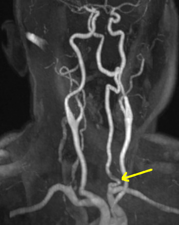
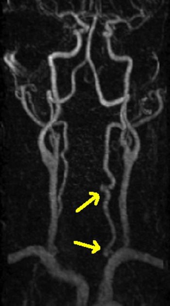
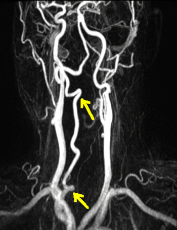
When the 37 patients identified by MRA as suffering from cervicogenic vertigo were evaluated, vertebral arterial loop formation was observed at only 1 level in 26 patients and at multiple levels in 9 patients (Figures 1–3). Vertebral arterial loop formation was observed in V1 in 78.3 per cent of cases (n = 29) (24.3 per cent (n = 9) on the right, 45.9 per cent (n = 17) on the left and 8.1 per cent (n = 3) bilateral) and in V2 in 21.6 per cent of cases (n = 8) (2.7 per cent (n = 1) on the right and 18.9 per cent (n = 7) on the left). Vertebral arterial loop formation was not observed in V3 or V4.
Magnetic resonance angiogram showing left vertebral arterial loop formation on the first segment (arrow).

Magnetic resonance angiogram showing left vertebral arterial loop formation on the first and second segments (arrows).

Magnetic resonance angiogram showing right vertebral arterial loop formation on the first and second segments (arrows).

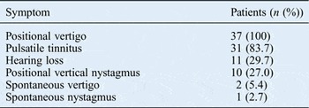
In our study, the symptoms of patients with vertebral arterial loop formation included: positional vertigo involving the neck, 100 per cent (n = 37); pulsatile tinnitus, 83.7 per cent (n = 31); hearing loss, 29.7 per cent (n = 11); positional vertical nystagmus, 27.0 per cent (n = 10); spontaneous vertigo, 5.4 per cent (n = 2); and spontaneous nystagmus, 2.7 per cent (n = 1) (Table I).

Discussion
There is no accepted definition, no ‘gold standard’ diagnostic test and no definitive treatment for cervicogenic dizziness.Reference Reid and Rivett 1 , Reference Han, Song and Kim 2 , Reference Furman and Whitney 4 , Reference Bracher, Almeida, Almeida, Duprat and Bracher 6 , Reference Wrisley, Sparto and Whitney 7 There are several approaches for determining the differential diagnosis of cervicogenic dizziness, and the diagnosis should not be based on only the combination of neck pain and dizziness.Reference Bracher, Almeida, Almeida, Duprat and Bracher 6 Cervicogenic dizziness may be defined as the combination of: dizziness, imbalance related to cervical spine position or movement, and painful or stiff neck.Reference Lystad, Bell, Bonnevie-Svendsen and Carter 3 Cervicogenic dizziness is generally accepted as a diagnosis of exclusion.Reference Wrisley, Sparto and Whitney 7 Dizziness secondary to vertebrobasilar insufficiency may generally be accompanied by visual hallucinations, sudden attacks of dizziness, loss of vision or visual field, diplopia, and headache.Reference Han, Song and Kim 2
A detailed history, comprehensive clinical examination, vestibular function tests and, potentially, radiological assessments provide input for diagnosis.Reference Han, Song and Kim 2 , Reference Wrisley, Sparto and Whitney 7 The clinical examination should address oculomotor function, posture, the active and passive range of motion of the cervical spine, instability of the cervical spine, mobility of the cervical vertebrae, compression of cervical spine, palpation of the cervical spine and pain; neurological examinations should also be conducted.Reference Han, Song and Kim 2 , Reference Olszewski and Repetowski 5 – Reference Wrisley, Sparto and Whitney 7 Although it is not specific to cervicogenic dizziness, a neck torsion and nystagmus test is widely used.Reference Han, Song and Kim 2 , Reference Furman and Whitney 4 , Reference Wrisley, Sparto and Whitney 7 In addition to the provocation of symptoms, development of nystagmus is an indicator of a positive result.Reference Han, Song and Kim 2 A smooth pursuit test can be used for diagnostic purposes during neck rotation; however, it requires special equipment, such as infrared illumination and Frenzel goggles.Reference Wrisley, Sparto and Whitney 7 A reduction in symptoms during traction may indicate a cervical component.Reference Han, Song and Kim 2 The Spurling test may be used to evaluate cervical compression. Patients are assessed individually for treatment purposes, and the intervention is determined according to clinical signs.Reference Han, Song and Kim 2 , Reference Furman and Whitney 4 The use of manual therapy techniques, such as mobilisation and manipulation, is supported for the treatment of cervicogenic dizziness.Reference Lystad, Bell, Bonnevie-Svendsen and Carter 3
Tortuous blood vessels are commonly seen in the cerebral arteries. While mild tortuosity is not commonly associated with clinical symptoms, severe tortuosity can lead to vertigo.Reference Zhang, Zhang, Zhang and Zhang 11 The aetiology and pathogenesis of tortuous vessels are poorly understood.Reference Cosar, Yaman, Eser, Songur and Ozen 12 Tortuosity has been used as a broad term, and it has been indicated that there are subgroups of looping, twisting and curving.Reference Ranganatha Sastry and Manjunath 13 In our study, we accepted sharper curling as looping. The classic diagnostic criteria go hand in hand with Epstein and colleagues’ description of this pathological entity, as they mentioned that vertebral arterial loop formation or other vascular malformations are displayed as signal void tubular structures on MRI, which necessitates magnetic resonance angiography to differentiate these vascular lesions.Reference Epstein and Silvergleid 9
Vertebral arterial loop formation was originally described by Hadley.Reference Hadley 14 Hadley identified cervical vertebrae bone erosion related vertebral arterial loop formation in 4 of 21 cadavers.Reference Hadley 14 Paksoy et al. reported the incidence of vertebral arterial loop formation as 7.5 per cent in their study of 173 patients with cervicobrachial pain symptoms.Reference Paksoy, Levendoglu, Ogün, Ustün and Ogün 15 However, the aetiology of vertebral arterial loop formation remains unclear. Oga et al. reported that vertebral arterial loop formation changed in parallel with cervical spondylotic changes.Reference Oga, Yuge, Terada, Shimizu and Sugioka 16 Sakaida et al. suggested that vertebral arterial loop formation developed as a result of vertebral artery elongation along with constriction in disc space.Reference Sakaida, Okada and Yamamoto 17
In this study, we investigated vertebral arterial loop formation in patients with cervicogenic vertigo only. We could not find any studies that reported the vertebral arterial loop formation ratio in asymptomatic cases. Most of the studies have been carried out on patients with: cervicobrachial pain symptoms,Reference Paksoy, Levendoglu, Ogün, Ustün and Ogün 15 cervical spondylotic alterations,Reference Oga, Yuge, Terada, Shimizu and Sugioka 16 constriction in disc spaceReference Sakaida, Okada and Yamamoto 17 or vertigo. Haemodynamic conditions, such as hypertension and atherosclerosis, have also been suggested as potential causes.Reference Sakaida, Okada and Yamamoto 17 Some authors have suggested that cervical trauma may contribute to the development of vertebral arterial loop formation.Reference Bruneau, Cornelius, Marneffe, Triffaux and George 18 Surgical treatments of vertebral arterial loop formation reported in the literature include microvascular decompression, foraminotomy of the compressed root sections and vascular reconstruction.Reference Epstein and Silvergleid 9
Lee et al. classified ‘rotational vertebral artery occlusion syndrome’ into four types according to location and pathology.Reference Lee, Lee and Yang 19 Kuether et al. reported certain results in relation to this classification in their patient series.Reference Kuether, Nesbit, Clark and Barnwell 20 In type 1, occlusion develops due to compression of the vertebral artery by the medial contour of the anterior scalene muscle and thyrocervical trunk when the patient turns his or her head. Although the cause of the vertebral artery occlusions associated with type 2 usually involves osteophytes, it may also involve fibrous bands and, rarely, soft tissues. Type 3, the most frequently observed, is caused by osseous or dural abnormalities. Symptoms emerge when the head is turned in the reverse direction. Type 4 is due to emboli in the intracranial section, and the patient's complaints continue when their head is in the neutral position. These embolic symptoms may be reversible.Reference Hadley 14
Psillas et al. reported that cases of subclavian steal syndrome presenting with symptoms of vertigo, loss of hearing and balance disorder had positional vertical nystagmus; however, they did not have spontaneous vertigo.Reference Psillas, Kekes, Constantinidis, Triaridis and Vital 21 The patients in our study were most frequently observed to have positional vertigo (100 per cent) and pulsatile tinnitus upon neck movement (83.7 per cent; Table I).
In our study, no occlusion was seen in the vertebral artery, and there was no discontinuation in flow; however, the frequency of vertebral arterial loop formation was highest on the proximal side, with a gradual decrease toward the distal side. The highest frequency was at V1 (78 per cent), and it was observed approximately twice as frequently on the left than on the right side of the segments in which it was present. This is because the vertebral artery originates from the subclavian artery on both sides, and the subclavian artery emerges directly out of the aortic arch on the left side. However, it emerges from the brachiocephalic trunk on the right side. Thus, the pressure and flow rate may be higher on the left side in the vertebral artery, and it may be higher on the proximal side than the distal side. This may have been why the incidence of vertebral arterial loop formation was higher in the proximal and left sides in the vertebral artery.
-
• This paper presents the frequency of vertebral artery loop formation in patients with cervicogenic vertigo
-
• Cervicogenic vertigo is characterised by dizziness related to cervical spine position
-
• Vertebral arterial loop formation is a rare anatomical variation that may result in bone erosion
-
• Magnetic resonance angiography is useful for diagnosis and imaging of vertebral arterial loop formation
-
• There is no accepted definitive diagnostic test or treatment for cervicogenic dizziness
Although there have been several case reports related to vertebral arterial loop formation, our study brought a novel approach to the literature in that we demonstrated an association with cervicogenic dizziness and classified it according to the level in which it was present. Our research, which comprised a small sample, should be considered preliminary; additional studies with larger samples are needed to confirm our findings.
Conclusion
In this study, we used magnetic resonance angiography to classify vertebral arterial loop formation associated with cervicogenic vertigo according to its level on the vertebral artery. Loop formation on the vertebral artery decreased in frequency from proximal to distal sides, and it was twice as frequent on the left side as the right side. The most frequently observed symptoms were positional vertigo and pulsatile nystagmus. The relationship between vertebral arterial loop formation and other types of co-existing dizziness should be investigated in further studies. This will elucidate the association between the presence of vertebral arterial loop formation and cervicogenic vertigo.