The root-ectoparasitic nematodes belonging to the genus Hemicriconemoides Chitwood & Birchfield, 1957, also known as sheathoid nematodes, have been reported to damage numerous crops all over the world (Geraert Reference Geraert2010; Sikora et al. Reference Sikora, Coyne, Hallmann and Timper2018; Van den Berg et al. Reference Van den Berg, Tiedt, Inserra, Stanley, Vovlas, Rius, Castillo and Subbotin2014). Many important crops and fruit trees, such as mango, litchi, citrus, grapevine, maize, pineapple, banana, rice, small millets, and tea, have been found infested by Hemicriconemoides spp., causing serious symptoms, e.g., leaf yellowing, root galls, root necrosis, root malformation, stunting, and wilting (Geraert Reference Geraert2010; Inserra et al. Reference Inserra, Stanley, Ochoa, Schubert, Subbotin, Crow and McSorley2014; Nguyen et al. Reference Nguyen, Nguyen, Linh Le and Trinh2020, Reference Nguyen, Trinh, Nguyen, Le and Bert2023b; Sikora et al. Reference Sikora, Coyne, Hallmann and Timper2018).
In terms of taxonomy and systematics, Hemicriconemoides was initially categorized under Criconematinae by Raski and Luc (Reference Raski and Luc1987). However, Siddiqi (Reference Siddiqi2000) and Geraert (Reference Geraert2010) later reclassified it as belonging to Hemicriconemoidinae, a classification supported by molecular approaches (Subbotin et al. Reference Subbotin, Sturhan, Chizhov, Vovlas and Baldwin2006; Subbotin et al. Reference Subbotin, Vovlas, Crozzoli, Sturhan, Lamberti, Moens and Baldwin2005). Within the genus Hemicriconemoides, there are currently 54 valid species documented worldwide (Geraert Reference Geraert2010; Maria et al. Reference Maria, Cai, Castillo and Zheng2018). However, the taxonomic status of some species, e.g., H. litchi, H. mangiferae, and H. strictathecatus, has been a subject of debate for many years due to their overlapping morphological traits (Nguyen et al. Reference Nguyen, Nguyen, Linh Le and Trinh2020). Traditional morphological identification of Hemicriconemoides spp. is challenging due to high intraspecific variability and the relatively large number of described species within the genus. Molecular data, specifically sequences of rDNA and mtDNA genes, have become valuable tools in nematode identification and phylogenetic analyses in general (Chen et al. Reference Chen, Ni, Yen and Tsay2007; van Megen et al. Reference van Megen, van den Elsen, Holterman, Karssen, Mooyman, Bongers, Holovachov, Bakker and Helder2009; Powers et al. Reference Powers, Harris, Higgins, Mullin, Sutton and Powers2011; Subbotin et al. Reference Subbotin, Vovlas, Crozzoli, Sturhan, Lamberti, Moens and Baldwin2005). Remarkably, recent studies applying molecular approaches have revealed the presence of cryptic species within the genus Hemicriconemoides (Van den Berg et al. Reference Van den Berg, Tiedt, Inserra, Stanley, Vovlas, Palomares-Rius, Castillo and Subbotin2015). Therefore, to address the need for accurate identification of these sheathoid nematodes, a combination of morphological and molecular approaches should always be applied.
In Vietnam, five Hemicriconemoides species have been reported, including H. brachyurus, H. cocophilus, H. litchi, H. microdoratus, and H. strictathecatus (Nguyen et al. Reference Nguyen, Nguyen, Linh Le and Trinh2020, Reference Nguyen, Trinh, Nguyen, Le and Bert2023b; Nguyen and Nguyen Reference Nguyen and Nguyen2000). However, molecular data of Hemicriconemoides spp. in Vietnam are only available for H. litchi (Nguyen et al. Reference Nguyen, Nguyen, Linh Le and Trinh2020). Given the importance of accurate identification for agricultural management and regulatory purposes, this study focuses on morphological and molecular characterizations of a new species belonging to the genus Hemicriconemoides associated with rose (Rosa gallica L.) in Vietnam, named Hemicriconemoides vietnamensis sp. n. The research aims to shed light on the taxonomy and phylogenetic relationships of this species, contributing to a better understanding of the diversity and distribution of the genus Hemicriconemoides.
Materials and methods
Sampling and extraction
Samples were obtained from the cultivation areas of rose in Vinh Phuc, Vietnam. The collection process involved clearing the detritus layer and then collecting soil (1 kg) and root (20 g) samples from the upper 30 cm soil layer. For taxonomical study, nematodes were extracted from the soil and the root using the modified Baermann tray method, following Whitehead and Hemming (Reference Whitehead and Hemming1965).
Morphological characterization
The extracted nematodes were preserved and fixed in TAF solution (91 ml H2O; 7 ml formalin 40%; 2 ml triethanolamine) before being transferred to anhydrous glycerine, following the protocol of Seinhorst (Reference Seinhorst1959). Permanent slides were prepared, and photomicrographs were taken, including measurements, using a Carl Zeiss Axio Lab.A1 microscope equipped with a digital camera.
Specimens designated for scanning electron microscopy were processed using a gradual dehydration protocol. Individuals were transferred through an ascending ethanol series (5%, 10%, 20%, 40%, 80%, 95%, and 98%), remaining in each concentration for 20 min. This was followed by three additional rinses in absolute ethanol, each lasting 10 min, to ensure complete dehydration. Subsequently, specimens were dried using a critical-point drying apparatus with liquid CO2 as the transitional fluid. After drying, nematodes were positioned on aluminium stubs using conductive double-sided carbon tape, taking care to orient diagnostically relevant regions appropriately. The mounted material was coated with a 10 nm gold layer using a sputter coater (120 s at 15 mA) to enhance surface conductivity. Observations were conducted with a JSM-840 scanning electron microscope (JEOL Ltd., Japan) at an accelerating voltage of 12 kV (Nguyen et al., Reference Nguyen, Nguyen, Le and Trinh2023a).
Molecular characterization
For molecular characterization, individual nematodes were sectioned into 2–3 pieces and placed in Eppendorf tubes containing 20μl of WLB solution (50 mM KCl;10 mM Tris pH 8.3; 2.5 mM MgCl2; 0.45% NP 40 (Tergitol Sigma); 0.45% Tween 20). The samples were then incubated at −20°C for at least 10 minutes. Following the incubation, 1 μl of proteinase K (1.2 mg ml−1) was added, and the samples were subjected to further incubation in a PCR machine at 65°C for 1 hour and at 95°C for 10 minutes. Subsequently, the samples were centrifuged at 14,000 rpm for 1 minute and stored at −20°C until they were used for PCR analysis, following the protocol described by Nguyen et al. (Reference Nguyen, Trinh, Couvreur, Nguyen and Bert2021).
The ITS, D2-D3 of 28S rDNA regions, and COI genes were amplified using the primers Vrain2R/Vrain2F (5′-CTTTGTACACACCGCCCGTCGCT-3′/5′-TTTCACTCGCCGTTACTAAGGGAATC-3′), D2A/D3B (5′-ACAAGTACCGTGGGGAAAGTTG-3′/5′-TCGGAAGGAACCAGCTACTA-3′), and JB3/JB4 (5′-TTTTTTGGGCATCCTGAGGTTTAT-3′/5′-TAAAGAAAGAACATAATGAAAATG-3′) (Nguyen et al. Reference Nguyen, Trinh, Couvreur, Nguyen and Bert2021, Reference Nguyen, Nguyen, Le, Trinh and Bert2022). Consensus sequences were assembled from both forward and backward sequences using Geneious R11. To construct the phylogenetic trees, closely related sequences were identified through Blast in GenBank (Altschul et al. Reference Altschul, Madden, Schäffer, Zhang, Zhang, Miller and Lipman1997). Multiple alignments were performed using Muscle in Geneious R11, and Bayesian phylogenetic trees were constructed with the best-fit models selected using Mega 7 (Nguyen et al. Reference Nguyen, Trinh, Couvreur, Nguyen and Bert2021).
Molecular species delimitation analyses were conducted utilizing three different methodologies: Bayesian implementation of Poisson tree processes (bPTP) (Zhang et al. Reference Zhang, Kapli, Pavlidis and Stamatakis2013), generalized mixed-Yule coalescent (GMYC) (Pons et al. Reference Pons, Barraclough, Gomez-Zurita, Cardoso, Duran, Hazell, Kamoun, Sumlin and Vogler2006), and Assemble Species by Automatic Partitioning (ASAP) (Puillandre et al. Reference Puillandre, Brouillet and Achaz2021). Ultrametric trees were prepared for bPTP and GMYC methods using BEAST v 1.10.424 with default parameters (1 × 10⁷ generations with subsampling every 1 × 10³ generations) following Nguyen et al. (Reference Nguyen, Nguyen, Le, Trinh and Bert2022). The final trees were generated utilizing TreeAnnotator 1.10.4, with a removal of 20% (2000 samples) designated as burn-ins. These final trees were then submitted to an online server (https://species.h-its.org/) to derive species delimitation results (Zhang et al. Reference Zhang, Kapli, Pavlidis and Stamatakis2013). For the ASAP method, aligned sequences in fasta format were uploaded to an online server (https://bioinfo.mnhn.fr/abi/public/asap/asapweb.html), and the best results with the smallest ASAP scores for each dataset were selected (Puillandre et al. Reference Puillandre, Brouillet and Achaz2021).
Results
Hemicriconemoides vietnamensis sp. n.
The female of Hemicriconemoides vietnamensis sp. n. from Vietnam. A: Entire body; B: pharyngeal region; C: en face view; D: annuli at mid-body; E, F: tail region.

Figure 1. Long description
A multi-panel figure containing six micrographs labeled A through F.
* Panel A: A light micrograph of the entire body, showing a curved, elongated nematode with a scale bar of 100 micrometers.
* Panel B: A high-magnification light micrograph of the anterior pharyngeal region, showing the internal stylet and esophageal structure. A scale bar indicates 10 micrometers.
* Panel C: A SEM en face view of the head, showing concentric rings of the cuticle surrounding the oral opening. A scale bar indicates 5 micrometers.
* Panel D: A light micrograph of the mid-body, highlighting the transverse cuticle annuli. A scale bar indicates 10 micrometers.
* Panel E: A SEM micrograph of the posterior tail region, showing the tapering end and distinct cuticular rings. A scale bar indicates 10 micrometers.
* Panel F: A light micrograph of the tail region, showing internal cellular structures and the pointed terminus. A scale bar indicates 10 micrometers.
Drawings of Hemicriconemoides vietnamensis sp. n. from Vietnam. A: Entire body; B: pharyngeal region; C: en face view; D: annuli at mid-body; E, F: tail region.

Figure 2. Long description
A multi-panel line drawing showing six anatomical views labeled A through F.
Panel A shows the entire body of the nematode, which is elongated and slightly curved. A scale bar indicates 100 micrometers.
Panel B provides a detailed view of the pharyngeal region at the anterior end. It shows a long, dark stylet with basal knobs and the surrounding pharyngeal bulb. A scale bar indicates 10 micrometers.
Panel C is an en face view of the head, showing a central oral opening surrounded by concentric circular layers and labial structures. A scale bar indicates 5 micrometers.
Panel D displays the annuli at the mid-body, showing the thick, transverse cuticle rings. A scale bar indicates 10 micrometers.
Panel E shows the tail region in a ventral view, highlighting the tapering end and the arrangement of the cuticle annuli. A scale bar indicates 10 micrometers.
Panel F shows the tail region in a lateral view, revealing internal reproductive structures and the pointed terminus of the tail. A scale bar indicates 10 micrometers.
Measurements
Measurements of Hemicriconemoides vietnamensis sp. n. from Vietnam and other countries. All measurements are in μm (except for ratio) and in the form: mean ± s.d. (range)

Table 1. Long description
The table consists of three columns: Character, Holotype female (n = 1), and Paratype females (n = 10). Measurements are in micrometers (mean plus or minus standard deviation and range).
Key morphological data includes:
* Body length (L): Holotype 531; Paratypes 527 plus or minus 28 (468–562).
* V percent: Holotype 93; Paratypes 92 plus or minus 1 (90–93).
* Lip width: Holotype 10.3; Paratypes 11.8 plus or minus 0.7 (10.3–12.7).
* Lip height: Holotype 5.2; Paratypes 5.2 plus or minus 0.3 (4.7–5.5).
* Knob height: Holotype 3.2; Paratypes 3.6 plus or minus 0.25 (3.2–4).
* Stylet: Holotype 64; Paratypes 65 plus or minus 2 (62–68).
* D G O: Holotype 2.9; Paratypes 2.7 plus or minus 0.5 (2.1–3.4).
* Anterior end to secretory-excretory pore: Holotype 115; Paratypes 123 plus or minus 4.6 (115–130).
* Anterior end to nerve ring: Holotype 84; Paratypes 88 plus or minus 3 (84–94).
* Anterior end to end of Pharynx: Holotype 100; Paratypes 104 plus or minus 3 (100–109).
* Max body diam. (M B D): Holotype 28; Paratypes 31 plus or minus 1 (28–32).
* V L: Holotype 35; Paratypes 38 plus or minus 4 (33–47).
* V B D: Holotype 24; Paratypes 25 plus or minus 1 (24–27).
* A B D: Holotype 20; Paratypes 20 plus or minus 0.7 (19.5–21).
* Tail length: Holotype 23; Paratypes 24 plus or minus 3 (18–29).
* R: Holotype 131; Paratypes 125 plus or minus 4 (120–131).
* R s t: Holotype 21; Paratypes 19 plus or minus 1 (18–21).
* R pharynx: Holotype 31; Paratypes 28 plus or minus 2 (25–31).
* R e x: Holotype 35; Paratypes 32 plus or minus 2 (31–35).
* R V: Holotype 11; Paratypes 11 plus or minus 1 (10–14).
* R v a n: Holotype 4; Paratypes 4 plus or minus 1 (3–5).
* R a n: Holotype 7; Paratypes 7 plus or minus 1 (6–9).
* a: Holotype 18.8; Paratypes 17.1 plus or minus 0.8 (16.2–18.8).
* b: Holotype 5.3; Paratypes 5.0 plus or minus 0.3 (4.4–5.4).
* c: Holotype 23; Paratypes 23 plus or minus 3.8 (18.4–30).
* c prime: Holotype 1.2; Paratypes 1.2 plus or minus 0.2 (0.9–1.4).
* V L / V B: Holotype 1.5; Paratypes 1.5 plus or minus 0.2 (1.3–1.8).
Description
Females: Body slightly curved ventrally (Figs. 1A, 2A). Sheath closely fitting to body, occasionally extending anteriorly over lip area. Lip region with two annuli, first one protruding outward. Lip region comprised of dorsoventrally oriented oral disc with slit-like opening and two prominent amphidal apertures (Figs. 1C, 2C). Stylet long, distinct, mostly straight. Stylet knobs rounded, posteriorly sloped, or sometimes laterally directed (Figs. 1B, 2B). Dorsal pharyngeal gland opening 2.1–3.4 μm posterior to stylet base. Pharyngeal lumen forming loop within median bulb, connected to refractive valve. Isthmus narrow, short, encircled by nerve ring. Distinct basal pharyngeal bulb present. Excretory pore distinct, 5–7 annuli posterior to pharynx end. Hemizonid and hemizonion not observable (Figs. 1B, 2B). Anastomoses absent (Fig. 2D). Vulva slit-like, lacks vulval flaps. Vagina oblique, extends half body diameter (Fig. 1E, F, 2F). Spermatheca rounded to oblong, contains oval sperm. Anus indistinct, 3–5 annuli posterior to vulva (Fig. 2E). Body gradually narrows from vulval region to tail tip, which is bluntly conical to pointed conical with rounded tip (Figs. 1E, F, 2E, F).
Male: Not found.
Etymology
The epithet refers to the name of the country where it has been found.
Molecular characterization and phylogenetic relationship
Characterization of ITS rDNA region
Three ITS rDNA sequences from Hemicriconemoides vietnamensis sp. n. were obtained in this study and submitted to the GenBank database under accession numbers PX957616–PX957618. The ITS rDNA sequences of Hemicriconemoides vietnamensis sp. n. from Vietnam are 99.5–99.8% similar to each other. They are closest to the sequences of H. strictathecatus with only 93.5–93.7% similarity (60–62 bp difference). The sequences of Hemicriconemoides vietnamensis sp. n. and H. strictathecatus formed a single clade with the maximum Posterior Probability support (Fig. 3). All the ITS sequences of Hemicriconemoides species recovered in this study were recognized as a new species using three molecular species delimitation methods (Fig. 3).
BI phylogenetic tree generated from ITS rDNA sequences (GTR + G + I model, 1 × 10⁶ generations, 20% Burn-in). Bayesian posterior probabilities are given next to each node. Sequences of Hemicriconemoides vietnamensis sp. n. from Vietnam are in bold font.

Figure 3. Long description
A horizontal cladogram with a scale bar of 0.06 at the bottom. The tree originates from an outgroup containing Criconema silvum MF6 83 23 6 and Criconema paraannuliferum ON7 05 09 7.
Moving from top to bottom, the main clades include:
* A top cluster featuring H. gaddi, H. silvaticus, H. parataiwanensis, H. californianus, H. chitwoodi, H. paracamelliae, H. parasinensis, and H. varionodus. Bayesian posterior probabilities at nodes range from 0.69 to 1.
* A middle cluster containing H. stricthecatus and a highlighted red bold group of Hemicriconemoides vietnamensis (P X9 57 61 6, PX9 57 61 7, PX9 57 61 8). This group is nested near H. californianus KF8 56 55 9 and H. fujianensis. A vertical blue bracket to the right of this section is labeled H. litchi.
* A lower large cluster including H. alexis, H. kanayaensis, H. minutus, H. wessoni, H. rosae, H. cocophillus, H. ortonwilliamsi, H. brachyurus, H. pseudobrachyurus, H. promissus, and H. macrodorus.
On the far right, three vertical colored bars (red for GMY C, blue for bPT P, and purple for ABGD) indicate species delimitation results. Most species are marked by all three bars, though some segments are broken or missing for specific taxa. In the top left corner, a legend identifies these three methods: GMYC in red, bPTP in blue, and ABGD in purple.
Characterization of D2-D3 of 28S rDNA region
Two sets of D2-D3 regions of 28S rDNA sequences from Hemicriconemoides vietnamensis sp. n. were acquired (accession numbers: PZ321692 and PZ321693). These sequences varied in length from 681 to 699 bp. These sequences are identical. The closest similarity was found between these sequences and those of H. phoenicis, sharing a similarity range of 97.6% to 97.7%, differing by 15 to 16 bp. Within the phylogenetic tree based on the D2-D3 regions, the sequences from Hemicriconemoides vietnamensis sp. n. were also found to be closest to the sequences of H. phoenicis, forming a sister relationship with 0.82 PP support. The sequences of Hemicriconemoides vietnamensis sp. n., H. phoenicis, and H. strictathecatus formed a single clade with maximal Posterior Probability (Fig. 4). The application of three molecular species delimitation methods (bPTP, GMYC, and ABDG) consistently identified the D2-D3 sequences of Hemicriconemoides vietnamensis sp. n. as a new species (Fig. 4).
BI phylogenetic tree generated from D2-D3 of 28S rDNA sequences (GTR + G + I model, 1 × 10⁶ generations, 20% Burn-in). Bayesian posterior probabilities are given next to each node. Sequences of Hemicriconemoides vietnamensis sp. n. from Vietnam are in bold font.

Figure 4. Long description
A rectangular cladogram rooted on the left with Crossonema civellae and Criconema mutabile. The tree branches rightward into several major clades of Hemicriconemoides.
* The top major clade includes species such as H. paracamelliae, H. chitwoodi, H. parataiwanensis, H. parasinensis, H. varionodus, H. gaddi, and H. silvaticus.
* The middle section contains a cluster including H. phoenicis and the target species Hemicriconemoides vietnamensis (P Z 3 2 1 6 9 2 and P Z 3 2 1 6 9 3) highlighted in red bold text. This is followed by a large group of H. strictathecatus and H. californianus.
* The lower section includes clades for H. alexis, H. fujianensis, H. kanayaensis, H. ortonwilliamsi, H. cocophillus, H. brachyurus, H. wessoni, H. minutus, H. rosae, H. promissus, and H. macrodorus.
Bayesian posterior probability values are positioned at each node, ranging from 0.59 to 1. To the right of the species names, three vertical bars represent different species delimitation methods: GMYC (red), bPTP (blue), and ASAP (purple). A scale bar at the bottom indicates 0.03 substitutions per site.
Characterization of COI mtDNA region
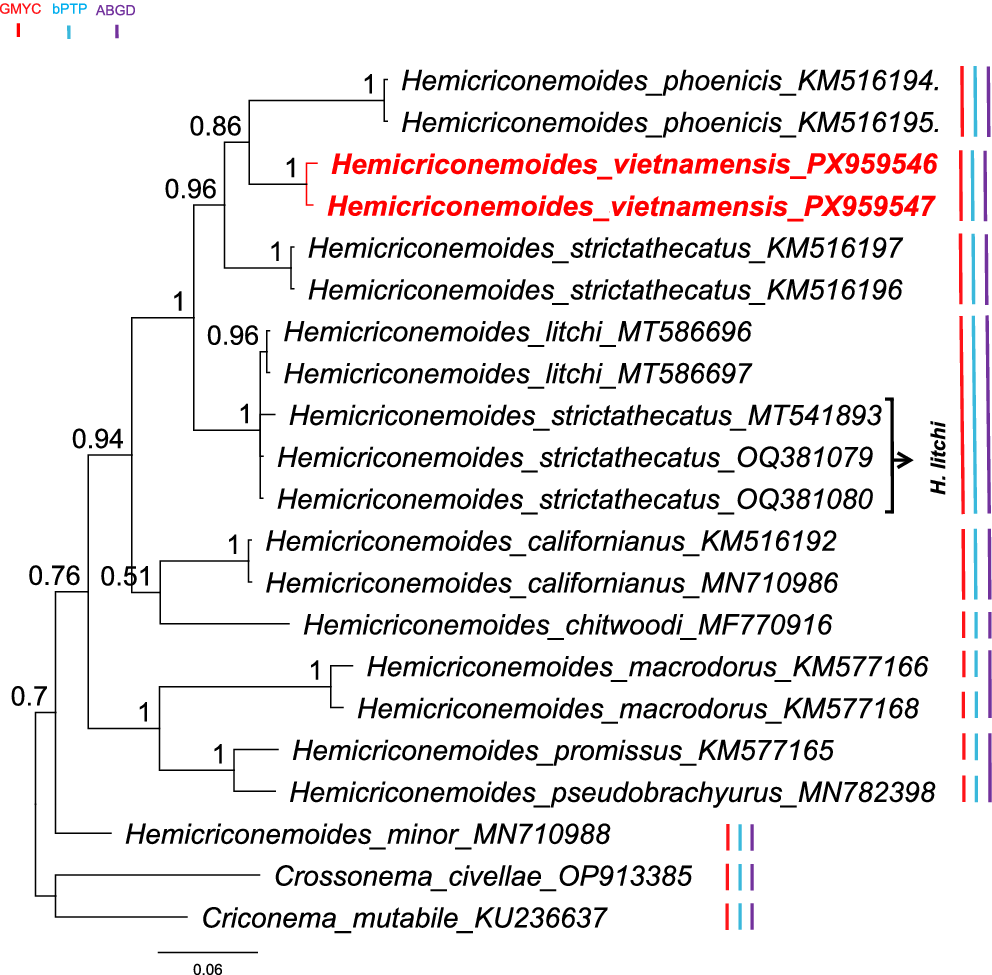
Two COI mtDNA sequences were obtained, which varied from 431 to 432 bp (accession numbers: PX959546 and PX959547). The COI mtDNA sequences of Hemicriconemoides vietnamensis sp. n. from Vietnam are 99.26% similar (3 bp difference) to each other. The COI mtDNA sequences of Hemicriconemoides vietnamensis sp. n. from this study are most similar (92.2–92.5% similarity; 28–29 bp difference) to the sequence of H. strictathecatus (accession numbers: KM516196 and KM516197). The phylogenetic tree based on COI mtDNA sequences showed that two sequences of Hemicriconemoides vietnamensis sp. n. were placed in a maximally supported clade, and this clade has a sister relationship to the sequences of H. phoenicis (accession numbers: KM516194 and KM516195) and H. strictathecatus (accession numbers: KM516196 and KM516197) (Fig. 5). Consistent with the aforementioned analyses, three molecular species delimitation methods confirmed the distinct species status of all COI sequences of Hemicriconemoides vietnamensis sp. n.
BI phylogenetic tree generated from COI mtDNA sequences (GTR + G model, 1 × 10⁶ generations, 20% Burn-in). Bayesian posterior probabilities are given next to each node. Sequences of Hemicriconemoides vietnamensis sp. n. from Vietnam are in bold font.

Figure 5. Long description
A rectangular cladogram showing evolutionary relationships.
* Root and Main Nodes: The tree originates from a root on the left with a posterior probability of 0.7. It splits into two main lineages. The lower lineage leads to Criconema mutabile K U 236637 and Crossonema civellae O P 913385. The upper lineage branches further into several clades.
* Top Clade: Contains Hemicriconemoides phoenicis (K M516194, KM516195) and the target species Hemicriconemoides vietnamensis (P X959546, PX959547) highlighted in bold red text. These share a node with a value of 0.86.
* Middle Clades: Includes Hemicriconemoides stricthecatus and Hemicriconemoides litchi. A bracket groups three H. stricthecatus sequences (M T541893, OQ381079, OQ381080) pointing to the label H. litchi.
* Lower Clades: Includes Hemicriconemoides californianus, Hemicriconemoides chitwoodi, Hemicriconemoides macrodorus, Hemicriconemoides promissus, Hemicriconemoides pseudobrachyurus, and Hemicriconemoides minor.
* Support Values: Numerical values between 0.51 and 1 are placed at each branching node.
* Species Delimitation Bars: To the right of the taxon names are three vertical colored lines: red (G MYC), light blue (b PTP), and purple (A BGD). These bars indicate species clusters identified by different methods. A legend for these colors is located at the top-left corner.
* Scale: A horizontal scale bar at the bottom represents 0.06 substitutions per site.
Diagnosis and relationships
Hemicriconemoides vietnamensis sp. n. can be diagnosed by the following distinct characteristics in its females: the body exhibits a slight ventral curvature, with the sheath closely adhering to the body, without anastomoses. The stylet is long and mostly straight, with rounded, posteriorly sloped, or sometimes laterally directed stylet knobs. The excretory pore is well-defined, positioned 5–7 annuli posterior to the end of the pharynx. Hemizonid and hemizonion are not visible. The reproductive system includes a slit-like vulva lacking vulval flaps, an oblique vagina extending for half the body diameter, and a rounded to oblong spermatheca containing rounded sperm. The anus is not distinctly visible. The body gradually narrows from the vulval region towards the tail tip, which is either bluntly conical or pointed conical in shape with a rounded tip.
Based on the identification key of Geraert (Reference Geraert2010) and morphological comparison with recently discovered species (Van den Berg et al. Reference Van den Berg, Tiedt, Inserra, Stanley, Vovlas, Palomares-Rius, Castillo and Subbotin2015; Maria et al. Reference Maria, Cai, Castillo and Zheng2018), Hemicriconemoides vietnamensis sp. n. is most similar to H. litchi, H. rotundoides, and H. strictathecatus. However, the new species can be distinguished from these species by distinct characteristics as below.
Hemicriconemoides vietnamensis sp. n. differs from H. litchi by the tail (bluntly conical to pointed conical in shape vs bluntly rounded), slightly larger body length (527 (468–562) vs 480 (450–500) μm), slightly smaller R value (120–131 vs 128–133), and larger c value (18.4–30 vs 19–23). Hemicriconemoides vietnamensis sp. n. can also be distinguished from H. litchi (H. litchi is a valid species following the division of Van den Berg et al. (Reference Van den Berg, Tiedt, Inserra, Stanley, Vovlas, Palomares-Rius, Castillo and Subbotin2015) and Nguyen et al. (Reference Nguyen, Nguyen, Linh Le and Trinh2020)) by molecular data of D2-D3 of 28S rDNA region (5.5–7% or 38 bp difference), ITS rDNA region (7–8.2% or 68–72 bp difference), and COI mtDNA region (8.1–8.8% or 34–36 bp difference). Additionally, three molecular species delimitation methods differentiated Hemicriconemoides vietnamensis sp. n. from H. litchi.
Hemicriconemoides vietnamensis sp. n. can be differentiated from H. rotundoides by the characterizations of the females, including the position of the excretory pore (located 5–7 vs 4–5 annuli posterior to the end of pharynx), the tail (bluntly conical to pointed conical in shape vs tail dorsally convex with the bluntly rounded terminus), the cuticular sheath (not well separated at the tail terminus at most of the specimens vs the cuticular sheath is loose at the tail terminus), and the higher RV value (10–14 vs 8–12 annuli). Molecular data of H. rotundoides is not available in GenBank.
Hemicriconemoides vietnamensis sp. n. can be separated from H. strictathecatus by the stylet knob (rounded, posteriorly sloped or sometimes laterally directed vs anchor-shaped), shorter stylet (62–68 vs 73–83 μm), smaller R value (120–131 vs 127–152), smaller Ran value (6–9 vs 9–11), and smaller VL/VB ratio (1.3–1.8 vs 1.5–2.0). Hemicriconemoides vietnamensis sp. n. is also different from H. strictathecatus by molecular data as indicated above.
Type host and locality
The new species was recovered from the rhizosphere of rose in Vinh Phuc, Vietnam (GPS coordinates 21°12′56″N 105°29′08″E).
Type material
Holotype female (slide number: R7472-01) and ten female paratypes (slide numbers: R7472-02 and R7472-03) were deposited at the Biology, Vietnam Academy of Sciences and Technology, Vietnam. The new species has been registered in accordance with the International Code of Zoological Nomenclature (ICZN), and the associated Life Science Identifier (LSID) is: urn:lsid:zoobank.org:act:11744DAB-39F5-4536-8E1C-3674252DA337.
Discussion
The concordance between morphological and molecular evidence provides robust support for the recognition of Hemicriconemoides vietnamensis sp. n. as a distinct species. Although morphologically closest to H. litchi, H. rotundoides, and H. strictathecatus, the new species is consistently separable by a discrete combination of characters. These traits, which are often subject to overlap in Hemicriconemoides, form a stable diagnostic set when considered collectively rather than individually. Molecular divergence further substantiates this distinction, with all three markers (ITS, D2–D3 of 28S rDNA, COI) showing levels of sequence differentiation exceeding typical intraspecific ranges reported for criconematids. The phylogenetic reconstruction places H. vietnamensis sp. n. in a well-supported lineage sister to H. phoenicis and H. strictathecatus, yet clearly separated from both, indicating recent but independent evolutionary divergence. Notably, the full agreement among bPTP, GMYC, and ASAP analyses reduces the likelihood that the observed genetic structuring reflects population-level variation or sampling artefacts. Instead, it supports species-level separation, highlighting the value of combining multiple loci with complementary delimitation frameworks. Taken together, the evidence suggests that reliance on single-character morphology or single-marker datasets would likely underestimate species boundaries within the genus, particularly in groups with subtle morphological differentiation.
Remarkably, three species delimitation methods revealed potential misidentification or mislabelled sequences in our phylogenetic trees. For instance, most species delimitation methods recognized Hemicriconemoides promissus as conspecific with H. pseudobrachyurus across three gene regions (Figures 2, 3, and 4). This finding concurs with the conclusion of Etongwe et al. (Reference Etongwe, Singh, Bert and Subbotin2020) to synonymize H. promissus with H. pseudobrachyurus. Similarly, in the ITS phylogeny, H. californianus (EU180057) was consistently recovered as conspecific with H. chitwoodi (KJ934162) by all three species delimitation methods, thereby supporting the reassignment of this sequence to H. chitwoodi as proposed by Van den Berg et al. (Reference Van den Berg, Tiedt, Inserra, Stanley, Vovlas, Rius, Castillo and Subbotin2014). Besides, the two sequences of H. californianus (EU180057 and KF856559) were well separated from each other, indicating they represent two different species. On the other hand, the intraspecific variation between sequences of a single species can result in an overestimation of species by different delimitation methods. This phenomenon is observed with species such as H. alexis, H. fujianensis, H. cocophillus, and H. minutus in the 28S tree, and H. cocophillus in the ITS tree. These findings align with previous studies indicating that species delimitation methods often recover more species than actually exist (Nguyen et al. Reference Nguyen, Nguyen, Le, Trinh and Bert2022; Singh et al. Reference Singh, Karssen, Couvreur, Subbotin and Bert2021).
The phylogenetic topology recovered in this study corroborates the multi-clade structure of the genus previously established by Van den Berg et al. (Reference Van den Berg, Tiedt, Inserra, Stanley, Vovlas, Rius, Castillo and Subbotin2014) and Van den Berg et al. (Reference Van den Berg, Tiedt, Inserra, Stanley, Vovlas, Palomares-Rius, Castillo and Subbotin2015), reinforcing the hypothesis that molecular lineages within Hemicriconemoides are strongly congruent with lip region morphology. Our results refine existing systematic hypotheses by demonstrating that while H. vietnamensis sp. n. is morphologically similar to H. rotundoides; its evolutionary trajectory is most closely aligned with the H. strictathecatus-H. litchi-H. phoenicis complex. This sister-group relationship with H. phoenicis in the 28S tree (0.82 PP) and its close proximity to H. strictathecatus in the COI tree (92.2–92.5% similarity) support the validity of these species as distinct but closely related evolutionary units.
Furthermore, our molecular findings contribute to resolving the long-standing taxonomic debate surrounding the “litchi-mangiferae-strictathecatus” complex. By utilizing multiple species delimitation methods (bPTP, GMYC, and ASAP), our study provides robust evidence that H. vietnamensis sp. n. is a distinct taxon, separate from H. litchi and H. strictathecatus, despite the overlapping measurements often found in this group. This placement reinforces the shift toward a polyphasic approach in nematology, where molecular markers are used to substantiate or challenge traditional morphometric delimitations that have historically led to synonymy disputes. For instance, while some authors previously proposed synonymizing H. litchi and H. mangiferae with H. strictathecatus based on morphological variability, our data support the more recent interpretation that these are distinct species within a closely related cluster (Nguyen et al. Reference Nguyen, Nguyen, Linh Le and Trinh2020; Van den Berg et al. Reference Van den Berg, Tiedt, Inserra, Stanley, Vovlas, Rius, Castillo and Subbotin2014, Reference Van den Berg, Tiedt, Inserra, Stanley, Vovlas, Palomares-Rius, Castillo and Subbotin2015).
Acknowledgements
This research was financially supported by the Institute of Biology under project code CSCL09.01/25–26.