The association of aortic atresia with an aortopulmonary communication is rare, with very few cases currently reported.Reference Rosenquist, Taylor and Stark1–Reference Atik, Cury and Albuquerque5 In all described thus far, there was associated interruption of the aortic arch. The finding of a solitary arterial trunk is most frequently found when the trunk itself is a common structure, supplying directly the systemic, pulmonary, and coronary arterial circulations. Unlike the situation when aortic atresia exists in isolation, however, the combination with an aortopulmonary window is associated with a well developed ascending aorta, which facilitates subsequent surgical management.Reference Rosenquist, Taylor and Stark1, Reference Redington, Rigby and Ho2, Reference Kutshe and Van Mierop6
Case report
We were presented with a neonate aged 20 days, born to non-consanguineous parents after an uneventful antenatal and natal history, who had suffered for 2 weeks with rapid breathing and difficulties in feeding. On examination, he was tachycardic and tachypnoeic, with arterial saturations of 85% in the right arm, and 80% in the right leg. The pulses in the right arm were bounding, whereas those in the left arm and legs were diminished.
Cardiovascular examination revealed a hyperdynamic precordium, with a normal first heart sound and a single loud second heart sound. There was an ejection systolic murmur graded at 2 from 6 audible at the left upper sternal border and left infraclavicular area. The liver was palpable 2 centimetres below the right costal margin. Mild cardiomegaly and increased pulmonary vascular markings were noted on the chest radiogarph, whilst the electrocardiogram revealed a QRS axis of 90° and right ventricular hypertrophy. The baby was stabilized with mechanical ventilation, and an infusion of prostaglandin E1 was commenced presuming the possibility of a ductal dependent lesion.
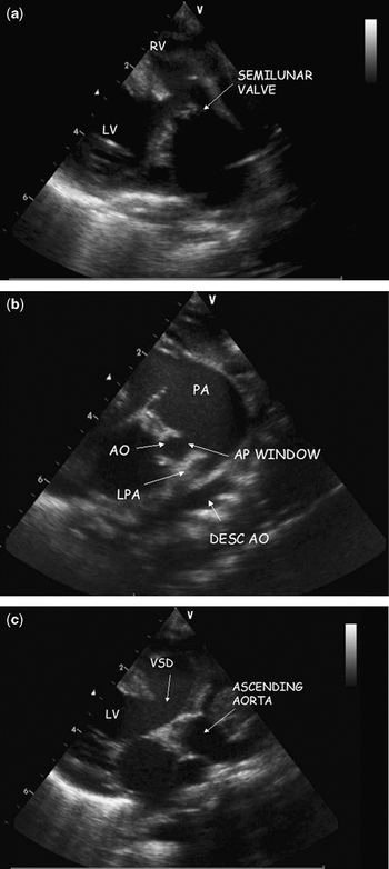
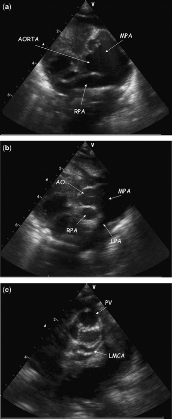
Echocardiogram revealed usual arrangement of the atrial appendages and thoraco-abdominal organs, with concordant atrioventricular connections. A solitary arterial trunk was visualized leaving the ventricular mass in all views, along with a large subarterial ventricular septal defect (Fig. 1a). The arterial trunk was interpreted as giving rise to the ascending aorta, and then bifurcating into the right and left pulmonary arteries. The initial diagnosis, therefore, was common arterial trunk. Findings contraindicating this diagnosis, however, were that the trunk bore an odd orientation relative to the ascending aorta. Further inspection revealed that the trunk arose from the morphologically right ventricle, and terminated at its bifurcation to the right and left pulmonary arteries. Careful study of the sections obtained in the parasternal long and short axes showed that the aorta and pulmonary trunk were normally related, and that a large communication existed between the intrapericardial components of the arterial trunks, the aortic valve being atretic (Figs 1b and c, 2a and b). The fact that the coronary arteries arose from the ascending aorta supported still further our final diagnosis of aortic atresia with aortopulmonary window (Fig. 2c). The aortic arch was interrupted distal to the right brachiocephalic artery, with a restrictive arterial duct supplying the descending aorta, and feeding the left common carotid and subclavian arteries in retrograde fashion.
Parasternal long axis echocardiographic view (a) with an anterior tilt showing a solitary arterial valve and trunk looking suspiciously like a common trunk arising from the right ventricle. Note the subarterial ventricular septal defect. Panel b shows the communication between the pulmonary trunk and the aorta, while panel c shows the normal relation of the ascending aorta, but with an atretic origin (RV = right ventricle, LV = left ventricle, PA = pulmonary trunk, AO = aorta, LPA = left pulmonary artery, AP window = aortopulmonary window, VSD = ventricular septal defect).

The parasternal short axis view at the base of the heart, in panels a & b, shows the pulmonary valve and trunk dividing to give rise to the right and left pulmonary arteries, and communicating with the aorta through an aortopulmonary window. Panel c shows the trifoliate pulmonary valve, and the main stem of the left coronary artery arising from the atretic aorta rather than the pulmonary trunk. MPA = pulmonary trunk, RPA = right pulmonary artery, PV = pulmonary valve, LMCA = main stem of left coronary artery.

There was no improvement in the clinical condition subsequent to the infusion of prostaglandin E1, so we proposed surgical intervention, but the parents decided against further treatment.
Discussion
Our initial echocardiographic diagnosis was common arterial trunk, with origin of the pulmonary arteries through a confluent segment arising from the left lateral aspect of the common trunk, the coronary arteries also presumed to be arising from the common trunk. Our subsequent interrogation revealed that the presumed common trunk continued as the pulmonary trunk, with the ascending aorta arising from its rightward and posterior component. Furthermore, the coronary arteries did not arise from the presumed common trunk. These findings indicated that, in reality, the arterial trunks were normally related, with an atretic ascending aorta supplied through a large aortopulmonary window, the pulmonary trunk itself arising exclusively from the right ventricle.
Aortic atresia is most commonly encountered in the setting of an intact ventricular septum and hypoplastic left ventricle. Cases are well recognised, nonetheless, in the setting of a ventricular septal defect and well-developed left ventricle,Reference Thiene, Gallucci and Macartney7 which was also the situation in our patient. The ascending aorta, however, is usually also hypoplastic in the setting of aortic atresia. In our patient, the well-developed ascending aorta is presumably the consequence of the presence of the large aortopulmonary window.
Interruption of the aorta between the brachiocephalic artery and the left common carotid artery is equally rare. It could result from an absolute decrease in the left ventricular output to the ascending aorta, in consequence of the aortic atresia, or may relate to primary deficiency of migration from the neural crest to the aortic arches.
Current embryological hypotheses explain separation of the aorta and pulmonary arteries on the basis of growth of a spiral septum, which allegedly divides the common embryonic outflow tract into the separate intrapericardial arterial trunks, albeit that more recent studies cast doubt on this concept.Reference Anderson, Webb, Brown, Lamers and Moorman8 It is certainly difficult to explain the association of aortic atresia and an aortopulmonary window when using the classical hypothesis.
To date, to the best of our knowledge, aortopulmonary window has not been encountered when neural crest tissue is removed experimentally.Reference Kirby, Gale and Stewart9 In our patient, the aortopulmonary window was the sole source of arterial supply to the coronary arteries and the brachiocepahlic artery. It could equally be that the anomalies, though involving the same region of the heart, were embryologically unrelated.
It is difficult clinically to distinguish common arterial trunk from aortic atresia with aortpulmonary window. The surgical management, paradoxically, would have been the same as for common arterial trunk with interruption of the aortic arch. Such management would have been made easier in our patient by the presence of a well developed ascending aorta. The surgeon would have baffled the left ventricular outflow tract through the ventricular septal defect to the pulmonary valve, using this channel to feed the systemic circulation having disconnected the pulmonary arteries, re-establishing continuity between the right ventricle and the pulmonary arteries with a homograft.
Our experience emphasises the importance of establishing precisely the origin of coronary arteries in cases of suspected common arterial trunk. It also shows the importance of considering associated anomalies such as aortopulmonary window in the setting of aortic atresia and interruption of the aortic arch, a combination which can explain the well-developed ascending aorta, and help potentially in subsequent survival.