Necrotising enterocolitis is a significant cause of morbidity and mortality in infants with congenital heart disease. Prevalence of necrotising enterocolitis in infants with congenital heart disease is significantly higher than that of the normal newborn population, especially in infants with single ventricle physiology such as hypoplastic left heart syndrome.Reference Jeffries, Wells, Starnes, Wetzel and Moromisato 1 – Reference Abdel-Massih, Boudjemline and Bonhoeffer 3 Although there are likely multiple factors contributing to the development of necrotising enterocolitis in infants, altered systemic perfusion is of greatest concern in those with congenital heart disease.Reference Lin and Stoll 4
There are now multiple initial palliative surgical options for patients with hypoplatic left heart syndrome;Reference Galantowicz, Cheatham and Phillips 5 – Reference Cua, Galantowicz and Turner 7 regardless of the type of initial surgical palliation, however, the incidence of necrotising enterocolitis appears to be comparable between the procedures.Reference Jeffries, Wells, Starnes, Wetzel and Moromisato 1 , Reference McElhinney, Hedrick and Bush 2 , Reference Luce, Schwartz and Beauseau 8 – Reference del Castillo, Moromisato and Dorey 10 Previous studies have documented an association between reversed diastolic flow in the abdominal aorta and the development of necrotising enterocolitis in neonates with congenital heart disease.Reference McElhinney, Hedrick and Bush 2 , Reference Carlo, Kimball, Michelfelder and Border 11 Harrison et alReference Harrison, Davis and Reid 12 reported that superior mesenteric artery perfusion in infants with hypoplastic left heart syndrome was impaired both before and after palliation with the Norwood procedure. This same impairment has been reported for single ventricle neonates following systemic-to-pulmonary arterial shunt palliation.Reference Cheung, Ho and Cheng 13 There have, however, been no studies assessing mesenteric blood flow in patients undergoing the hybrid procedure, which consists of bilateral pulmonary artery banding, stenting of the ductus arteriosus, and balloon atrial septostomy.
The objective of this study was to document coeliac and superior mesenteric artery blood flow patterns in patients with hypoplastic left heart syndrome by pre- and post-hybrid procedure.
Materials and methods
The Institutional Review Board approved this prospective cross-sectional study conducted on patients with hypoplastic left heart syndrome at Nationwide Children’s Hospital. Patients were included in this study if they had the diagnosis of hypoplastic left heart syndrome defined as aortic atresia/mitral atresia, aortic atresia/mitral stenosis, aortic stenosis/mitral atresia, or aortic stenosis/mitral stenosis, and were scheduled to undergo a hybrid procedure as their initial palliative procedure. Patients were excluded if they had the diagnosis of hypoplastic left heart syndrome, but were not scheduled to have a hybrid palliation. Patients were also excluded if they had a diagnosis of a hypoplastic left heart syndrome variant, such as an unbalanced atrioventricular septal defect, double outlet right ventricle, heterotaxy, or unbalanced truncus arteriosus.
Baseline demographics were collected and included gender, gestational age, and birth weight. Age at pre-hybrid mesenteric blood flow analysis, age at hybrid procedure, and age at post-hybrid mesenteric blood flow analysis were also recorded. Incidence of necrotising enterocolitis, defined as Bell’s stage ≥2, was also recorded.
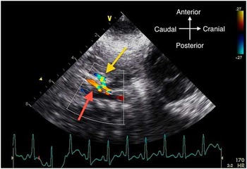
Pre- and post-mesenteric blood flow analysis was performed only when patients were breathing without invasive support and not receiving any continuous inotropic support. Ultrasounds were performed pre- and post-hybrid procedure for each patient at least 1 hour after feeding to make the studies as homogeneous as possible, unless the patients were getting continuously fed. Doppler examination of the superior mesenteric artery and coeliac artery was conducted by a single person (C.L.C.), and measurements were taken by a single observer (C.T.C.). A Vivid I ultrasound machine (GE Healthcare, Wauwatosa, Wisconsin, United States of America ) with an appropriate sized transducer (7–10 MHz) was used for image acquisition. The transducer was positioned in the mid-abdomen just under the sternum in the sagittal plane. Colour flow mapping was used to identify the appropriate vessels. The coeliac artery was defined as the first arterial branch arising off the descending aorta below the diaphragm, and the superior mesenteric artery was defined as the arterial vessel immediately caudal to the coeliac artery (Fig 1).
Colour analysis of coeliac and superior mesenteric artery blood flow. Red arrow, superior mesenteric artery; yellow arrow, coeliac artery.

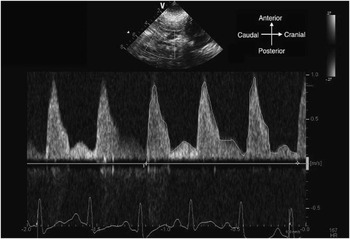
Sample volume was placed within 2–3 mm of the respective vessels for interrogation. Angle of insonation was <15° for all measurements. Pulse wave Doppler analysis was performed for both vessels. Doppler indices measured included antegrade velocity-time integral and retrograde velocity-time integral (Fig 2). Effective velocity-time integral was calculated as antegrade velodity-time integral–retrograde velocity-time integral. Retrograde velocity-time integral/antegrade velocity-time integral ratio was also determined. All measurements were made in triplicate.
Doppler analysis of coeliac blood flow.

Results are presented as means and standard deviations unless otherwise specified. Paired t-tests were used to test for significant differences in means between groups; p≤0.05 for a two-tailed test was considered statistically significant. All analyses were performed using Stata version 12 SE (StataCorp. 2011, Stata Statistical Software: Release 12; StataCorp LP, College Station, Texas, United States of America).
Results
A total of 13 infants met the inclusion criteria, 8 male and 5 female. Gestational age was 38.4±1.3 weeks, with a birth weight of 3.2±0.4 kgs. Age at pre-hybrid ultrasound assessment was 2.6±1.9 days, age at hybrid procedure was 4.7±1.9 days, and age at post-hybrid assessment was 10.5±6.1 days. Of the patients, three patients developed necrotising enterocolitis during their hospital course. At the time of ultrasound analysis, there was no significant difference in heart rates pre- and post-hybrid procedure (166+13 versus 159+11 bpm, respectively). Studies were conducted on two patients undergoing pre- and post-hybrid procedure while they were continuously being fed.
There was a trend towards a significant difference in the coeliac antegrade velocity-time integral pre- versus post-surgery values. There were no significant differences in the retrograde velocity-time integral and retrograde velocity-time integral/antegrade velocity-time integral ratio pre- and post-surgery. There was a significant increase in overall effective forward flow in the coeliac artery post-procedure (Table 1).
Coeliac artery values pre- and post-hybrid palliation.

ns=not significant; VTI=velocity-time integral
Similarly, there were significant differences in the superior mesenteric artert antegrade velocity-time integral and effective velocity-time integral pre- versus post-surgery values. There were no significant differences in the retrograde velocity-time integral or retrograde velocity-time integral/antegrade velocity-time integral ratio pre- versus post-surgery (Table 2).
Superior mesenteric artery values pre- and post-hybrid palliation.

ns=not significant; VTI=velocity-time integral
There were no differences in any of the Doppler indices between the coeliac and superior mesenteric artery pre-hybrid palliation (Table 3). There were, however, significant differences between the coeliac versus superior mesenteric artery antegrade velocity-time integral as well as effective velocity-time integral postoperatively (Table 4).
Coeliac artery versus superior mesenteric artery (SMA) pre-hybrid palliation.

ns=not significant; VTI=velocity-time integral
Coeliac artery versus with superior mesenteric artery (SMA) post-hybrid palliation.

ns=not significant; VTI=velocity-time integral
Discussion
Necrotising enterocolitis is a significant cause of morbidity and mortality in infants born prematurely as well as in neonates with congenital heart disease.Reference Jeffries, Wells, Starnes, Wetzel and Moromisato 1 , Reference McElhinney, Hedrick and Bush 2 , Reference Cozzi, Aldrink, Nicol, Nicholson and Cua 14 Altered systemic circulation, specifically mesenteric flow, remains one possible factor in the development of necrotising enterocolitis in this population.Reference Lin and Stoll 4 , Reference Harrison, Davis and Reid 12 , Reference Cheung, Ho and Cheng 13 , Reference Hebra, Brown and Hirschl 15 , Reference Johnson, Ansong and Li 16 Previous studies have evaluated mesenteric blood flow in patients with hypoplastic left heart syndrome undergoing the Norwood procedure with a Blalock–Taussig shunt or with right ventricle to pulmonary artery conduit;Reference del Castillo, Moromisato and Dorey 10 , Reference Harrison, Davis and Reid 12 , Reference Johnson, Ansong and Li 16 however, no data exist on patients undergoing the hybrid procedure for hypoplastic left heart syndrome. In this study, Doppler mesenteric indices of perfusion improved in patients with hypoplastic left heart after the hybrid procedure; however, there appeared to be preferential flow to the coeliac artery versus the superior mesenteric artery in these patients post-procedure.
Patients with hypoplastic left heart syndrome undergoing the various Norwood modifcations were not evaluated in this study; however, the post-surgical velocity-time integral values in this study of 12.9 cm for the coeliac artery and 10.5 cm for the superior mesenteric artery were remarkably similar to the values reported by del Castillo of 10.9 cm for both the coeliac and superior mesenteric artery in patients undergoing the various Norwood procedures.Reference del Castillo, Moromisato and Dorey 10 This suggests that, regardless of which initial palliative procedure is performed for these patients, the mesenteric flow characteristics are fairly comparable between procedures. This is consistent with previous echocardiographic data documenting that patients undergoing the hybrid procedure have values comparable with those undergoing a Norwood procedure with shunt versus conduit.Reference Birnbaum, Berger and Fenstermaker 17 – Reference Cozzi, Stines and Luce 19
Pre- and post-prandial changes in velocities were not evaluated in these patients, although one would assume that the changes in patients undergoing the hybrid procedure would be more comparable to patients undergoing the Norwood shunt versus the Norwood conduit. This assumption is based on the fact that both the hybrid procedure and the Norwood shunt still have persistent retrograde flow during diastole, whereas the Norwood conduit patients do not necessarily have retrograde diastolic flow.Reference del Castillo, Moromisato and Dorey 10 Only future studies will determine this issue. Regardless, it is unlikely that mesenteric flow changes in patients undergoing the hybrid procedure would be normal based on the previous studies on hypoplastic left heart syndrome patients.Reference del Castillo, Moromisato and Dorey 10 , Reference Harrison, Davis and Reid 12 , Reference Johnson, Ansong and Li 16
Theoretically, an increased antegrade velocity-time integral would signify increased perfusion through the blood vessel, whereas an increased retrograde velocity-time integral would signify decreased blood flow through the respective vessel analysed. Hence, an effective antegrade velocity-time integral was used as another marker for blood flow. Other studies on neonates with patent ductus arteriosus have used these parameters of antegrade or retrograde flow measurements or slight variations in these measurements to quantify blood flow to the mesenteric blood vessels and risk for necrotising enterocolitis.Reference Havranek, Rahimi, Hall and Armbrecht 20 – Reference Shimada, Kasai, Hoshi, Murata and Chida 22 The clinical usefulness of these various parameters for predicting necrotising enterocolitis, however, is still in debate for the premature infant or the infant with congenital heart disease.Reference Carlo, Kimball, Michelfelder and Border 11 , Reference Johnson, Ansong and Li 16 , Reference Cozzi, Stines and Luce 19 , Reference Havranek, Rahimi, Hall and Armbrecht 20 , Reference Louis, Mukhopadhyay, Sodhi, Jain and Kumar 23 , Reference Miller, Minich, Lambert, Joss-Moore and Puchalski 24 Even though the effectiveness of these values to predict necrotising enterocolitis is yet to be determined, the underlying concept of how these values measure blood flow through the vessels is undisputed.
As expected, there was a significant increase in both coeliac and superior mesenteric artery absolute flow characteristics after the hybrid procedure versus before the procedure. This is consistent with the clinical observation that systolic blood pressures increase after the hybrid procedure.Reference Galantowicz, Cheatham and Phillips 5 Despite this increase in flow, patients undergoing the hybrid procedure are still at risk for developing necrotising enterocolitis with rates similar to those undergoing the various Norwood procedures.Reference Luce, Schwartz and Beauseau 8 This study was underpowered to determine whether there was an association of necrotizing enterocolitis with mesenteric blood flow characteristics, but hopefully these data form a framework for further investigation.
As stated above, the mesenteric flow characteristics in patients undergoing hybrid procedure were similar to patients undergoing the various Norwood procedures;Reference del Castillo, Moromisato and Dorey 10 all patients with hypoplastic left heart syndrome, however, regardless of the procedure, have reportedly significantly decreased mesenteric flow characteristics compared with normal newborn infants.Reference Papacci, Giannantonio and Cota 25 – Reference Ilves, Lintrop, Talvik, Muug, Asser and Veinla 27 In addition, although there were no differences in velocity-time integral between the superior mesenteric artery and the coeliac artery pre-hybrid procedure, there appeared to be an absolute flow increase to the coeliac artery post-hybrid. This finding of greater blood flow in the coeliac artery versus the superior mesenteric artery appears to be a normal developmental occurrence.Reference Papacci, Giannantonio and Cota 25 – Reference Ilves, Lintrop, Talvik, Muug, Asser and Veinla 27 Nevertheless, theoretically this may place the region of superior mesenteric artery perfusion under greater risk for necrotising enterocolitis compared with the region perfused by the coeliac artery, considering absolute flow to both the arteries is already abnormal compared with normative data. This is exactly what is seen clinically in patients with congenital heart disease, with the small intestine being at most risk for necrotising enterocolitis in these patients.Reference Cozzi, Aldrink, Nicol, Nicholson and Cua 14 , Reference Hebra, Brown and Hirschl 15
There are several limitations to this study. The sample size was small, and no association could be drawn with mesenteric flow characteristics and incidence of necrotising enterocolitis owing to this issue. This study only evaluated patients undergoing the hybrid procedure and comparisons with patients undergoing the various Norwood procedures were based solely on reported data. In addition, there was no control population of healthy neonates without heart disease to verify that the mesenteric flow changes seen in this study are indeed a normal developmental occurrence. This latter issue is because of the fact that we did not have readily available access to healthy newborn infants, because we are a tertiary referral centre and the healthy newborns are cared for elsewhere. Standard techniques and images were performed to obtain the measurements, but slight differences concerning how these values were obtained between institutions cannot be excluded. Technically, blood flow output is estimated non-invasively as heart rate×velocity-time intergral×cross-sectional area. However, measuring the radii of the vessels accurately and precisely was difficult. Small errors in measuring the radius of the blood vessels would lead to large errors in output calculations; therefore, the assumption was made that the radii of the blood vessels did not change significantly pre- and post-hybrid procedure.
In conclusion, Doppler mesenteric indices of perfusion improve in patients with hypoplastic left heart syndrome after the hybrid procedure; however, there appears to be preferential flow to the coeliac artery versus the superior mesenteric artery in these patients post-procedure. Further studies are needed to determine whether this preferential flow is a risk factor for necrotising enterocolitis in this high-risk group.
Acknowledgement
We would like to acknowledge Dr Kevin Kollins for technical support.
Financial Support
This research received no specific grant from any funding agency, commercial or not-for-profit sectors.
Conflicts of Interest
None.
Ethical Standards
The authors assert that all procedures contributing to this work comply with the ethical standards of the relevant National Guidelines on Human Experimentation and with the Helsinki Declaration of 1975, as revised in 2008, and has been approved by the institutional committee of Nationwide Children’s Hospital.