Introduction
Laryngopharyngeal reflux (LPR), an atypical variant of gastroesophageal reflux, is the back-flow of the contents of the stomach to the upper parts of the upper oesophageal sphincter without gagging or vomiting. Laryngopharyngeal reflux may cause non-specific symptoms such as inflammation and mucosal lesions in the larynx, trachea, pharynx and oral cavity due to contact with the acid and pepsin contained in the stomach.
White blood cell count and its subtypes are known to be classic inflammatory markers and have been widely studied, particularly in cardiovascular disease.Reference Bhat, Teli, Rijal, Bhat, Raza and Khoueiry 1 In recent studies, the neutrophil-to-lymphocyte ratio and platelet-to-lymphocyte ratio have been introduced as potential markers of inflammation in non-cardiac disorders.Reference Atan, İkincioğulları, Köseoğlu, Özcan, Çetin and Ensari 2 – Reference Turkmen, Erdur, Ozcicek, Ozcicek, Akbas and Ozbicer 4 Mean platelet volume is another parameter generated as part of routine complete blood count tests such as neutrophil, lymphocyte and platelet counts, and has traditionally been overlooked by clinicians.Reference Sandhaus and Meyer 5 Previous studies have shown mean platelet volume to have inverse correlations with the activity of some diseases, such as inflammatory bowel diseases, rheumatoid arthritis and ankylosing spondylitis.Reference Kapsoritakis, Koukourakis, Sfiridaki, Potamianos, Kosmadaki and Koutroubakis 6 – Reference Yazici, Yazici, Erer, Erer, Calik and Ozhan 8
In the literature, we were unable to find any study delineating the correlation between LPR and inflammatory parameters such as neutrophil-to-lymphocyte ratio, platelet-to-lymphocyte ratio and mean platelet volume. This study aimed to examine the relationships between neutrophil-to-lymphocyte ratio, platelet-to-lymphocyte ratio, mean platelet volume and LPR, and determine whether these inflammatory parameters change in response to treatment.
Materials and methods
The study included consecutive patients diagnosed with LPR between 2010 and 2012, and age- and sex-matched asymptomatic healthy controls. Reflux symptom index,Reference Belafsky, Postma and Koufman 9 reflux finding scoreReference Belafsky, Postma and Koufman 10 and double-channelled 24-hour pH monitoring were used for the diagnosis of LPR. Participants with systemic disease such as diabetes mellitus, hypertension, bronchial asthma, thyroid dysfunction, acute or chronic infective inflammatory disease, other laryngeal diseases and malignant diseases, and those taking continuous medications such as theophylline, anticholinergics, calcium channel blockers, acetyl salicylic acid, anti-inflammatory drugs or oral contraceptives, were excluded.
The reflux symptom index and reflux finding score were used to quantitatively evaluate patients' symptoms and signs. The reflux symptom index is composed of nine questions, with responses graded on a scale from 0 to 5, with 0 denoting no symptoms and 5 indicating very severe symptoms.Reference Belafsky, Postma and Koufman 9 Indirect laryngoscopy was performed using a 90° endoscope (Karl Storz, Tuttlingen, Germany). Images were saved and evaluated by two otorhinolaryngologists who were totally blind to the study. In order to calculate reflux finding score, the presence or absence of pseudosulcus vocalis, ventricular obliteration, erythema, laryngeal oedema, vocal fold oedema, diffuse laryngeal oedema, hypertrophy, and granulation or the presence of thick endolaryngeal mucosa in the interarytenoid area were considered.Reference Belafsky, Postma and Koufman 10 Patients with a reflux symptom index of more than 10 and a reflux finding score of more than 7 were included in our study.
Manometric examination of the oesophageal trunk and lower oesophageal sphincter was performed using a water-perfused manometry system (Medical Measurement Systems, Enschede, the Netherlands) and an eight-channelled Dent-Sleeve catheter (Mui Scientific, Ontario, Canada).
Patients then underwent double-channelled 24-hour pH monitoring using a disposable double-sensor pH catheter with a 15 cm interval (Comfort Tec; Sandhill Scientific, Milwaukee, Wisconsin, USA) calibrated with solutions with pH values of 1 and 7. The pH probe was placed 5 cm over the lower oesophageal sphincter and the second sensor was placed 15 cm proximal to the first. During pH monitoring, participants were forbidden to consume foods containing gas, acid, spices, or hot or spicy food or beverages. Participants' oral intake was noted during the measurement period.
Data were transferred to a computer and analyses were performed using Polygram software (Medtronic, Copenhagen, Denmark). The following calculations, made while the patient was in the supine and upright positions, were taken separately: the number of times and duration of time in which the pH level was less than 4.0 at the distal and the proximal probes, reflux of more than 5 minutes in total, and the reflux index. Total reflux index was calculated as the summation of these indices. Reflux at the proximal and/or distal oesophagus was diagnosed if the total reflux index was 1 per cent or greater at the proximal and 4 per cent or greater at the distal part of the oesophagus. The distal channel DeMeester score was also calculated and scores of 14.7 or greater were considered reflux.Reference Dobhan and Castell 11
Reflux symptom indices and reflux finding scores obtained after one and three months of treatment were compared. Thirty milligrams of lansoprazole was given for medical therapy for three months. The study group therefore involved both pre- and post-treatment groups.
Complete blood count parameters were analysed using a haematology analyser (Cell-Dyn Ruby Hematology System; Abbott Diagnostics, Lake Forest, Illinois, USA). Haemoglobin, erythrocyte, leukocyte, neutrophil, lymphocyte and platelet counts, and mean platelet volume data, were evaluated using the samples, of all study participants, obtained before the proton pump inhibitor treatment and at the end of the three-month treatment. Neutrophil-to-lymphocyte ratio and platelet-to-lymphocyte ratio values were then calculated.
Statistical analysis
Statistical analyses were performed using Predictive Analytics Software (PASW, Chicago, Illinois, USA), version 18.0 for Windows. Data were tested for normal distribution using the Kolmogorov–Smirnov test. The Mann–Whitney U test was used to investigate the differences between groups, the Spearman correlation coefficient was used for the correlation analysis and the chi-square test was utilised for examination of categorical variables. Statistical significance was defined as p < 0.05. Wilcoxon analysis was performed for the comparison of data before and after treatment.
Sample size
The sample size required for the study was calculated based on the platelet-to-lymphocyte ratio values. Power analyses revealed that a total sample size of 58 patients would be required for a power of 80 per cent at a 5 per cent significance level. We included a total of 80 patients with a power of 85 per cent.
Results
The study included 45 patients diagnosed with LPR (9 men (20 per cent); mean age ± standard deviation (SD), 37.4 ± 11.6 years) and 35 asymptomatic healthy controls (7 men (20 per cent); mean age ± SD, 38.6 ± 8.9 years). There were no significant differences between the study and control groups in terms of gender or age (p = 0.9 and p = 0.6, respectively).
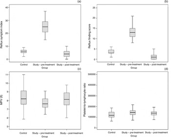
In the study group, the mean proximal reflux index score was 1.6 ± 1.2, the mean distal reflux index score was 26.5 ± 5.1 and the mean DeMeester score was 17.6 ± 16.9, based on the double-channelled 24-hour pH monitoring. The mean reflux symptom index was 25.2 ± 5.4 in the study group and 6.0 ± 1.7 in the control group, and the mean reflux finding score was 13.4 ± 3.4 in the study group and 3.5 ± 1.4 in the control group (p < 0.001 for both). In the study group, the pre-treatment versus post-treatment reflux symptom indices were 25.2 ± 5.4 versus 4.3 ± 3.1 (p < 0.001), and the reflux finding scores were 13.4 ± 3.4 versus 1.3 ± 1.8 (p < 0.001) (Figures 1a and 1b).
(a) Reflux symptom index, (b) reflux finding score, (c) mean platelet volume (MPV) and (d) platelet-to-lymphocyte ratio data for control and study (pre-treatment and post-treatment) groups.

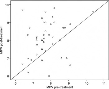
Haemogram data, and neutrophil-to-lymphocyte and platelet-to-lymphocyte ratios, are shown in Table I. Mean platelet volume was found to be significantly lower and platelet-to-lymphocyte ratio was found to be significantly higher in the pre-treatment study group than in the control group (Figures 1c and 1d). Although platelet-to-lymphocyte ratios did not change after treatment (155.2 ± 61.8 in the pre-treatment group vs 147.6 ± 54.1 post-treatment; p = 0.10), mean platelet volume values increased significantly at the third month of treatment (7.4 ± 0.8 vs 8.1 ± 1.0; p = 0.047) (Figure 2).
Pre- versus post-treatment mean platelet volume (MPV) values for the study group.

Haemogram data, and neutrophil-to-lymphocyte and platelet-to-lymphocyte ratios of study and control groups

Data represent mean (standard deviation) values, unless indicated otherwise. *p < 0.05 is statistically significant
Correlations were observed between platelet-to-lymphocyte ratio and: reflux symptom index (Spearman's ρ = 0.25, p = 0.025) and reflux finding score (Spearman's ρ = 0.28, p = 0.013). However, there were no correlations between neutrophil-to-lymphocyte ratio and: reflux symptom index (Spearman's ρ = −0.032, p = 0.8) and reflux finding score (Spearman's ρ = −0.026, p = 0.9, respectively). In addition, there was a correlation between mean platelet volume increase and treatment response in terms of decreasing symptom scores in the first and third months of treatment (Spearman's ρ = 0.31, p = 0.04, and Spearman's ρ = 0.33, p = 0.03 respectively) in the study group.
There were no correlations between neutrophil-to-lymphocyte ratio and double-channelled 24-hour pH monitoring parameters (proximal reflux index (Spearman's ρ = −0.1, p = 0.4), distal reflux index (Spearman's ρ = 0.04, p = 0.8) or DeMeester scores (Spearman's ρ = 0.08, p = 0.6)) in the study group. Similarly, no correlations were observed between platelet-to-lymphocyte ratio and double-channelled 24-hour pH monitoring parameters (proximal reflux index (Spearman's ρ = −0.1, p = 0.1), distal reflux index (Spearman's ρ = −0.1, p = 0.4) or DeMeester scores (Spearman's ρ = −0.2, p = 0.2)) in the study group.
Discussion
Laryngopharyngeal reflux disease is caused by the contact of gastric content – which passes over the upper oesophageal sphincter – with the larynx, pharynx, trachea and oral mucosa. An inflammatory process has been suggested to play a role in the aetiopathogenesis of LPR due to acidic content exposure. In addition to the acidity of gastric juice, pepsin, bile salts, bacteria and pancreatic proteolytic enzymes may cause tissue damage to the upper airway level. Pepsin in particular can damage extragastric tissues at a pH of up to 6, with receptor-mediated endocytosis to laryngeal epithelial cells.Reference Johnston, Wells, Blumin, Toohill and Merati 12 Proton pump inhibitors are used in the treatment of the disease, to reduce the amount of acid, along with different therapeutic approaches based on the patients' clinical conditions.Reference Carroll, Fedore and Aldahlawi 13 , Reference Chiba, Kudara, Abiko, Endo, Suzuki and Sugai 14
Neutrophil-to-lymphocyte ratio and platelet-to-lymphocyte ratio are among the laboratory markers introduced into clinical practice for the purpose of evaluating systemic and subclinical inflammation.Reference Balta, Demirkol, Unlu, Arslan and Celik 15 The neutrophil-to-lymphocyte ratio also provides information about neutrophils, lymphocytes and infiltrating leukocytes that significantly contribute to the tissue injury.Reference Motomura, Shirabe, Mano, Muto, Toshima and Umemoto 16 Increased neutrophil-to-lymphocyte ratio has been reported in hypertension, diabetes mellitus, metabolic syndrome, left ventricular dysfunction, acute coronary syndrome, valvular heart disease, impaired thyroid functions, renal and hepatic dysfunction, and malignancy.Reference Demir 17 , Reference Balta, Cakar, Demirkol, Arslan and Akhan 18 Platelet-to-lymphocyte ratio has been suggested to be a better indicator of chronic inflammation than neutrophil-to-lymphocyte ratio.Reference Turkmen, Erdur, Ozcicek, Ozcicek, Akbas and Ozbicer 4 In this study, neutrophil-to-lymphocyte ratio values were not significantly different in the study and control groups because patients in the study group did not have any morbidities such as obesity, hypertension or diabetes mellitus. However, we found higher levels of platelet-to-lymphocyte ratio in LPR patients, supporting the presence of chronic inflammation in LPR. Higher platelet-to-lymphocyte ratio values in the LPR group are probably a result of the difference in platelet count between the study and control groups. Chronic inflammation in LPR may increase platelet count and increase platelet-to-lymphocyte ratio.Reference Jenne and Kubes 19
While contact of the mucosa with acids and pepsins occurs in both LPR and gastroesophageal reflux, there are other physiopathological differences. Protective mechanisms of the oesophagus such as mucosal bicarbonate production and primary peristalsis prevent acid damage in the oesophagus, unlike in the larynx and the pharynx.Reference Koufman 20 A recent study reported that the neutrophil-to-lymphocyte ratio was not higher in patients with reflux oesophagitis than in control participants, as was found in our study, while platelet-to-lymphocyte ratio was not reported.Reference Ateş, Yaraş, Sarıtaş, Altıntaş, Sezgin and Orekici 21 To our knowledge, this is the first time that platelet-to-lymphocyte ratios have been reported as higher in LPR patients.
Mean platelet volume is measured as part of a routine complete blood count test and is one of the platelet function indices that reflect the platelet production rate.Reference Briggs 22 Previous studies have shown inverse correlations between mean platelet volume and inflammation activity in some diseases such as inflammatory bowel diseases, rheumatoid arthritis and ankylosing spondylitis.Reference Kapsoritakis, Koukourakis, Sfiridaki, Potamianos, Kosmadaki and Koutroubakis 6 , Reference Kisacik, Tufan, Kalyoncu, Karadag, Akdogan and Ozturk 23 In the current study, mean platelet volume levels were lower in LPR patients and these levels increased after treatment. This finding might reflect the active inflammatory process in LPR patients before treatment. More importantly, we also found correlations between mean platelet volume and symptom recovery post-treatment. Mean platelet volume increased significantly in concordance with symptom improvement in LPR patients after treatment. Thus, we can conclude that mean platelet volume can be used as a simple marker of LPR disease and disease activity in the follow-up period (after medical treatment).
-
• Platelet-to-lymphocyte ratio was significantly higher and mean platelet volume was significantly lower in laryngopharyngeal reflux (LPR) patients
-
• Mean platelet volume increase during treatment was significantly associated with LPR symptom improvement
-
• Thus, mean platelet volume can be used as a simple marker of disease activity during LPR treatment
Our study has several limitations. The cut-off values for neutrophil-to-lymphocyte ratio, platelet-to-lymphocyte ratio and mean platelet volume could not be calculated. Determination of cut-off values to predict diagnosis and treatment response might have been possible with a larger study group. Secondly, we did not use the ‘gold standard’ multichannel intraluminal impedance testing for diagnosis of LPR. However, this study did employ the reliable methods of reflux symptom index, reflux finding score and double-channelled 24-hour pH monitoring for diagnosis.
Conclusion
The platelet-to-lymphocyte ratio – a marker of chronic inflammation – was found to be significantly higher in LPR patients. Moreover, LPR patients with increased mean platelet volume values at follow up were found to have a better response to treatment. Both measurements are inexpensive and easy to obtain, and should therefore be used as diagnostic and follow-up tools in patients with LPR disease.