The long-term prognosis of children who underwent surgery for a secundum-type atrial septal defect does not differ from the normal population with regard to mortality, morbidity, and exercise tolerance.Reference Meijboom, Hess and Szatmari 1 – Reference Meyer, Korfhagen, Covitz and Kaplan 5
In some echocardiographic studies, however, right ventricular enlargement was found to persist for a long time after closure.Reference Meijboom, Hess and Szatmari 1 , Reference Roos-Hesselink, Meijboom and Spitaels 2 , Reference Meyer, Korfhagen, Covitz and Kaplan 5 In contrast, recent observations using optimal imaging techniques such as magnetic resonance imaging point towards normalisation of right ventricular size.Reference Bolz, Lacina, Buser, Buser and Guenthard 6 These observations need further confirmation in direct comparison with references, as they may have implications for – the timing of – treatment strategies.
Studies in the 1980s and 1990s have reported arrhythmias as a major concern for the long term.Reference Meijboom, Hess and Szatmari 1 , Reference Morris and Menashe 7 – Reference Rostad and Sorland 11 Relatively scarce information is available on the rhythm status of patients with secundum-type atrial septal defect operated on more recently.Reference Bolz, Lacina, Buser, Buser and Guenthard 6
Information on the mid-term to long-term follow-up of patients operated on for secundum-type atrial septal defect remains important for comparison with non-surgical closure results, and to evaluate the results of current surgical management.
The aim of our study was to assess mid-term prognosis of right ventricular dimensions, rhythm status, and exercise capacity in patients operated for a secundum-type atrial septal defect.
We therefore performed a cross-sectional study in a homogeneous group of patients with secundum-type atrial septal defect. The results of magnetic resonance imaging and exercise testing were compared with those of matched healthy reference groups.
Materials and methods
The study population consisted of all 30-day survivors after surgical closure of a secundum-type atrial septal defect performed at Erasmus Medical Centre between 1990 and 1995. Patients were identified from the medical records of the Departments of Cardio-thoracic Surgery and Paediatric Cardiology. Patients were eligible for prospective analysis if they met the following inclusion criteria: secundum type of atrial septal defect without other cardiac malformations, residence in the Netherlands, Dutch speaking and no underlying syndrome with mental retardation. From the medical records, pre-, peri-, and post-operative parameters were collected.
In all, 78 consecutive patients, who had been operated upon for a secundum-type atrial septal defect, were identified. Of those, seven of them were completely lost to follow-up, as they had moved abroad post-operatively, and 13 patients suffered from Down's syndrome. Of the remaining 58 survivors, 16 refused to participate in the cohort evaluation because of practical reasons. Ultimately, 42 patients agreed to participate in the prospective cross-sectional part of the study. Between 2005 and 2006, all patients visited the outpatients’ clinic for medical history, physical examination, echocardiography, exercise testing, standard 12-lead electrocardiography, 24-hour ambulatory electrocardiography, and magnetic resonance imaging. The study was approved by the national Medical Ethics Review Board in the Netherlands. Written informed consent was obtained from all patients, references, and their parents.
Reference group
A reference group of healthy schoolchildren and adolescents matched with the patients for gender and body surface area was selected to undergo magnetic resonance imaging. Another reference group of healthy schoolchildren and adolescents matched for gender, age, and height was selected to undergo a bicycle exercise test. Different groups were used after consultation with the institutional review board, in order to minimise the burden of participation for healthy children. Children or adolescents involved in organised extracurricular sports activities for more than 6 hours a week were excluded. This was done because the normal physical activity level in the relevant population is mainly determined by travel-related physical activity – a high percentage of children or adolescents in the relevant age group cycle to school or work – school-related physical education, and organised sports activity. Hours spent during extracurricular organised sports activity hardly ever exceeds 6 hours.Reference Arvidsson, Slinde, Hulthen and Sunnegardh 12
Clinical evaluation
Standard electrocardiogram
Standard 12-lead electrocardiograms were performed on a Cardio Perfect 4.0 machine (CardioControl, Northampton, England). Electrocardiograms were interpreted by a single observer (WBdK). We compared results according to criteria described by Rijnbeek et al.Reference Rijnbeek, Witsenburg, Schrama, Hess and Kors 13
Twenty-four hour ambulatory electrocardiogram
A three-channel recorder (Philips, Andover, Massachusetts, United States of America) was used on a day with usual activities. Arrhythmias were judged according to Garson's criteria.Reference Garson 14
Echocardiography
Conventional echocardiography was carried out, consisting of transthoracic M-mode and two-dimensional echocardiography, as well as pulsed-wave and continuous-wave Doppler measurements, using an ultrasound Philips Sonos 5500 (Philips Medical Systems, Best, The Netherlands). A fractional shortening of greater than 28% was regarded as normal. Pulmonary arterial hypertension was diagnosed if a tricuspid regurgitation with a maximal flow of 2.8 metres per second was detected. Measurements by Schnittger were used for reference values.Reference Schnittger, Gordon, Fitzgerald and Popp 15
Magnetic resonance imaging
Magnetic resonance imaging was performed on a GE 1.5-T Signa CV/I scanner (GE Healthcare, Milwaukee, Wisconsin, United States of America) using dedicated phased-array coils. All images were acquired during breath-holds at end-expiration. A multi-phase, multi-slice volumetric data set was acquired using a fast two-dimensional cine scan employing steady-state free precession. Imaging parameters were as previously reported.Reference van den Berg, Hop and Strengers 16
Flow measurements were performed perpendicular to flow using a standard two-dimensional flow-sensitised scan. Scans were retrospectively gated, starting the acquisitions on the R-wave. Temporal resolution was approximately 40 milliseconds per cardiac phase, and 24 phases were reconstructed retrospectively. Imaging parameters were as previously reported.Reference van den Berg, Hop and Strengers 16 , Reference van den Berg, Wielopolski and Meijboom 17 Ventricular size was normalised to body surface area as previously reported.Reference Robbers-Visser, Boersma and Helbing 18
Maximal exercise capacity
A maximal exercise bicycle test was performed using a stepwise incremental protocol on a Jaëger Oxycom Champion System (Viasys Healthcare GmbH, Hoechberg, Germany) that allowed breath-by-breath ergometry. Workload was increased every minute by 10, 15, or 20 Watts according to the Godfrey protocol.Reference Godfrey 19 The respiratory quotient was used to objectify maximal performance. Exercise performance was regarded as maximal when subjective exhaustion combined with a respiratory quotient is greater than or equal to 1.10 at peak exercise was reached. The following parameters were obtained: maximal achieved workload (Watt), maximal oxygen uptake (VO2-max, millilitre per kilogram per minute), peak heart rate (beats per minute), and respiratory quotient. Results were compared with values from the references.
Statistical methods
Continuous data were presented as mean with standard deviation and ranges. Student's t-tests were used to compare the two groups. Chi-square test and, if appropriate, Fisher's exact test were used for the comparison of categorical variables. In all analyses, the two-sided level of significance was set at p-value less than 0.05.
Associations between clinically relevant parameters and magnetic resonance imaging – ventricular dimensions and function – or exercise test results were assessed by paired samples t-test.
Shapiro–Wilk test was used to test for normality.
SPSS V17 was used as the statistical package.
Results
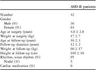
Baseline characteristics from the chart review showed differences between the group of 58 survivors and the group of 42 patients on duration of follow-up (10.9 ± 3.6 versus 16.5 ± 3.3; p = 0.02 and age at last follow-up (6.1 ± 2.3 versus 11.8 ± 1.8; p = 0.02). This can be explained by the fact that some of the survivors were dismissed from continued surveillance at short term after surgery. Data of the 42 patients that were included in the prospective cross-sectional study are given in Table 1.
Baseline characteristics.Footnote *

ASD = atrial septal defect
* Data are given as mean ± standard deviation
Data were sufficiently distributed for parametric testing.
Operation
The operative procedure included median sternotomy and complete cardiopulmonary bypass. The defects were closed by direct suturing in 40 patients and by a pericardial patch in two patients. In one patient, lateral thoracotomy was performed on cosmetic grounds. The mean perfusion time was 39 plus or minus 20 (21–188) minutes; aortic cross-clamp time 15 plus or minus 8 (6–35) minutes; and lowest nasal temperature 31°C plus or minus 3 (17–33) °C. Between 1990 and 1995, 78 patients have been operated on for secundum-type atrial septal defect. No peri-operative deaths were recorded. However, one patient required mediastinal re-exploration because of post-operative haemorrhage. During follow-up, no late re-interventions were needed and none of the patients died.
Standard 12-lead electrocardiogram
In all, 40 patients (95%) were in sinus rhythm and two patients (5%) showed junctional rhythm. Another patient had a complete right bundle branch block and one other patient had a first-degree atrioventricular block, blocks that have not been recorded pre-operatively. No ventricular hypertrophy was detected electrocardiographically.
Ambulatory 24-hour electrocardiogram
A total of 38 patients (90%) were examined. In four of them, assessment did not succeed because of technical problems with the recorder. No cardiac symptoms were reported during the registration. In all, 20 patients (48%) were in continuous sinus rhythm and one patient in a continuous junctional rhythm. The others (50%) showed sinus rhythm with short periods of non-sinus, atrial rhythm. No signs of atrial arrhythmias – fibrillation, flutter, ectopic, tachycardia – were noted. In four patients (10%), a first-degree atrioventricular block was seen continuously on the recordings, in spite of the absence of this block at 12-lead electrocardiogram in three of them.
Echocardiography
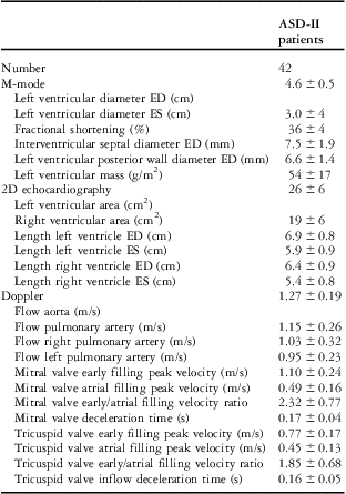
None of the 42 patients had a residual atrial septal defect. Echocardiographically, dimensions of both ventricles were within normal limits (Table 2).Reference Schnittger, Gordon, Fitzgerald and Popp 15 Fractional shortening of the left ventricle in all but one patient was normal. Semi-quantitative assessment of the right heart did not reveal dilatation of the right ventricle. Trivial tricuspid regurgitation was observed in six patients, and mean peak flow was 2.2 plus or minus 0.3 (1.8–2.5) metres per second. Trivial mitral regurgitation was observed in one patient; nine patients displayed trivial pulmonary regurgitation, and mean peak flow was 1.5 plus or minus 0.4 (0.8–2.2) metre per second. In all, 32 patients showed normal ventricular septal motion; in 10 patients, decreased motion of the septum was detected and no patients displayed a paradoxical pattern of the septum. No haemodynamically significant abnormalities were identified. Mean mitral and tricuspid E/A ratios, parameters for diastolic ventricular function, were 2.32 and 1.85, respectively (Table 2), and similar to the reference group.
Echocardiographic results.Footnote *
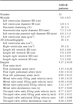
ASD = atrial septal defect; ED = end diastolic; ES = end systolic
* Data are given as mean ± standard deviation
Magnetic resonance imaging
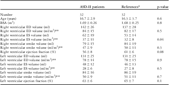
In 32 of the 42 patients, magnetic resonance imaging was performed. Of the 10 patients, four of them refused to undergo the examination and six of them did not complete the examination because of claustrophobia. The results of magnetic resonance imaging are given in Table 3. The mean right ventricular end-systolic volume was significantly larger in patients. In 25% of patients, the right ventricular end-systolic volume was larger than the 95th percentile. Left ventricular dimensions in patients did not differ from those in references.
Magnetic resonance imaging dimensions and flows (mean ± S.D.).
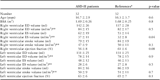
ASD = atrial septal defect; BSA = body surface area; ED = end diastolic; ES = end systolic
*Gender and BSA matched
**Indexed to BSA
The stroke volumes of the left and right ventricle (83 ± 16 millilitres versus 80 ± 15 millilitres; p = 0.5) were similar in patients and references; stroke volumes across the pulmonary and aortic valves (86 ± 19 millilitres versus 89 ± 21 millilitres; p = 0.2) were similar as well.
No association was found between ventricular dimensions and function (all p-values >0.4). In addition, no association with age or weight at surgery could be established (p = 0.7 and p = 0.8, respectively).
Exercise testing
Maximal exercise capacity was assessed in 38 patients. Owing to exhaustion, four patients failed to perform. The mean exercise capacity was 169 plus or minus 43 (80–300) Watt, the mean maximal oxygen uptake was 38 plus or minus 8.2 (20–55) millilitres per minute per kilogram, and the mean maximal heart rate was 162 plus or minus 17 (155–209) beats per minute. All three scores were slightly less than those in references (Table 4), but there were no significant differences (p-values 0.8, 0.1, and 0.4, respectively). No correlation between exercise capacity and age at surgery or follow-up duration could be demonstrated.
Exercise testing.Footnote *

ASD = atrial septal defect
* Data are given as mean ± standard deviation (range)
** Gender and age matched
Comment
This study is the first to provide a direct comparison of cardiac dimensions between young patients with secundum-type atrial septal defect and a matched reference group, using the current technique of choice for assessment for right ventricular dimensions, cardiac magnetic resonance imaging. We found that, compared with a healthy reference population, right ventricular end-systolic volume was significantly increased after a mean follow-up duration of 12 years. Other cardiac dimensions and biventricular ejection fraction were similar to those in the reference group. Exercise tolerance was normal, as expected. None of the patients demonstrated symptomatic arrhythmias. No residual atrial septal defects or other relevant sequelae were detected, and none of the patients used cardiac medication. No major cardiac events during follow-up could be demonstrated. Quality of life, reported on earlier in this population, was good.Reference Spijkerboer, Utens, De Koning, Bogers, Helbing and Verhulst 20 These data provide further evidence of excellent mid-term to long-term outcome of surgical closure of secundum-type atrial septal defect. This provides a basis for comparison with non-surgical closure follow-up data.Reference Du, Hijazi, Kleinman, Silverman and Larntz 21
A remarkable observation from our study is the enlargement of the right ventricle in a young population with secundum-type atrial septal defect. Compared with the healthy reference population, the right ventricle was enlarged in 25% of the patients.Reference Robbers-Visser, Boersma and Helbing 18 In patients operated on at older age, right ventricular dilatation has been a relatively consistent finding at echocardiography. Few data exist on the myocardial basis of right ventricular remodelling after prolonged right ventricular volume overload. Factors influencing persisting right ventricular dilatation after closure of a secundum-type atrial septal defect may be related to the duration of volume overload (age at operation) and closure technique. We did not establish a relation between age at operation and right ventricular dilatation. Earlier studies reporting on surgical closure of secundum-type atrial septal defects in children showed residual right ventricular dilatation 5 years after closure.Reference Meyer, Korfhagen, Covitz and Kaplan 5 More recent studies in children suggest normalisation of right ventricular size, as assessed with magnetic resonance imaging at 20 years’ follow-up.Reference Bolz, Lacina, Buser, Buser and Guenthard 6 However, in that study by Bolz and co-workers, in children operated at a mean age of 7 years, magnetic resonance imaging volumes were compared with reported normal values, which may have included data acquired with different magnetic resonance imaging sequences.Reference Rominger, Bachmann, Pabst and Rau 22 After device closure, rapid normalisation of right ventricular size has been reported using echocardiographic assessment of right ventricular size.Reference Eyskens, Ganame, Claus, Boshoff, Gewillig and Mertens 23 In children, too few data are available to demonstrate a relationship between age and right ventricular dimensional changes after device closure.Reference Santoro, Pascotto and Caputo 24 , Reference Veldtman, Razack and Siu 25 In adults, results of studies on this relationship have been equivocal. The rate of remodelling may be influenced by the technique of closure, which points towards the potential effects of myocardial protection and interventional technique during closure.Reference Pawelec-Wojtalik, Wojtalik, Mrowczynski, Surmacz and Quereshi 26
The observed dilatation of the right ventricle in our study raises several questions: First, what is the functional significance of this observation and second, should this observation lead to a reconsideration of the age of elective closure of secundum-type atrial septal defect?
The functional significance of the dilated right ventricle seems limited. Global systolic performance of the right ventricle and left ventricle was maintained, as was the biventricular diastolic function. Exercise capacity of the patients was normal; none of the patients used cardiac medication, and arrhythmias were all asymptomatic. This is in accordance with studies in patients operated on at older ages and with longer follow-up, in whom the effects of dilatation of the right ventricle, as assessed echocardiographically, seemed limited. The clinical relevance of the observed increase in the number of patients with right ventricular dilatation over time in some studies remains to be established.Reference Roos-Hesselink, Meijboom and Spitaels 2 On the other hand, significant right ventricular dilatation resulting from prolonged volume overload has been suggested to have a negative effect not only on right ventricular function, but also on left ventricular function.Reference Walker, Moran, Gauvreau and Colan 27 This may be a reason for measures in order to prevent right ventricular dilatation. Currently, elective surgical closure of moderate to large size secundum-type atrial septal defect is usually performed between 4 and 6 years of age, and it can be speculated that earlier closure of secundum-type atrial septal defect might prevent long-term right ventricular dilatation.Reference Kirklin, Brown and Bryant 28 Although the results of cardiac surgery at young age in general are excellent, the main argument to delay surgery is spontaneous closure or reduction of size below levels with haemodynamic relevance. In defects smaller than 6 millimetres, closure rates up to two-third of the patients have been reported.Reference Kirklin, Brown and Bryant 28 The natural history of larger defects has been less well assessed. McMahon and co-workers studied the change in maximal diameter retrospectively, in the group with moderate-size secundum-type atrial septal defect. They found that 10% of this group showed a significant reduction in size (to below 6 millimetres) during a 3-year follow-up. In the group with a secundum-type atrial septal defect larger than 12 millimetres, no spontaneous reduction in size occurred.Reference Kirklin, Brown and Bryant 28 On the basis of these data, it could be argued that for a secundum-type atrial septal defect with important shunt size, judged not to be candidates for device closure, delaying surgery to 3–6 years of age does not have any advantages, and may contribute to long-term right ventricular dilatation. However, on the basis of the reports of rapid reduction of right ventricular size after device closure and the potential avoidance of cardiopulmonary bypass, candidates for device closure should be carefully selected.
Arrhythmias, particularly of supraventricular origin, have been a major concern in the late follow-up after surgical closure of atrial septal defects.Reference Meijboom, Hess and Szatmari 1 , Reference Morris and Menashe 7 , Reference Kammeraad, van Deurzen and Sreeram 9 , Reference Rominger, Bachmann, Pabst and Rau 22 , Reference Bolger, Sharma and Li 29 With the improvements of surgical techniques, these problems have been reduced considerably.Reference Kammeraad, van Deurzen and Sreeram 9 In a recent series of patients operated on at older age, Roos-Hesselink et alReference Roos-Hesselink, Meijboom and Spitaels 2 reported 6% of symptomatic supraventricular tachyarrhythmias at a mean follow-up of 26 years. Few reports of arrhythmias after operation in young children have been published recently.Reference Bolz, Lacina, Buser, Buser and Guenthard 6 Our results confirm the general impression that arrhythmias have become rare after early surgical closure of secundum-type atrial septal defect. We did not note any signs of clinically relevant arrhythmias. The same is true for pulmonary hypertension, of which we found no evidence in this study. This was in contrast to earlier studies that found percentages of 13% and 28% pulmonary arterial hypertension.Reference Murphy, Gersh and McGoon 3 , Reference Cherian, Abraham, Uthaman, Sukumar, Krishnaswami and Bhaktaviziam 30
Limitations
Major limitation of this study is the relatively small number of patients, limiting statistical power. However, this is one of the largest homogeneous groups of atrial septal defect type II patients. The lack of separate evaluation of regional ventricular function, for example using tissue Doppler techniques, may be considered a limitation.
In the reference population, children or adolescents involved in organised extracurricular sports activities for more than 6 hours a week were excluded. This was done to exclude a very small number of extremely physically active children. If these children would not have been excluded, the exercise performance data provided as reference might have been skewed towards somewhat higher values than currently reported.
The use of separate reference groups for magnetic resonance imaging and exercise testing for ethical reasons could be considered a limitation. However, as these groups were randomly selected from the same larger general normal population, we consider this to be a minor limitation.
Conclusion
This study confirms the generally excellent condition of young patients at mid-term after surgical closure of a secundum-type atrial septal defect. However, with magnetic resonance imaging, the current ‘gold standard’ technique for assessment of right ventricular size, an enlargement of right ventricular end-systolic volume, was noted. These findings have no clinically relevant influence on rhythm status or exercise capacity at this stage of follow-up, but may have implications for the timing of surgery or the technique of closure. Longer follow-up is required to reveal the functional consequences of the findings.