Introduction
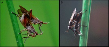
Very few studies have been conducted on the sensilla of the antenna of Sciomyzidae (Knutson and Vala Reference Knutson and Vala2011). Having seen photographs (Figs. 1A, 1B) of mating in two Sepedon Latreille (Diptera: Sciomyzidae) species, it occurred to J.G.S. that the female might be receiving sensory input regarding the male via her antennae during copulation because the male appositions her antennae with his forelegs. It also seemed possible that any sensilla present, their number and/or their location might differ between the sexes because males receive no corresponding input during copulation. On the basis of these observations, it was decided to examine the antennae for mechanosensilla to determine if they differed between the sexes.
(A) Mating pair of Sepedon fuscipennis collected at Clay Pond (Great Falls), Maryland, United States of America, by Karolyn Darrow (Smithsonian Institution). Note the male is appositioning his forelegs to the female's styli. The female is seen in the process of bubbling (see arrow); (B) Male of Sepedon sp., possibly Sepedon aenescens Wiedemann (Berg and Valley Reference Berg and Valley1985a), in Taiwan appositioning his forelegs to the stylus of the female while she feeds on the nuptial gift (photographed by S.-Y. Lee, Taiwan Agricultural Research Institute; available from http://taipei.tzuchi.org.tw/tzquart/2006su/qs2.htm [accessed 28 November 2012]).

Materials and methods
Collecting and maintaining flies
This study was conducted during the summers of 2009 and 2010. In 2009, live Sepedon fuscipennis Loew were collected in a cattail (Typha latifolia Linnaeus) marsh adjacent to the running track at the University of Massachusetts in Amherst, Massachusetts, United States of America (42°37.849′N, 72°5.403′W). In 2010, W.L.M. collected live S. fuscipennis in a broadleaf cattail (T. latifolia) marsh in Fishers, Indiana, United States of America (39°55.981′N, 86°3.022′W) and shipped them in 80% ethanol to J.G.S. (see the section “Taxonomy”). Specimens of Sepedon testacea Loew examined during the summer of 2010 were collected in South Africa with Ray Miller (KwaZulu-Natal, South Africa).
Flies were captured either by use of a sweep net or with a clear plastic cup (473 cc) used in conjunction with a 30.5 cm square of plastic nylon mesh. The cup and mesh were positioned on either side of a cattail leaf, trapping the fly inside. A plastic lid, in which a 7.6 cm hole had been cut and over which mesh had been glued, was affixed to the cup. Until taken to the laboratory, flies were kept in screened fly cages (20.3 cm per side) with one side covered with a clear plastic sheet. Droplets of a mixture of yeast and honey were placed on the inner sides of the cage. Water was available to the flies from plastic cups (266 cc) containing dental wicks (Absorbal®, Wheatridge, Colorado, United States of America).
In the laboratory, flies were kept in a metal cage (30.5 cm per side) with clear plastic sides to allow better observation. The cage floor was lined with cotton that was moistened daily to replicate the natural environment of the flies. Wooden applicator sticks were positioned in the cage to provide resting and oviposition sites. Flies isolated for observation were held in a clear plastic cup (473 cc) with a mesh top, similar to the container in which they had been caught. They were supplied with droplets of a mixture of yeast, powdered milk, honey, and water that was coloured with red food dye dabbed on the cage wall. Cages were cleaned weekly to prevent mould infestation. After several days of observations, flies were killed by freezing, placed in 80% ethanol, and prepared for the scanning electron microscope (SEM).
Taxonomy
The nominative S. fuscipennis is widespread throughout the United States of America and Canada (Knutson et al. Reference Knutson, Orth, Fisher and Murphy1986). Three forms of the S. fuscipennis group are known: S. f. fuscipennis (from just west of 100° longitude in southern Texas to just north of 40° latitude in Illinois and Indiana, east to southern New England, south to Florida, United States of America); S. f. nobilis Orth (Alaska and Northwest Territory east to Newfoundland, south to New York, west to New Mexico and California in the United States of America and Canada); and S. f. “intermediate” (narrow distribution band from Kansas northeast through Illinois, Indiana, south Ohio, Pennsylvania, New York, to Massachusetts, United States of America) (Orth Reference Orth1986). Two forms are known from Massachusetts (S. f. fuscipennis and S. f. “intermediate”). To ensure collection and subsequent study of a single form, on 6 June 2010, W.L.M. collected 6♂ and 4♀ S. f. fuscipennis directly into 80% ethanol at an isolated stand of broadleaf cattail (T. latifolia) in Fishers, Indiana, a site from which only S. f. fuscipennis is known to occur. He shipped them to J.G.S. on 8 June. As expected, all 10 specimens exhibited characters diagnostic for S. f. fuscipennis – distinct velvety black parafrontal spots and, in the males, the anterior process of the aedeagus relatively large and long relative to the posterior process.
Observing the mating behaviour
Flies collected during the summer of 2009 were kept in small screened cages as described above. Flies were observed periodically for mating behaviour. Flies collected during the summer of 2010 were killed immediately in the field.
Preparation for SEM
Using SEM to observe any type of sensilla on the antennae, especially the styli, of flies collected in 2009 was difficult because the antennae were completely covered by compacted masses of adhering pollen. Therefore, the procedures of Cuperus (Reference Cuperus1985) and Albert (Reference Albert2003) to clean and remove any debris on the antennae were followed. Specimens received from W.L.M. in 80% ethanol (summer 2010) were removed from the 80% ethanol and blotted dry, with care being taken not to break any parts. Once dry, they were placed in a beaker of boiling carbon tetrachloride for one minute. The specimens were then run through three changes of fresh, boiling carbon tetrachloride for one minute each, air dried, and placed onto stubs for SEM.
SEM
During the summer of 2009 some of the flies were examined in low vacuum SEM requiring only mounting the fixed flies on stubs. Later in 2009 and in 2010 flies were prepared for high vacuum conventional SEM. Flies were placed in 80% ethanol until critical point dried by use of liquid CO2. Specimens were blotted dry using Kimwipes® (Roswell, Georgia, United States of America), mounted on stubs, sputter coated with ∼15 nm gold, and examined in an FEI Quanta 200 SEM (FEI Company, Hillsboro, Oregon, United States of America), at 10 kV.
Results
Mating behaviour and bubbling
The main focus of the behavioural studies was to determine if the male always appositioned his forelegs to the styli of the female during copulation (Figs. 1A, 1B). In all of our observations (2009) (n = 10), males always appositioned their forelegs to the styli of the females during copulation. Once flies of both sexes had fed on a honey–yeast-powdered milk mixture coloured red using food dye, they immediately began to “bubble” (Fig. 1A). The colour of the bubble produced by bubbling flies always took on the colour of the red food dye and was produced from food previously ingested into the crop (conclusion supported by dissections).
SEM preparation
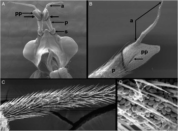
During the first summer of research (2009), it was impossible to observe the sensilla on the antennal styli of most specimens because of two factors: (1) the orientation of the specimen and the antennae on the stubs (Figs. 2A, 2B) and (2) the large clusters of pollen adhering to and between the spinelike setae of the styli (Figs. 2C, 2D).
(A) Head of male Sepedon fuscipennis showing position of the antennae. The unlabelled arrows point to the outer portion of the antennae, and the same orientation applies to the styli (a). The scape (s), pedicel (p), postpedicel (pp), and styli (a) are present (bar = 500 μm); (B) Portion of the antenna showing the pedicel (p), postpedicel (pp), and stylus (a). Note the arrow pointing to the postpedicular groove (bar = 200 μm); (C) Antenna of male showing masses of pollen stuck between the spinelike chaetae or setae of the stylus and obscuring underlying sensilla (bar = 50 μm); (D) Higher magnification of C showing the masses of adhering pollen stuck between the spinelike setae (bar = 10 μm). Specimens from 2009 and were not cleaned as in “Materials and methods”.

Styli mechanosensilla
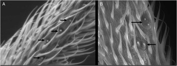
After cleaning the antennae with boiling carbon tetrachloride (2010), it was possible to observe the sensilla on the styli of every specimen. SEM clearly showed a groove (Fig. 2B) on the postpedicel of the antenna. However, the main interest of the study was the mechanosensilla, thus SEM results focussed on them. The number and arrangement of the mechanosensilla differed between the sexes. In both sexes they were always located on the medial surface of the stylus. Females had more sensilla than did males, singly arranged in a linear fashion of four or more campaniform sensilla (Fig. 3A), situated on the 3rd segment of the styli on the “elongate, acuminate apical” portion (Stuckenberg Reference Stuckenberg1999) and about halfway between the base and the tip of the stylus (Fig. 2B). In contrast, males had two patches of mechanosensilla, with two campaniform sensilla for each patch (Fig. 3B). As in females, mechanosensilla in males were located about halfway between the base and the tip of the stylus.
(A) Inner portion, about halfway between the base and the tip, of the stylus of a female Sepedon fuscipennis showing the arrangement of the mechanosensilla (four are visible here – see arrows) tucked between the numerous spinelike chaetae or setae (bar = 10 μm); (B) In contrast to the female, the male has two groups of mechanosensilla arranged in pairs and in a nonlinear arrangement (bar = 10 μm). Specimens from 2010 and cleaned as stated in the “Materials and methods”.

Discussion
Suggestions for specimen preparation
It is imperative that future studies of antennal sensilla or of any other cuticular structure of adult species of Sepedon follow the cleaning procedure reported by either Cuperus (Reference Cuperus1985) or Albert (Reference Albert2003) and used in the summer 2010 study prior to placement of specimens on stubs. This procedure is essential in removing debris such as pollen from the structures being examined. It is also imperative to mount the head so as to allow observation of the medial surface of the stylus of the antenna, where the mechanosensilla are located.
Mechanosensilla on the antennal styli and their function
The suggestions of Stuckenberg (Reference Stuckenberg1999) have been followed here. He stated, “It is proposed that the term arista be reserved for the cyclorrhaphous antennal mechanoreceptor”. Knutson and Vala (Reference Knutson and Vala2011) recommend using stylus for the Sciomyzidae. The only known study of sensilla on the antenna of a species of Sepedon is by Gaponov et al. (Reference Gaponov, Khitzova and Sergeev2006), who made no mention of mechanosensilla on the stylus of any of the Sciomyzidae species examined; they reported only olfactory sensilla. Mechanosensilla are classified as Type I mechanoreceptors because they are associated with the cuticle and, based on function, respond to cuticular distortion or compression (i.e., they are strain sensors) (McIver Reference McIver1985). The mechanosensilla caps of insects can be level with the cuticle or elevated above the cuticle. In S. fuscipennis, the campaniform sensilla (Figs. 3A, 3B) are associated with the cuticle, the sensilla are dome shaped and raised above the cuticle. On the basis of their structure, it is hypothesised that these sensilla should respond to cuticular distortion or compression as the stylus is bent medially during mating.
Knutson and Vala (Reference Knutson and Vala2002) noted that, “Except for the morphology of the postabdomen, body size, and development of setae on the ventral surface of the hind femur in many genera, there is little sexual dimorphism amongst Sciomyzidae”. The presence of sexually dimorphic mechanosensilla on the styli of S. fuscipennis adds to this list of sexually dimorphic characters. The fact that the mechanosensilla show sexual dimorphism (i.e., differences in number and arrangement) (Figs. 3A, 3B) suggests that they serve different functions for each sex. It was difficult for us to count the numbers of mechanosensilla because the antennal styli of the Sciomyzidae are heavily covered with chaetae or setae (e.g., Sepedon) (Figs. 3A, 3B) or long dense setae (e.g., Coremacera Rondani) (Gaponov et al. Reference Gaponov, Khitzova and Sergeev2006). Berg and Valley (Reference Berg and Valley1985a) provided a detailed account of mating in Sepedon species; however, their focus was on production and importance of the nuptial gift produced by the male. They made no mention in their paper concerning the position of the male during copulation and the apposition of his forelegs to the styli of the female. A photograph taken by entomologist Ernie Bay of Washington State, United States of America of a pair of Sepedomerus macropus (Walker) mating also shows the male appositioning his forelegs to the styli of the female (photograph in the possession of W.L.M.). According to Lloyd Knutson, Gaeta, Italy (personal communication), this behaviour among the Sciomyzidae is unique to the genus Sepedon, but based on the photograph of S. macropus, this behaviour may apply to other members of the genus. In a brief study of copulatory behaviour of Sepedon neavei Steyskal, S. testacea Loew, and Sepedon pleuritica Loew, Barraclough (Reference Barraclough1983) did not report the male tarsi contacting the female's antennae. We did not examine the styli of those species of Sciomyzidae, such as Tetanocera Duméril species, where the male mates with their tarsi over the female eye. Also, we have no information on whether males of other members of Sepedon, besides S. fuscipennis and S. macropus, also apposition the female antennae during mating. Unless one specifically and carefully examines mating behaviour, as done by Tobin and Stoffolano (Reference Tobin and Stoffolano1973), minute details such as where the foretarsi are placed can be missed. Interesting, however, is the report of Barnes (Reference Barnes1979) for several species of Neolimnia Tonnoir and Malloch: “During copulation the male's fore tarsi rest on the female's frons, and frequently his claws are hooked over her anterior frontal margin between the antennae and eyes”. No mention, however, is made of the male touching the female's styli or antennae.
Based on observations in this study, personal communication with Knutson, and Figures 1A, 1B, it is suggested that the mechanosensilla on the antennal styli of female Sepedon provide feedback to the female regarding the males appositioning his forelegs to her styli. This may be an inherent, behavioural component of the mating ritual that the female uses as an important signal of the correct species and, if not present, will not accept the male's copulatory attempt. The position of the sensilla on the medial surface of the styli and their linear arrangement correlates with the bending of her antennae medially as the male appositions his forelegs to the lateral surface. This agrees with the idea of Johnson et al. (Reference Johnson, Bonser and Jeronimidis2009), who also noted that arrangement and location of mechanosensilla influence the stress placed upon them, which in this case would be from the lateral surface medially. This also agrees with Johnson et al. (Reference Johnson, Bonser and Jeronimidis2009), who further noted that, “The oval shape of a campaniform sensillum means that it can be deformed more readily in one direction than the other, providing a level of directional sensitivity to the strain”.
Another suggested function of these sensilla in both sexes is monitoring any contact the adult has while obtaining nectar from flowers. This hypothesis is based on the large packs of pollen found adhering to the antennal styli (Figs. 2C, 2D) and on the “bubbling” behaviour observed in our laboratory during the summer of 2009 (i.e., flies took nutrients, presumably nectar, into the crop, but later used these fluids to produce bubbles) as shown in Figure 1A. Stuckenberg (Reference Stuckenberg1999) also suggested that “…the emergence of the Brachycera from nematocerous ancestors involved coevolution of antennal transformation and pseudotracheate labella, linked to a new feeding mode appropriate in new floras that appeared in the Triassic and Jurassic”.
Even though bubbling behaviour has been reported in some Diptera (Stoffolano et al. Reference Stoffolano, Acaron and Conway2008), no specific reference in the literature for the Sciomyzidae was found. Mating trophallaxus, however, has been reported for several sciomyzids (Barraclough Reference Barraclough1983; Knutson and Vala Reference Knutson and Vala2011) and this behaviour involves regurgitation of liquids and, possibly salivary gland secretions from the crop (Stoffolano and Haselton, Reference Stoffolano and Haselton2013) (S. testacea is mentioned in the paper by Barraclough Reference Barraclough1983). Bubbling behaviour in the Sciomyzidae, unlike that shown in other Diptera for getting rid of excess water in the diet (Stoffolano et al. Reference Stoffolano, Acaron and Conway2008), may also be involved in the production of the froth “nuptial gift” presented to the female during courtship and mating (Green Reference Green1977; Berg and Valley Reference Berg and Valley1985a, Reference Berg and Valley1985b). Prasad et al. (Reference Prasad, Knutson and Ghorpadé2010) mentioned trophallaxis in the India species of Sepedon they studied, but did not mention bubbling.
During SEM examination of male S. testacea in Cape Town, South Africa (their summer 2010), with Ray Miller, large packs of pollen on the styli were found. Thus, both our study of S. fuscipennis and observations in South Africa with S. testacea suggest that adult Sepedon obtain carbohydrates from flowers. In fact, Berg and Knutson (Reference Berg and Knutson1978) reported that adults have been collected while feeding on flowers. A study on honey bees (Apis mellifera Linnaeus; Hymenoptera: Apidae) by Scheiner et al. (Reference Scheiner, Schnitt and Erber2005) demonstrated that antennal mechanosensilla are mainly involved in surface discrimination and not in shape discrimination of flowers. The mechanosensilla on the antennal styli of both sexes of Sepedon may serve a similar function.
Future studies and questions
The Sciomyzidae are of considerable interest in regard to biological control programs to halt or lessen the impact of schistosomiasis (Berg Reference Berg1973; Appleton et al. Reference Appleton, Miller and Maharaj1994) and fascioliasis (McDonnell Reference McDonnell2004), plus gastropod pests of agriculture and horticulture (Barker et al. Reference Barker, Knutson, Vala, Coupland and Barnes2004), yet very little research has been conducted on either adult sensory physiology or on their selection of flowers from which they obtain carbohydrates. Both areas should be explored further.
Judd (Reference Judd1964) reported three species of Sciomyzidae feeding on marsh marigold (Caltha palustris Linnaeus; Ranunculaceae); one of the species was S. fuscipennis, which confirms that this species feeds on floral resources in the vicinity of marshes and which helps explain the numerous pollen grains observed on the antennal styli of S. fuscipennis during the 2009 study and the South African research by J.G.S. Studies describing the manner and/or type of flowers on which S. fuscipennis feeds are needed. Examination of the literature for the presence and use of chemoreceptors on the antennae of anthophilous flies failed to produce any results. Thus, finding chemoreceptors responsive to sugars on the antennae of Chrysops fuliginosa Wiedemann (Diptera: Tabanidae) (Liscia et al. Reference Liscia, Angioy, Crnjar, Pietra and Stoffolano1982) supports the suggestion that many flies use their antennae in perceiving nectar sugars. A closer look at the Sciomyzidae is needed. Feeding on flowers should account for the large quantities of pollen found on the styli in this study. Histological and ultrastructural studies also should be made of the entire antennal sensilla with a focus on the postpedicel groove. According to Stuckenberg (Reference Stuckenberg1999), the Sciomyzidae fit into the category of antennal evolution that he called individuals having a “compound flagellar base” or postpedicel; the segment that should possess most of the olfactory sensilla, which in Sepedon may be inside the postpedicular groove (Fig. 2B). In a cleared postpedicel of Sepedon sp. and, using a compound light microscope, David McAlpine, Australian Museum, Sydney, Australia (personal communication) found a major sacculus opening into the groove and up to seven smaller secondary sacculi or pits. In this study, we did not examine the sacculus. Stuckenberg (Reference Stuckenberg1999) also noted that the enlargement of the postpedicel segment permitted for an increase and diversification of sensilla with invaginated pits (here the postpedicular groove) and that this adaptation related to life history feeding strategies such as anthophily, nectarivory, saprophagy, and coprophagy. For the Sciomyzidae, it probably was driven by adults feeding on snails and various types of eggs for protein and nectar for carbohydrates. Both are essential in their diet as shown by McDonnell et al. (Reference McDonnell, Mulkeen and Gormally2005). Finally, more studies need to be conducted on sensory inputs between the sexes during mating and what effect, if any, ablation of the female's antennal styli might have on mating.
Acknowledgements
This research was supported by the Massachusetts Agricultural Experiment Station (MAS-00983 to J.G.S.) and is published as Contribution No. 3482 from the Massachusetts Agricultural Experiment Station. Funds from the Crampton Fund from the Division of Entomology, Department of Plant, Soil, and Insect Sciences, helped defray the cost of publication. Thanks to Ray Miller, Pietermaritzburg, University of KwaZulu-Natal, Scottsville, South Africa, for collecting and assisting in getting the SEM of S. testacea. Appreciation is extended to Elyssa Longo and Emily Errico for collecting and maintaining flies during the summer of 2009. Thanks to Lloyd Knutson for encouraging this study and to Rory McDonnell and Amnon Freidberg for reading the manuscript and making suggestions. Finally, thanks to all reviewers for important suggestions and editorial comments.