Introduction
Multi-catheter interstitial brachytherapy (MIB) for accelerated partial breast irradiation (APBI) is an alternative for a select group of women with early breast cancer after breast-conserving surgery.Reference Strnad, Ott and Hildebrandt1 Large prospective studies have shown the safety of APBI using MIB in terms of long-term local control as compared to whole breast irradiation (WBI).Reference Antonucci, Wallace and Goldstein2,Reference Ott, Strnad and Hildebrandt3 Moreover, in these studies, the APBI approach had superior cosmetic outcomes compared to WBI. Recently, in the 5-year update of The Groupe Européen de Curiethérapie and the European Society for Radiotherapy & Oncology (GEC-ESTRO), it was observed that the cumulative incidence of late skin toxicity was lower (10·7 versus 6·9%) in the APBI arm (p = 0·02).Reference Polgár, Ott and Hildebrandt4
Cosmetic outcome and sequelae such as infection, pigmentation, fibrosis, atrophy, telangiectasia and fat necrosis following brachytherapy are impacted mainly by the doses received by the skin and subcutaneous structure.Reference Vargo, Verma and Kim5 The American Brachytherapy Society (ABS) recommends a dose maximum (Dmax) of the skin surface <100% (60–70% preferred) of the prescription dose for breast brachytherapy.Reference Shaitelman, Amendola and Khan6 In cancer of the cervix, the ABS Guideline recommends that the dose received by 2 cc volume of bladder and rectum should be reported in case of 3D planning for brachytherapy.Reference Viswanathan, Beriwal and Jennifer7 Logically like bladder and rectum, volumetric dose calculation of the skin structures is also likely to be more clinically relevant than point doses on the body surface. This is reflected in the recently published GEC-ESTRO guidelines which state that the dose to 0·2 cc (D0·2) and 1 cc (D1) to the skin volume (defined as a 5-mm shell below the body contour) should be restricted to <100 and <90% respectively of the prescribed dose.Reference Strnad, Major and Polgar8 However, for breast brachytherapy, skin sub-volumes generated along the breast tissue is likely to give a more accurate estimation of the skin doses than the external contour drawn over the entire body surface. Previous investigators have reported dosimetric parameters for skin structures that were contoured external or internal to body surface and reported that doses to small volumes are robust measures of breast skin dose instead of Dmax.Reference Hilts, Halperin and Morton9 Similarly, a study by Kim et al. suggested an objective method to measure the maximal dose to the true skin by reporting the indices for a structure expanded by a few millimetre in three dimensions external to the breast in order to avoid errors resulting from a manual selection of dose point.Reference Kim and Trombetta10
The current study was carried out to evaluate correlation outcomes between dosimetric parameters of various skin structures to the development of acute sequelae like wound infection or dehiscence and late sequelae like cosmesis, tumour bed fibrosis, fat necrosis, atrophy and telangiectasia in patients treated with MIB for APBI. This is the first report of the clinical correlation with the skin dosimetry attempting to derive a robust measure for the estimation of skin doses.
Material and Methods
Sixty-two consecutive patients who underwent APBI using twice-daily fractions of 4 Gy each to a total dose of 32 Gy with MIB from December 2011 to April 2016 were included in this observational retrospective study. The study being of a retrospective nature, local ethics committee approval was not mandatory for the study as per the local standards.
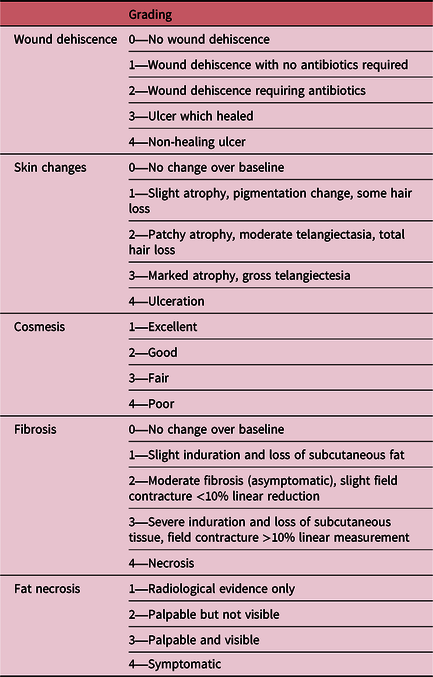
All patients underwent computerised tomography-based treatment planning on Oncentra version 4.5.2.152 by Nucletron B.V. from the Netherlands and the TG-43 algorithm was used for dose calculation.Reference Rivard, Coursey and DeWerd11 The inclusion criteria, technique, planning and dosimetric data about the dose and volume indices have been reported in detail in our previous publication.Reference Gurram, Wadasadawala and Joshi12 All patients underwent open cavity surgery without any oncoplastic procedure. The interstitial brachytherapy procedure was done either intra-operatively (n = 41) or post-operatively (n = 21). Patients were examined weekly for 4 weeks after the completion of brachytherapy treatment and monthly thereafter up to 3 months for the assessment of acute toxicities such as erythema, oedema, infection and wound dehiscence. Follow-up protocol after the above was 6-monthly for the first 5 years and annually thereafter. Wound dehiscence, skin changes in terms of pigmentation, atrophy, telangiectasia and ulceration, were classified based on the severity as shown in Table 1. Radiation Therapy and Oncology Group (RTOG) criteria were used for grading late sequelae like fibrosis and fat necrosis.Reference Cox, Stetz and Pajak13 Cosmesis was graded using the Harvard scale of subjective assessment which was done by the physician in the outpatient clinic during the follow-up visit.Reference Bear, Anderson and Brown14
RTOG grading of acute and late toxicitiesReference Gurram, Wadasadawala and Joshi12

RTOG, Radiation Therapy Oncology Group.
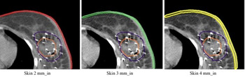
The body was delineated using the automated segmentation tool (magic wand) in the treatment planning system. The target volumes defined in the original treatment plans by the treating physician were used without any change. The tumour bed was delineated taking into account the seroma, clips and post-operative changes. An additional clinical target volume (CTV) of 1–1·5 cm was grown and cropped anteriorly from the body by 5 mm inside and posteriorly was limited to the pectoral muscle. For the current study, various thickness of the skin was retrospectively contoured over the treated breast by one of the co-authors and cross-verified by the physicist. Firstly, the treated breast was contoured. In every patient, contouring was done including skin thickness of 2 mm internal to the body, 3 mm internal to the body and 4 mm internal to the body. These were referred to as skin 2 mm_in, skin 3 mm_in and skin 4 mm _in, respectively. For obtaining 2-mm skin thickness inside the body, the external contour was subtracted by 2 mm followed by extraction from breast contour. Similarly, for 3-mm and 4-mm thickness, the external contour was subtracted by 3 mm or 4 mm respectively followed by extraction from the breast. This has been depicted in Figure 1.
Depiction of skin contours (Skin 2 mm_in, Skin 3 mm_in, Skin 4 mm_in) as defined in the study.

The dose received by 0·1 cc, 0·2 cc, 1 cc, 2 cc, 5 cc of all the defined skin structures were evaluated. Similarly, the volume of each defined skin structure receiving 33% (V33), 50% (V50), 66% (V66) and 100% (V100) of the prescribed dose was calculated based on dose–volume histogram parameters.
Statistical analysis
The median values have been reported for continuous variables while proportions have been reported for categorical variables while describing the patient characteristics. The univariate analysis for studying the correlation of all the dose and volume variables of each skin structure with the development of acute and late sequelae was done using the Mann–Whitney U test. A two-sided p-value of <0·05 was used as the cut-off for significance level. A receiver-operating characteristic (ROC) curve was used for calculating the cut-off for the significant results obtained in the univariate analysis. All results of the cross-tabulations of the dose and volume parameters significant on univariate analysis were studied using the chi-square test and risk estimate was reported as odds ratio with 95% confidence interval (95% CI) taking the cut-offs derived from the ROC analysis. All analysis was done in SPSS version 23.
Results
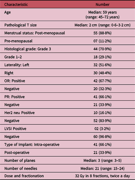
The clinico-pathological details of the patients included in the study have been described in Table 2. All the implants were of good quality and the median coverage for the cavity, homogeneity index and conformity were 0·94, 0·73 and 0·69, respectively. The median follow-up of the cohort was 54 months (range: 28–86 months). Acute and late sequelae were assessed by the physician at each follow-up. For the purpose of analysis, we have evaluated the late sequelae at 2 years and at last follow-up.
Patient, tumour and implant characteristics

LVSI, lympho-vascular space invasion; OR, oestrogen receptor; PR, progesterone receptor.
Acute sequelae
Only two patients (3·2%) developed Grade 3 wound dehiscence in the form of the superficial ulcer which healed with scar formation in 4 weeks but without requiring any re-suturing. Another six patients developed Grade 1–2 wound dehiscence. None of the dose–volume metrics correlated with the development of acute sequelae post-MIB.
Late sequelae
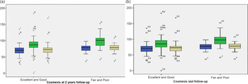
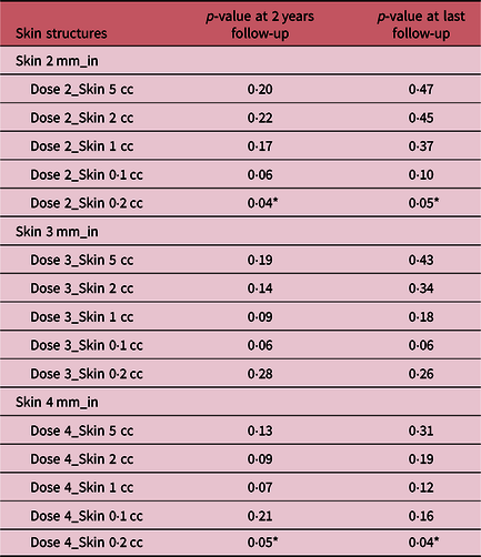
At 2 years follow-up asymptomatic fat necrosis which was picked up on mammography was observed in 10 (16·1%) patients. Skin changes in the form of Grade 2 atrophy and telangiectasia were seen in 5 (8%) patients. None of the patients had fibrosis outside the tumour bed. Twenty-five (40·3%) patients had no tumour bed fibrosis at last follow-up. Only 2 (3·2%) patients had Grade 2 fibrosis while the remaining 35 (56·4%) patients had Grade 1 skin fibrosis. Cosmetic results were dichotomised as good/excellent and fair/poor for the purpose of analysis. While comparing with baseline cosmesis 10 (16·1%) patients had a worsening of cosmesis. Forty-one (66·1%) patients had good to excellent cosmesis at 2 years follow-up while the remaining 21 (30·7%) patients had fair/poor cosmesis. As the majority of the patients underwent intra-operative brachytherapy, it was not possible to study the effect of surgery on cosmetic outcome separately in the immediate post-operative period due to the retained catheters in the implanted breast. Moreover, as this is a retrospective study, the assessment of cosmetic outcome was not done after implant removal or at 4 weeks post-brachytherapy. The mean dose received by 0·2 cc of skin 2 mm_in was 72% (range: 31·9–135%), skin 3 mm_in was 80% (range: 7–324%) and skin 4 mm_in was 90·8% (range: 0–183%). The correlation between dose received by 0·2 cc of skin 2 mm_in, 3 mm_in and 4 mm_in with cosmesis at 2 years and last follow-up is depicted in Figure 2a and 2b. A significant correlation was observed between dose received by 0·2 cc of skin 4 mm_in with cosmesis at 2 years (p = 0·05) as well as last follow-up (p = 0·04). However, skin 2 mm_in (p = 0·04) also correlated with the only cosmesis at 2 years follow-up as shown in Table 3.
(a) Box plot suggesting a correlation between cosmesis at 2 years follow-up and dose received by 0·2 cc of Skin 2 mm_in (blue), Skin 4 mm_in (green) and Skin 3 mm_in (yellow). (b) Box plot suggesting a correlation between cosmesis at last follow-up and dose received by 0·2 cc of Skin 2 mm_in (blue), Skin 4 mm_in (green) and Skin 3 mm_in (yellow).

Univariate analysis of dosimetric factors affecting cosmesis at 2 years and at last follow-up using the Mann–Whitney U test

* Statistical significance.
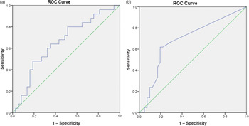
The ROC analysis provided the cut-off value for dose received by 0·2 cc of skin 4 mm_in as 90 and 92% of the total prescribed doses for prediction of excellent/good cosmesis at 2 years follow-up and last follow-up, respectively. The sensitivity was 64 and 61·9% respectively while specificity was 64·9% for both endpoints as shown in Figure 3a.
ROC curves suggesting the threshold dose and volume to defined skin thickness for excellent and good cosmesis. (a) Dose to 0·2 cc of skin 4 mm_in at last follow-up: 90%. (b) Volume receiving 100% of the total prescribed dose to skin 3 mm_in (V100) at last follow-up: 0·05 cc.

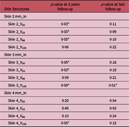
The volume of skin enclosed by 100% of the dose prescription is an important surrogate for the quality of implant. The mean volume of skin 2 mm_in, skin 3 mm_in, skin 4 mm_in receiving 100% of the prescribed dose was 0·06 cc (range: 0·0–2·1 cc), 0·14 cc (range: 0·0–4·6 cc), 0·27 cc (range: 0·0–6·8 cc), respectively. With respect to the volumetric indices, the significant correlation was observed between V100 of skin 3 mm_in with cosmesis at 2 years follow-up, as well as at last follow-up (p = 0·00 and 0·01 respectively) as shown in Table 4. For excellent or good cosmesis, the ROC cut-off for volume of skin 3 mm_in receiving 100% of prescribed dose is 0·05 cc (sensitivity = 66·7% and specificity = 70·7% and area under the curve = 0·87, OR = 1·74) as shown in Figure 3b. None of the other skin metrics showed any significant correlation with the development of other late sequelae.
Univariate analysis of volumetric factors affecting cosmesis at 2 years and at last follow-up using the Mann–Whitney U test

* Statistical significance.
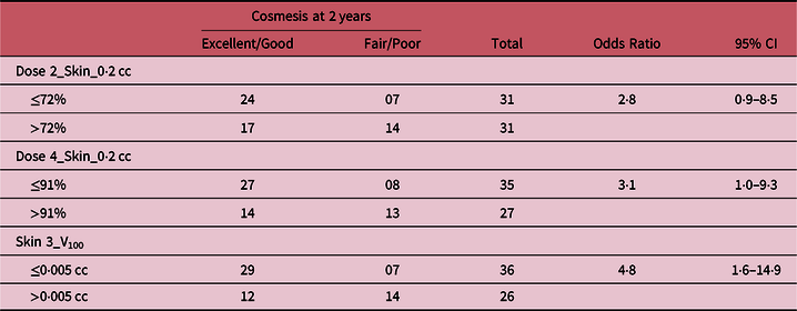
The results of the cross-tabulations done for the four-dose and volume parameters found significant on univariate analysis have been shown in Table 5. It can be noted that dose to 0·2 cc of skin 4 mm_in and V100 of skin 3 mm_in have the highest odds ratio for prediction of cosmesis at 2 years.
Odds ratio using the chi-square test for predicting cosmesis at 2 years for various dose and volume parameters

Discussion
The role of MIB in early breast cancers has been evaluated in many prospective trials. The skin dose received by this technique is significantly less as compared to other APBI methods.Reference Major, Niehoff and Kovács15 Hence, this study was done to evaluate the correlation between dose received by various skin thicknesses with the development of acute and late sequelae in an Indian cohort. The thickness of the skin (with no prior radiation) is approximately 1·5–2 mm.Reference Huang, Boone and Yang16 Hence, it would be appropriate to evaluate doses to this thickness.
Before we discuss the study results, it is important to keep in mind certain important factors pertaining to interstitial brachytherapy. The development of late sequelae post-brachytherapy as discussed in the introductory section is related to the dose received by the overlying skin. With respect to breast brachytherapy, the factors that are likely to affect skin dosimetry include the distance of the implanted catheters from the skin, the proximity of the CTV to the skin, the policy of cropping the CTV from the skin, the volume of the target volume and the brachytherapy technique. Additionally, the ABS recommendations suggested reporting of the dose maximum of the skin. Recently, the GEC-ESTRO published guidelines for reporting of the dose received by the organ at risk based on dose–volume parameters. It mentions that a 5-mm thickness below the body should be defined as skin volume.Reference Strnad, Major and Polgar8 However, apart from a dosimetric study by Hilts et al., authors have not come across any clinical report studying the impact of dose–volume parameters of skin volumes of variable thickness on the cosmetic outcome or other acute or late sequelae.Reference Hilts, Halperin and Morton9
We observed the significant correlation of the dose received by 0·2 cc of skin contoured 4 mm inside (skin 4 mm_in) with cosmesis at 2 years follow-up and at last follow-up with the threshold of 90% for prediction of excellent or good cosmesis. Thus, supporting the GEC-ESTRO guideline which recommends dose to 0·2 cc (D0·2) of skin <90% of the prescribed dose.Reference Strnad, Major and Polgar8 Similarly, a significant correlation was observed between the volume of skin 3 mm_in receiving 100% of the prescribed dose and cosmesis at 2 years and at last follow-up with the threshold of 0·05 cc. None of the guidelines to date suggest reporting of volumetric parameters. The study by Hilts et al. showed large variability between the volumetric parameters (V33 and V66) as well as Dmax and small dose volumes (D0·1 cc) irrespective of the skin definition. However, it should be noted that 3-mm skin was not used by Hilts et al. and the dosimetric study involved only 10 patients with MIB. In the current study, the additional skin structure that was contoured was 3-mm sub-surface.
The exact volumetric structure for the skin (3–4 mm in the current study or 5 mm as recommended by GEC-ESTRO guidelines) that should be contoured routinely in breast brachytherapy should be validated in a larger study with respect to clinical endpoints. As the majority of the work was completed before the publication of GEC-ESTRO guidelines, 5-mm skin volume was not contoured.
The conventional dose calculation algorithm based on the TG-43 brachytherapy dose calculation formalism is limited by their inability to accurately calculate dose at the air-tissue interface and hence could have affected the dose calculations of the skin sub-surfaces studied here. Recently, Monte Carlo-based algorithms as well as inverse planning optimisation algorithms have been developed but still not in routine clinical use. But these algorithms are still not available in our institute as well, hence the study was carried based on the TG-43 formalism which is also proposed by the GEC-ESTRO guidelines.Reference Strnad, Major and Polgar8
This is the first report in an Indian cohort in which the skin metrics have been correlated with post-brachytherapy sequelae. We did not observe any significant correlations between doses received by all defined thickness of skin and the other acute and late sequelae such as wound dehiscence, tumour bed fibrosis, telangiectasia and skin atrophy and fat necrosis. This may be due to the small sample size and a low number of events which can be inadequate for multiple testing. We intend to continue this study accruing a greater number of patients and collect prospective data on dosimetry with the additional 5-mm skin structure that is recommended by GEC-ESTRO. Nonetheless, the results from the current study provide additional guidance for evaluating skin dose in breast brachytherapy.
Conclusion
The reporting of skin doses in MIB has not been practiced uniformly due to the lack of clear consensus on the delineation of the skin contour as well as reporting of the various dose–volume statistics. We have attempted to evaluate the skin dose–volume effects with respect to acute and late skin sequelae. Doses received by 0·2 cc of skin and V100 seem critical for the cosmetic outcome. Prospective studies with longer follow-up and larger sample sizes are required for evaluation of other sequelae and validation of these results.
Acknowledgements
None.
Financial support
This research received no specific grant from any funding agency, commercial or not-for-profit sectors.
Conflicts of interest
None.
Funding support
None.