INTRODUCTION
Many marine invertebrates have complex life histories that include planktonic larval forms. The growth, behaviour and morphology of these larvae are often not well studied, even for ecologically or economically important species. Laboratory rearing allows for description of the morphology of ontogenetic stages and collection of developmental data critical for phylogenetic, ecological and applied fisheries research.
The larval and early juvenile development of king crabs (Anomura: Paguroidea: Lithodidae) has been the subject of several recent studies. Much of this work has focused on economically important members of this group including: Lithodes aequispinus (e.g. Shirley & Zhou, Reference Shirley and Zhou1997); Lithodes santolla and Paralomis granulosa (e.g. Calcagno et al., Reference Calcagno, Anger, Lovrich, Thatje and Kaffenberger2004); Paralithodes camtschaticus (e.g. Epelbaum et al., Reference Epelbaum, Borisov and Kovatcheva2006) and Paralithodes platypus (e.g. Stevens et al., Reference Stevens, Persselin and Matweyou2008). Interest has also been generated by debate surrounding the hypothesized evolution of the Lithodidae from a pagurid hermit crab ancestor (reviewed in McLaughlin & Lemaitre, Reference McLaughlin and Lemaitre1997; McLaughlin et al., Reference McLaughlin, Lemaitre and Sorhannus2007). This hypothesis has recently been supported by both morphological (Richter & Scholtz, Reference Richter and Scholtz1994) and molecular evidence (Cunningham et al., Reference Cunningham, Blackstone and Buss1992; Morrison et al., Reference Morrison, Harvey, Lavery, Tieu, Huang and Cunningham2002; Zaklan, Reference Zaklan, Paul, Dawe, Elner, Jamieson, Kruse, Otto, Sainte-Marie, Shirley and Woodby2002; Tsang et al., Reference Tsang, Ma, Ahyong, Chan and Chu2008). While similarity between lithodid and pagurid zoeae has provided additional support for this hypothesis (MacDonald et al., Reference MacDonald, Pike and Williamson1957; Gould, Reference Gould1992), a pagurid ancestry for the Lithodidae has also been questioned by interpretations of lithodid post-larval abdominal development (McLaughlin & Paul, Reference McLaughlin and Paul2002; McLaughlin et al., Reference McLaughlin, Anger, Kaffenberger and Lovrich2003, Reference McLaughlin, Lemaitre and Tudge2004).
To date, complete descriptions of development have been published for 17 lithodid species in 9 genera. A summary of developmental studies can be found in Zaklan (Reference Zaklan, Paul, Dawe, Elner, Jamieson, Kruse, Otto, Sainte-Marie, Shirley and Woodby2002), and descriptions of development have been subsequently produced for Acantholithodes hispidus (Hong et al., Reference Hong, Perry, Boutillier and Kim2005) and Paralomis spinosissima (Watts et al., Reference Watts, Thatje, Clarke and Belchier2006). Epelbaum et al. (Reference Epelbaum, Borisov and Kovatcheva2006) expanded on basic morphological description to include colour, growth and behavioural data for Paralithodes camtschaticus reared in the laboratory. The inclusion of such data increases the applicability of developmental studies to ecological and oceanographic research.
In the genus Lopholithodes, development has been described only for Lopholithodes mandtii (Puget Sound king crab) (Zoea I by Haynes, Reference Haynes1993; all stages by Crain & McLaughlin, Reference Crain and McLaughlin2000a). Three distinct larval series obtained off the Oregon coast in 1970–1971 were also tentatively identified as Lopholithodes by Lough (Reference Lough1975). Lopholithodes foraminatus (Simpson, 1859) (brown box crab) occur sympatrically with L. mandtii along the north-west coast of North America, although L. foraminatus are generally deeper and live on soft rather than hard substrates (Jensen, Reference Jensen1995).
Lopholithodes foraminatus has been the subject of a small scale commercial fishery in Oregon since 1982 and has been investigated as a commercial species in British Columbia (Zhang et al., Reference Zhang, Workman and Phillips1999; Zhang, Reference Zhang2001). Box crabs are also caught as bycatch in shrimp and groundfish trawl fisheries (Zhang et al., Reference Zhang, Workman and Phillips1999). Commercial fishing activity has apparently been the cause of local stock collapses in the lithodids Paralithodes camtchaticus (Dew & McConnaughey, Reference Dew and McConnaughey2005), Paralithodes platypus (Stevens, Reference Stevens2006), Lithodes santolla and Paralomis granulosa (Calcagno et al., Reference Calcagno, Lovrich, Thatje, Nettelmann and Anger2005). Zhang et al. (Reference Zhang, Workman and Phillips1999) identified the lack of early life history data as an obstacle to the development of management strategies for L. foraminatus.
This study describes the morphological development of laboratory reared zoeae, glaucothoe, and early juvenile stages of Lopholithodes foraminatus. Data are also presented on behaviour, length and weight at stage, and variation in stage duration with temperature. The implications of selected developmental characters, in particular secondary lecithotrophy, to the biology of the Lithodidae are discussed. Key morphological features useful in the identification of the larvae and postlarvae of L. foraminatus are also described. These data will be applicable not only for species identification purposes but also for phylogenetic, ecological and fisheries research on lithodid crabs.
MATERIALS AND METHODS
Obtaining and rearing larvae
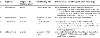
Zoeae released by 4 female L. foraminatus were reared for use in different aspects of this study (Table 1). Adult crabs were captured in rectangular Dungeness crab traps at approximately 120 m depth on the north side of Baker Passage in the northern Strait of Georgia, British Columbia, Canada (50°01′12″N 124°56′43″W). Adult crabs were maintained in the recirculating seawater systems at the University of Victoria. Females A and B began releasing larvae within 2 months of capture (Table 1). The mean water temperatures (±standard deviation) experienced by these females between the date of capture and completion of hatching were 10.5±0.1°C and 9.4±0.2°C respectively. Female C was captured already releasing larvae and female D was held in the laboratory for 1 year before the beginning of hatching. In spring 2006 female A released zoeae in a 170 l tank with a screened outflow. All suspended individuals were collected every morning with either a 5 mm diameter pipette or a fine mesh net. In subsequent seasons, females releasing larvae were maintained in partially covered 20 l plastic tubs supplied with a constant flow of seawater. Larvae passed through a 90° overflow pipe that ended in a T at the bottom of a 750 ml plastic container with a 400 µm Nitex® mesh bottom. This container was seated in a 1 l glass beaker. This arrangement allowed zoeae to swim to the top of the 750 ml container and avoid being damaged by turbulence. In all cases hatching occurred in the first hours of darkness and healthy zoeae for rearing experiments were collected the following morning. Virtually all zoeae remaining on the bottom of the tub in the morning had morphological abnormalities and were unable to suspend themselves in the water column. For this reason no effort was made to clean out these zoeae on a daily basis as in previous studies (e.g. Crain & McLaughlin, Reference Crain and McLaughlin2000a).
Sources of larvae and culture methodologies utilized in describing the development of Lopholithodes foraminatus.

F, vacuum filtered seawater (Pall glass fibre filter); UF, unfiltered seawater; GB (1), 1 l glass beaker; PB, 4 l plastic bucket; #/L, density of larvae per litre; D, daily water change; 2/D, water changed every 2nd day; P, daily partial water change; ST, vessels immersed in seatray; I, vessels in incubator or refrigerator on 12/12 light/dark cycle.
* Female C was releasing larvae when captured. She also exhibited complete reversed asymmetry, with a larger left cheliped and pleopods 2–5 on only the right hand side of her abdomen (a manuscript describing this reversed asymmetry is in preparation).
Seawater used for all rearing was taken from taps in the University of Victoria recirculating seawater system. Water in this system is collected from the ocean and is re-circulated through chillers, sand filters and UV sterilization tubes.
Zoeae were maintained in 1 l glass beakers or 4 l plastic buckets at densities of 15–50 individuals/litre in either glass-fibre filtered seawater or seawater drawn directly from the system (details of rearing methodologies in different aspects of this study are outlined in Table 1). Cultures were fed daily with a combination of newly hatched Artemia sp. nauplii and >24 hour old Artemia nauplii that had been fed with Isochrysis sp. paste (Reed Mariculture, Campbell, CA). An attempt was made to provide nauplii at a density such that a few would still be present on the subsequent day. When all individuals in a culture vessel had moulted to the glaucothoe stage, a piece of nylon mesh was provided as a settlement substrate. In 2006, glaucothoe were initially fed with Artemia. However; after experiments showed that this was a non-feeding stage (see below), no food was provided to glaucothoe during subsequent rearing work.
Juvenile crabs were maintained at varying densities in containers from 100 ml to 4 l with 400 µm Nitex® mesh bottoms. These containers were immersed in a seatray in the re-circulating system. In 2006, juveniles were initially fed every 48–72 hours with the encrusting bryozoan Membranipora membranacea and with Artemia nauplii which had been fed with powdered sea urchin test. This choice of a high calcium diet was based on anecdotal accounts that juvenile lithodids require a diet high in calcium salts (Jensen, Reference Jensen1995; S. Duff, personal communication, 2006). In subsequent years, better growth and survival were obtained with a diet of chopped frozen krill (Euphausia pacifica) every 48–72 hours, supplemented with Membranipora at least twice per week. Juvenile crabs of the same instar were maintained together and moved to new containers upon moulting. This allowed the stage of juveniles to be followed up to the fifth instar. Subsequently juveniles were maintained in bulk cultures of mixed instars.
Test for lecithotrophy and secondary lecithotrophy
Two 1 l cultures (15/l) of newly hatched zoeae were reared without food to determine whether larvae could develop lecithotrophically. To test for secondary lecithotrophy, 68 glaucothoe were collected from a 4 l mass culture of zoeae on the day of moulting to stage. Five 500 ml beakers of 6 glaucothoe and one of 4 glaucothoe were provided with Artemia nauplii every second day. Another set of cultures was not provided with nauplii. Culture temperature was ≈10°C and water was changed every second day. The mean number of days required to moult to the first crab instar and the arcsine transformed proportion surviving this moult were compared between treatments for the 6 replicates using Student's t-test.
Measurement
Two 1 l cultures containing 40 larvae each were reared for length and weight measurements. Measurements were made of 1st stage zoeae within 24 hours of hatching; 2nd and 3rd stage zoeae within 24 hours of moult to stage; 4th stage zoeae within 48 hours of moult to stage; glaucothoe within 72 hours of moult to stage, and 1st crab instars within 5 days post-moult.
All individuals to be measured were anaesthetized with Alka Seltzer® tablets dissolved in seawater and photographed in a drop of water on a microscope slide at 25 × magnification with a Sony PowerHAD digital video camera mounted on an Olympus SZX9 stereomicroscope. Zoeae were photographed in a lateral orientation while glaucothoe and juveniles were photographed dorsally. Measurements were made using Northern Eclipse software calibrated with a slide micrometer. For zoeae, the distance from the tip of the rostrum to the posterior mid-dorsal notch of the carapace was selected as the most accurate measurement since both of these points are in the same focal plane when a zoea is viewed laterally. This measurement is the same as used by Crain & McLaughlin (Reference Crain and McLaughlin2000a). No separation was made between carapace length and rostrum length as described by Epelbaum et al. (Reference Epelbaum, Borisov and Kovatcheva2006) for red king crab. This is because L. foraminatus zoeae lack the obvious notch that defines the base of the rostrum in red king crab zoeae (A. Epelbaum, personal communication, 2007). Glaucothoe and juveniles were measured from the tip of the rostrum to the mid-posterior point of the carapace and across the widest point of the carapace, not including spines.
After being photographed, the wet and dry weight of each individual was determined. Larvae were passed momentarily through distilled water, blotted on lens paper, and placed on pre-weighed pieces of tinfoil. Wet weight was measured with a microbalance. Specimens were then dried for at least 24 hours in a 60°C oven. At least 24 hours after removal from the oven the specimens were re-weighed on the microbalance to determine dry weight. This delay allowed specimens to equilibrate to a constant weight; weighing specimens immediately after removal from the oven was complicated by their rapid change in weight while on the balance.
Stage durations
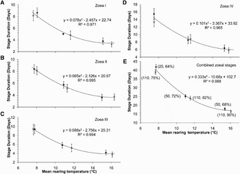
Stage duration was determined primarily from 3 groups of 110 larvae, hatched from female D on 22 February 2008 and reared in 4 l buckets in incubators set to 8°C, 12°C and 16°C with a 12:12 light–dark cycle. Temperature of the culture water was measured daily. Partial water changes were carried out on a daily basis and exuviae were counted (approximately) and removed. Mean date of moulting to stage was calculated as: Σ ((# of exuviae) × (day after hatching))/total number of exuviae counted for that stage. Mean stage duration was calculated as the difference between the mean date of moulting to the subsequent stage and the mean date of moulting to the stage in question± the standard deviation in the date of moulting to the subsequent stage. Due to slight temperature fluctuations in the incubator the mean temperature for each stage was calculated from daily temperature measurements rather than incubator presets. Stage duration data were also recorded using the same methodology for zoeae from females A and C reared in 1l glass beakers (see Table 1). Two beakers of 25 zoeae from female A were maintained in a seatray at ≈11°C. Two beakers of 25 zoeae from female C were maintained in a seatray at ≈16°C, and two beakers were maintained in a fridge at ≈8°C (data for only one of these beakers are presented due to complete mortality in the other). Second order polynomial regression equations were used to describe the relationship between mean zoeal stage duration and temperature for each stage, and for all four zoeal stages combined (Paul & Paul, Reference Paul and Paul1999). Regression equations were calculated using Microsoft Excel. Stage duration data for glaucothoe were recorded from beaker cultures maintained in seatrays at ≈11°C and ≈16°C.
Morphological description
Mortalities and healthy zoeae of each stage were fixed and preserved in 4% borate buffered formalin in seawater. Glaucothoe and juveniles were fixed for at least 24 hours in 4–5% borate buffered formalin in seawater and either preserved in the fixation solution or rinsed with 50% ethanol and preserved in 70% ethanol. Exuviae of all stages were preserved in 70% ethanol. Specimens and exuviae were dissected under a stereomicroscope and appendages were photographed in multiple focal planes using a DVC digital camera mounted on a Zeiss Axioskop compound microscope. Photographs of focal planes were imported sequentially as layers into Adobe Photoshop CS3. Drawings were made in an overlying layer by tracing structures in multiple focal planes using the pen tool. For whole animal drawings, live zoeae were held immobile on a slide using a coverslip supported by plasticine feet and photographed and drawn using the methodology described above. Using live zoeae for illustrations avoided the raised and distorted carapace typical of preserved material. Whole animal drawings of glaucothoe and drawings of juvenile crab carapaces and abdomens were produced from preserved exuviae. For clarity, illustrations of the lateral view of whole individuals show only the appendages on the near side.
All structures are illustrated and described for the first zoeal stage. For subsequent stages, only structures showing significant changes in morphology are illustrated. At least 5 specimens of each appendage for each stage were examined to confirm morphology and setal arrangement (Clark et al., Reference Clark, Calazans and Pohle1998). Care was taken to examine both the preserved healthy larvae and exuviae. Examination of exuviae allowed for observation of setae on the opposite side of appendages and observation of preserved whole specimens allowed for confirmation of appendage shape.
Terminology
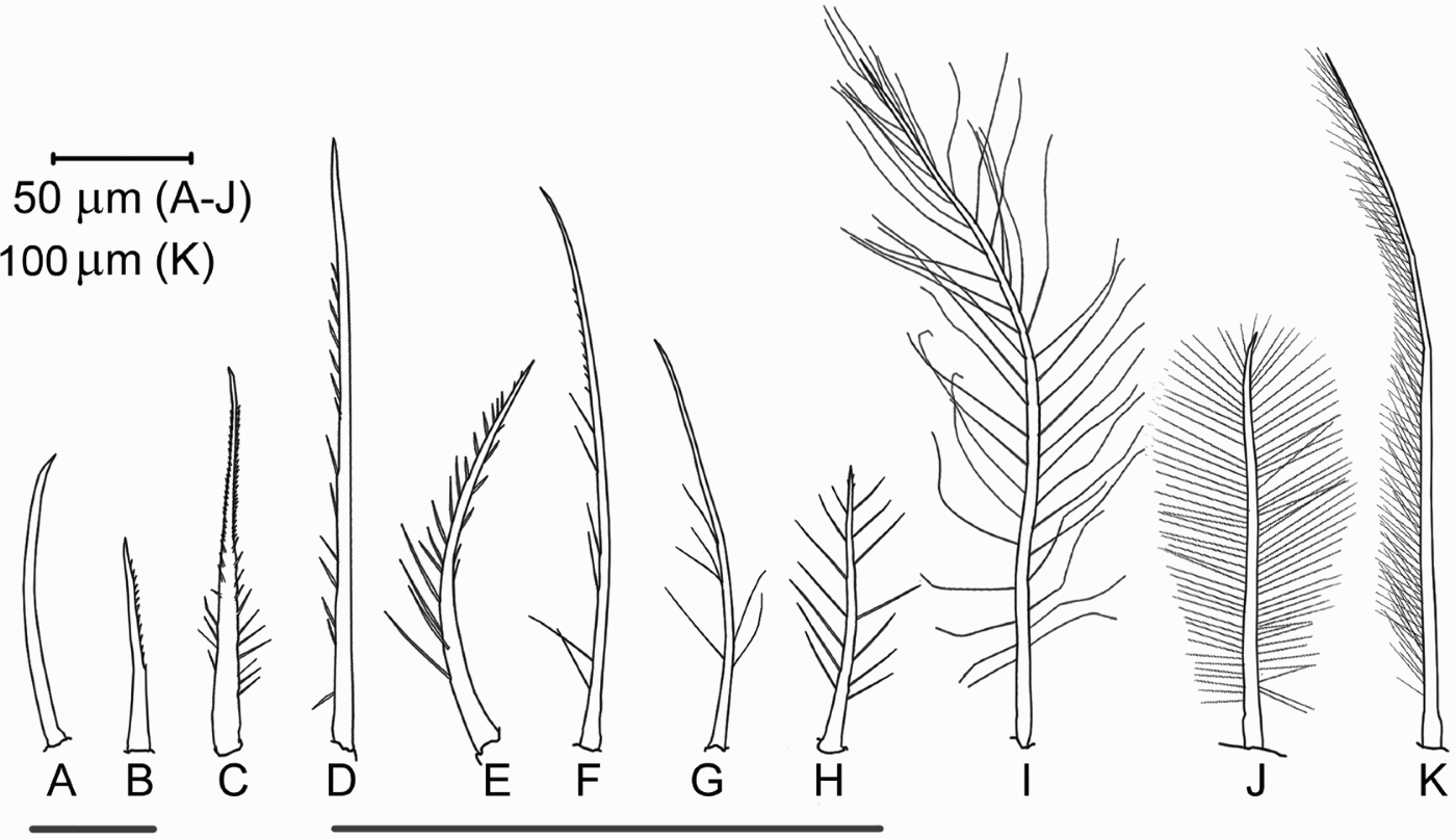
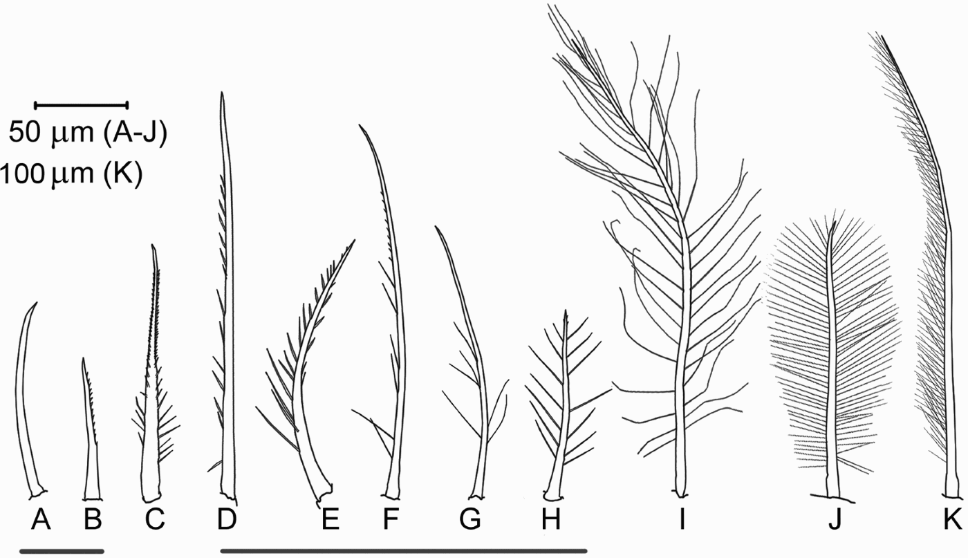
The terminology used to describe the setae of lithodid larvae and juveniles varies. Crain (Reference Crain1999) described 6 types of setae from the mouthparts of Placetron wosnessenskii following the system used by Lavalli & Factor (Reference Lavalli and Factor1992) for the mouthparts of larval lobsters. More recently, Epelbaum et al. (Reference Epelbaum, Borisov and Kovatcheva2006) adapted the terminology of Factor (Reference Factor1978) to describe 10 types of setae from Paralithodes camtschaticus larvae and postlarvae. Epelbaum & Borisov (Reference Epelbaum and Borisov2006) modified this scheme and reduced the number of setal types to 7. These systems were found to be problematic for Lopholithodes foraminatus due to the prevalence of setal types intermediate between described classifications. The present study uses a simplified scheme of setal terminology based on the works cited above (Figure 1). Setae with long setules growing primarily in 2 directions are referred to as plumose, or natatory when used for swimming. Those with sparse, long setules arranged at random are referred to as pappose. All setae bearing any combination of short stout setules, short fine setules, scales, or apparent serration are referred to as denticulate. This category includes the serrate and serrulate types of the above authors. Any denticulate setae that also bear long fine setules are referred to as plumodenticulate. Unarmed setae are referred to as simple, and spiniform processes are described individually. There is still some overlap between these setal types, partially due to the presence of minute setules at the limit of resolution with light microscopy.
Summary of setal types of Lopholithodes foraminatus larvae and post larvae: (A) simple; (B–D) denticulate; (E–F) plumodenticulate; (G) pappose; (H–J) plumose; (K) plumose (natatory). The grey bars indicate areas of significant overlap between types.
Appendage terminology largely follows that of Epelbaum et al. (Reference Epelbaum, Borisov and Kovatcheva2006). In the case of mouthparts, external refers to the surface of the appendage facing away from the mouth, while internal refers to the surface facing towards the mouth. Unless otherwise specified, illustrations show the external surface of appendages.
Samples of zoeal instars, glaucothoe, and 1st and 2nd crab instars were deposited in the invertebrate collection of the Royal British Columbia Museum in Victoria, BC Canada.
RESULTS
Behaviour
A prezoeal stage was observed in hatching vessels soon after lights were turned out in the evening. Prezoeae suspended themselves by a jackknife flexion of the abdomen and thorax. The prezoeal exoskeleton was apparently shed within minutes of hatching. At this point the first zoeal stage adopted the mode of swimming that would be used throughout zoeal development. Zoeae swam by vigorously beating the exopods of their maxillipeds while suspended in a head down orientation with the abdomen extended straight above them. Periodically they would flex the abdomen against the ventral surface of the thorax and drag the telson across the mouthparts. When not actively swimming, zoeae would sink rapidly.
Zoeae were positively phototactic and in a transparent culture vessel would tend to suspend more readily when placed on a dark coloured than a light coloured surface. Over the course of zoeal development larvae spent less time suspended. In the first days after hatching virtually all individuals would be suspended in the culture vessel while by the 4th zoeal stage less than 25% would generally be suspended at any given time.
Zoeae of all 4 stages exhibited a characteristic response to physical or chemical perturbation. When sucked into a pipette, grasped with forceps, or placed in fixative, the antennae were rapidly flexed laterally into a position where the scaphocerites were at 90° to the long axis of the body. This position would be maintained until the threat had passed.
Cannibalism by zoeae was almost never observed or inferred by larvae disappearing from cultures.
Swimming by glaucothoe was accomplished by beating the pleopods with the pereopods extended anteriorly and held stationary. Glaucothoe would suspend relatively frequently during the first one or two days after moulting but thereafter became less active. They would seek a substrate they could grasp and in a smooth bottomed container would end up clinging to each other in a ball of many individuals. When provided with a mesh substrate they would cling to it with their pereopods and would sometimes extend their abdomens and rhythmically beat their pleopods. Periodically the 5th pereopods could be seen through the translucent carapace being drawn back and forth across the gills. In response to stress, glaucothoe would tightly draw in their pereopods and tuck in their eyestalks in the adult manner. This response was maintained through the juvenile stages. Feeding and cannibalism by glaucothoe was not observed (see below).
Juvenile crabs no longer had the ability to swim and would walk slowly across the substrate. The first juvenile stage was relatively inactive and responded to food only if it was extremely close. Subsequent juvenile stages became more active and more proactive in walking towards a food source. Cannibalism was also more prevalent in later stages but seemed to be mostly restricted to predation on moulting or moribund individuals.
Test for lecithotrophy and secondary lecithotrophy
All first stage zoeae reared without food died by the 10th day after hatching. Some individuals shed scraps of exoskeleton or became stuck in the exuvium but none moulted successfully to the second zoeal stage.
Glaucothoe were observed to grasp Artemia nauplii with their chelae and pull them towards the mouthparts. However, in all cases the nauplii were then pushed away again by the claws or the respiratory current. Of the glaucothoe reared without food, 27 (79.4%) survived the moult to the first crab instar compared to 26 (76.5%) of those provided with food. There was no significant difference in either survival to the first crab instar (Student's t-test, t = −0.47, df = 10, P = 0.65) or the number of days before moulting (Student's t-test, t = 0.36, df = 10, P = 0.72) between glaucothoe that were provided with Artemia and those that were not.
Growth
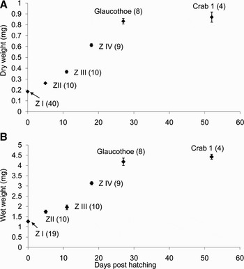
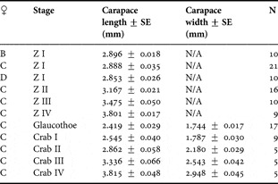
Length measurements for all larval stages are summarized in Table 2. Mean dry weights (±SE) for newly hatched zoeae from females B, C and D were 183±8 µg, 179±2 µg, and 199 ±2 µg respectively. This yields an overall mean dry weight for first stage zoeae of 185±3 µg (Figure 2A). The mean wet weights (±SE) for newly hatched zoeae from females C and D were 1.34±0.06 mg, and 1.19±0.02 mg respectively. This yields an overall mean wet weight for first stage zoeae of 1.26±0.03 mg (Figure 2B).
Relationship between mean dry (A) and wet (B) weights (±SE) and days post-hatching for Lopholithodes foraminatus zoeae, glaucothoe and first crab instars. Stages reared in 1 l beakers at an initial density of 40/l with daily changes of filtered seawater (every second day for glaucothoe). Rearing temperature for all cultures ≈11°C. Numbers in parentheses indicate sample size.

Length measurements of Lopholithodes foraminatus zoeae, glaucothoe and first stage juveniles. Zoeae were measured from the tip of the rostrum to the mid-dorsal notch in the posterior margin of the carapace. Carapace length of glaucothoe and juveniles was measured from the tip of the rostrum to the mid-point of the posterior carapace margin; carapace width was measured at the widest point not including spines.

NA, not applicable.
Stage duration and survival
The relationships between stage duration and temperature for each zoeal stage and all 4 zoeal stages combined are illustrated in Figure 3. Initial numbers of larvae in each treatment and per cent survival to the glaucothoe stage are presented in Figure 3E. The second order regression functions calculated for each zoeal stage, and for all 4 zoeal stages combined, level out at approximately 16°C.
Relationship between mean rearing temperature and mean stage duration (±SD) for Lopholithodes foraminatus zoeal stages (A) I, (B) II, (C) III, (D) IV and (E) all four zoeal stages combined. Open symbols indicate mean stage duration values for larvae cultured in 4 l buckets in incubators at a density of 27.5 individuals/l (50% unfiltered water changes daily); filled symbols indicate larvae cultured in beakers at a density of 25 individuals/l (complete filtered water changes daily). Beakers were immersed in flowing water (≈16°C and ≈11°C) or held in a fridge (≈8°C). Parentheses in 3E indicate initial number of larvae, % surviving to the glaucothoe stage.

The equation for the complete zoeal period (duration = 0.333 (temp °C)2−10.68 (temp °C)+102.7) gives degree day estimates for zoeal development of 308.6 at 8°C, 280.6 at 11°C, and 273.1 at 16°C. The duration of the glaucothoe stage (±standard deviation) was 13.7±1 days at 15.5°C (212 degree days, 34 glaucothoe, 85% survival to the first crab instar) and 19.0±1.7 days at 11.5°C (219 degree days, 36 glaucothoe, 72% survival to the first crab instar). Survival through subsequent crab stages was poor, with almost no individuals surviving for more than 1 year. When maintained in mesh containers in the seatray at approximately 9°C, the first individuals were moulting to the 5th crab instar by about 28 weeks post-hatching.
Morphological description
PREZOEA
Prezoeae were covered in an unarmed cuticle. The posterolateral spines were contained by this cuticle and folded down along the posterior margin of the carapace. The rostrum was folded ventrally. The prezoeal cuticle was elaborated into feathery projections where it covered the telson, antennules and antennal exopods.
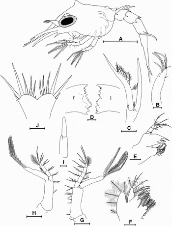
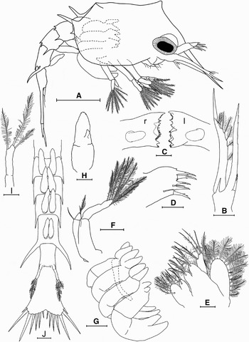
ZOEA I (FIGURE 4)
Colour: the pale orange colour of newly hatched zoeae became more intense during the first stage. This colour was due to orange chromatosomes in the carapace, rostrum, antennae and maxillipeds. When held in the dark, zoeae became much paler because pigment in the chromatosomes was concentrated; this remained true for subsequent zoeal stages. Chromatosomes with concentrated pigment appeared red rather than orange. There was a transparent patch dorsally in the vicinity of the heart. The abdomen and telson were also transparent. The digestive gland was pale orange-brown. When viewed with incident light the compound eyes were golden yellow.
Cephalothorax: the rostral spine constituted approximately half of the total length of the carapace. The posterolateral spines were pronounced and the posterior margin of the carapace was deeply notched. There was a distinctly peaked mid-dorsal carina with a generally steeper anterior than posterior slope. The lateral walls of the carapace were inflated and completely covered the coxae of the maxillipeds, the developing pereopods, and the majority of the first and second abdominal segments. The eyes were sessile.
Antennule (Figure 4B): the antennule projected anteriorly and dorsally and had a slight lateral curve. It was incompletely 2 segmented with a medial unarmed endopod bud. The putative protopod had 1–2 minute dorsal setae. Distally the antennule bore 6–7 aesthetascs and 3–4 simple setae. Throughout development the exact setal arrangement of the antennule was very difficult to positively determine due to the tendency of aesthetascs to kink or break and the positioning of minute setae between the bases of the aesthetascs.
Lopholithodes foraminatus: (A) zoea I (lateral); (B) antennule (left ventrolateral); (C) antenna (right ventral); (D) [r]ight (dotted line indicates anterior ridges of molar lobe) and [l]eft mandibles; (E) maxillule (right); (F) maxilla (right); (G) 1st maxilliped (left externolateral); (H) 2nd maxilliped (right externolateral); (I) 3rd maxilliped (right internal); (J) telson (ventral). Scale bars: A = 1 mm; B, E, F, I = 100 µm; C, G, H, J = 250 µm; D = 50µm.

Antenna (Figure 4C): ventrally the protopod had a large unarmed spine at the base of the exopod and a spinulose spine at the base of the endopod. The endopod was not delineated from the protopod and had a bifid tip. The exopod was slightly longer than the endopod and usually reached the tip of the rostral spine. Medially the exopod had a flattened ridge bearing 5 plumose setae which became smaller and sparser distally. This ridge terminated in a flattened lobe bearing 4 pappose and/or simple setae. The distal portion of the exopod was unarmed and acute.
Mandibles (Figure 4D): the mandibles were angled slightly anteriorly and the internal surfaces were held against, and partially covered by, the labrum. This positioning was maintained throughout zoeal development and is illustrated for the third zoeal stage in Figure 6B. Both the left and right mandibles had a large apical tooth at their medio-ventral angle. They were distinctly asymmetrical in the balance of their denticulation. The gnathal lobe of the left mandible consisted of the apical tooth and 2 acute denticulations; the molar lobe had 3 ridges of small denticulations. The gnathal lobe of the right mandible had a series of acute denticulations which were drawn out into a convex dorsal shield. The molar lobe consisted of 2 ridges internal to this shield and 2 dorsal teeth. Although the proportions of mandibular denticulations changed, the basic left right asymmetry was maintained through subsequent zoeal stages.
Maxillule (Figure 4E): the coxal endite had 1 short simple seta submarginally and 1 pappose, 1 denticulate and 4 plumodenticulate setae marginally. The plumodenticulate setae were strongly curved anteriorly and dorsally. The basial endite had 2 submarginal simple setae and 2 or 3 large marginal spines. Each of these spines had several internally projecting denticulations. The endopod was 3 segmented. The proximal segment bore 1 or 2 minute simple setae, the second segment 1 pappose seta, and the distal segment 3 terminal pappose setae.
Maxilla (Figure 4F): both the coxal endite and the basial endite were bilobed. The proximal lobe of the coxal endite had 1 pappose seta submarginally and 5 weakly plumose and 2 plumodenticulate setae marginally. The distal lobe of the coxal endite had 1 pappose seta submarginally and 3 plumodenticulate setae marginally. Both lobes of the basial endite had a submarginal pappose seta. The proximal and distal lobes had 3–4 and 3 marginal plumodenticulate and/or pappose setae respectively. The endopod had 2–3 pappose setae projecting from a ledge about half way along the medial margin and bore 1 subterminal and 3 terminal pappose setae. The exopod (scaphognathite) had 5 marginal plumose setae. Fine hair-like structures arose from all processes of the maxilla.
First maxilliped (Figure 4G): the coxa was usually armed with 1 small simple seta. The basis had 2 small simple setae basally; 5 simple and/or pappose setae along the medial margin; and 3 pappose and/or simple setae terminally. The endopod had 5 segments. The 3 proximal segments bore fine hair-like processes on their lateral surfaces. Progressing distally the first 4 segments of the endopod bore 3, 2, 1, and 2 pappose and/or simple setae respectively. The distal segment had 1 subterminal pappose seta, 1 subterminal plumodenticulate seta, 2 terminal plumodenticulate setae and 1 weakly plumose seta laterally at the base of the segment. The incompletely 2 segmented exopod had 2 subterminal and 2 terminal natatory setae.
Second maxilliped (Figure 4H): the coxa was unarmed. The basis bore 1 marginally denticulate seta about halfway along the medial surface and 1 pappose and 1 denticulate seta terminally. The endopod was 4 segmented and each segment had 1 pappose and 1 denticulate seta distally. In addition, the distal segment had a pair of long terminal denticulate setae and 1 plumose seta laterally at the base of the segment. The middle 2 segments of the endopod had fine hair-like processes on their lateral surfaces. The exopod was incompletely delineated into 2 segments and had 2 subterminal and 2 terminal natatory setae.
Third maxilliped (Figure 4I): the third maxillipeds were angled anteriorly, fitting between the bases of the second maxillipeds. The coxa was indistinct and the large basis bore an unarmed endopod bud. The exopod consisted of 1 segment and was also unarmed.
Abdomen: the first abdominal somite was unarmed. Somites 2 through to 5 had 4 posterodorsal spines: those on somite 2 were significantly smaller than on the other 3 somites. Somites 2 through to 4 bore a pair of short posterolateral spines while somite 5 bore a pair of long posterolateral spines that projected dorsally and were curved posteriorly. In some specimens, one or both of these spines had a bifid tip. The 6th abdominal somite was fused with the telson to form a single segment.
Telson (Figure 4J): the telson was broad and consisted of 2 convex lobes flanking a pronounced median cleft. Laterally each lobe had a short unarmed spine that was continuous with the exoskeleton (the balance of telsonal processes were clearly delineated). Immediately medial to this process was a minute anomuran hair which projected ventrally. Progressing towards the medial cleft, each lobe bore 5 spines armed with numerous minute denticulations. The second of these processes was the longest. In many of the first stage zoeae hatched from female A, a small 6th spine was present adjacent to the medial cleft on 1 or both lobes. However, in first stage zoeae hatched from other females this process was almost never observed. Between spines and in the median cleft the margin of the telson bore minute denticulations.
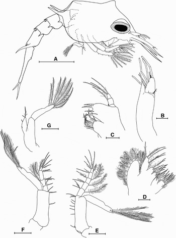
ZOEA II (FIGURE 5)
Colour: the orange pigmentation was considerably more intense than in the first stage. The mouthparts and bases of the antennae appeared intense red-orange. It was not clear whether this resulted from a higher density of orange chromatosomes or from the presence of chromatosomes with a darker pigment. The abdomen and telson were very slightly pigmented orange but still appeared transparent at low magnification.
Cephalothorax: the overall shape of the carapace was unchanged from the first stage. However, the posterolateral spines were proportionally smaller. The eyes were now stalked.
Antennule (Figure 5B): the antennule was now 2 segmented with a large endopod bud arising from the proximal segment. Two to 4 minute setae projected from the distal margin of the proximal segment. The distal segment had 4–6 subterminal aesthetascs, 1 to 3 terminal aesthetascs and 2–3 simple terminal setae.
Lopholithodes foraminatus: (A) zoea II (lateral); (B) antennule (right dorsomedial); (C) maxillule (left); (D) maxilla (right); (E) 1st maxilliped (left); (F) 2nd maxilliped (right externolateral); (G) 3rd maxilliped (left externolateral). Scale bars: A = 1 mm; B–D = 100 µm; E–G = 250 µm.

Maxillule (Figure 5C): the coxal endite now had 1 submarginal denticulate seta and 1 denticulate, 1 simple, 1 pappose and 4 large plumodenticulate marginal setae. The basial endite now bore 2 submarginal denticulate setae and 4 marginal spines with several pronounced denticulations. In some cases an additional small simple marginal spine was present.
Maxilla (Figure 5D): the scaphognathite now had 9 to 12 marginal plumose setae.
First maxilliped (Figure 5E): all segments of the endopod now had a plumose seta on their lateral surface; the hair-like processes present in corresponding locations in the first stage were absent. The exopod now bore 7 or 8 natatory setae.
Second maxilliped (Figure 5F): the 2nd and 3rd segments of the endopod now each had a plumose seta on their outer surface. The hair-like processes present in corresponding locations in the first stage were absent. The exopod now bore 7 or 8 natatory setae.
Third maxilliped (Figure 5G): the endopod was now proportionally larger than in the first stage but was still unsegmented and apparently did not articulate with the basis. It bore 2 denticulate and 1 plumose setae. The exopod now bore 7 or 8 natatory setae.
Telson: the telson was largely unchanged from the first stage. However, the small spiniform process present or absent from one or both sides of the median cleft in the first stage was now present on both sides.
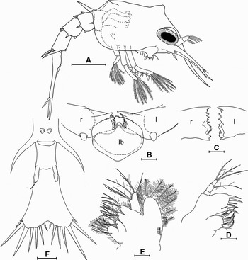
ZOEA III (FIGURE 6)
Colour: the orange colour of the carapace and thoracic appendages was very intense. The abdomen now had a diffuse orange pigmentation that was visible at low magnification; this pigment did not appear to be organized in chromatosomes.
Mandibles (Figure 6B, C): the mandibles now had small palp buds anteriorly. The asymmetry in dentition of the left and right mandibles agreed with that in the previous 2 stages.
Lopholithodes foraminatus: (A) zoea III (lateral); (B) [l]eft and [r]ight mandibles and labrum (lb) (ventral, in situ); (C) mandibles ([r]ight and [l]eft); (D) maxillule (right); (E) maxilla (left); (F) telson and abdominal segments 5 and 6 (ventral). Scale bars: A = 1 mm; B–E = 100 µm; F = 250 µm.

Maxillule (Figure 6D): the coxal endite now had 2 denticulate, 1 small plumodenticulate and 4 large plumodenticulate marginal setae. The basial endite now always bore 5 denticulate spines.
Maxilla (Figure 6E): the scaphognathite now bore 10 to 13 plumose marginal setae interspersed with up to 3 minute buds.
Abdomen (Figure 6F): somites 2–5 now bore minute pleopod buds and uropods were present on somite 6. The uropods consisted of a single segment with one or two simple terminal setae.
ZOEA IV (FIGURE 7)
Colour: the thoracic appendages and carapace were still bright orange due to the presence of chromatosomes. The diffuse orange colour of the abdomen extended into the developing pleopods. In exuviae the collapsed exoskeleton of the pereopods and pleopods and the exoskeleton of the mouthparts were visibly orange.
Cephalothorax: the carapace was markedly more globular than in the preceding stages. Large lipid droplets were now visible in the vicinity of the digestive gland; directly posterior to the eye and ventral to the heart. The pereopods (Figure 7F) were much larger than in previous stages and the developing chelae projected from under the edge of the carapace. The developing 5th pereopod lay under the other pereopods. Pairs of developing gills at the base of the pereopods were proportionally larger than in the third zoeal stage.
Lopholithodes foraminatus: (A) zoea IV (lateral); (B) antenna (right ventral); (C) mandibles ([r]ight and [l]eft); (D) basial endite of maxillule (right); (E) maxilla (left); (F) 3rd maxilliped (left externolateral); (G) pereopods (right lateral); (H) 2nd pleopod (left anterior); (I) uropod (right ventral); (J) abdomen (ventral). Scale bars: A = 1 mm; B, F, G, J = 250 µm; C–E, H–I = 100 µm.

Antennule: the exopod now had 6–7 subterminal aesthetascs, up to 1 subterminal seta, 2–3 terminal aesthetascs and 1–6 terminal setae.
Antenna: (Figure 7B): the exopod now had 6–7 plumose setae and 2–3 minute setae. The endopod had a scattering of minute denticulations and in some cases a minute subterminal seta. In some instances these endopod characters were partially developed in the previous 2 stages.
Mandibles (Figure 7C): the mandibular palp buds were slightly larger than in the third zoeal stage but still unarmed.
Maxillule: the basial endite (Figure 7D) now had an additional 2 small marginal denticulations: one at the distal angle of the endite and one between the 2nd and 3rd most distal spines. The setation was otherwise unchanged.
Maxilla (Figure 7E): one of the 3 terminal setae on the endopod was now weakly plumose, the other two remained pappose. The scaphognathite now bore 14–18 plumose marginal setae interspersed with 1–5 minute buds.
Third maxilliped (Figure 7F): the endopod now appeared 2 segmented. The proximal segment had 1 denticulate seta. The distal segment bore 1 plumose and 1 denticulate seta.
Abdomen (Figure 7J): somites 2–5 now bore pairs of incompletely 2 segmented, bilobed, unarmed pleopods (Figure 7H). The uropods on somite 6 were now marginally 2 segmented and had 2 or 3 plumose setae (Figure 7I). While the telson and 6th somite were now partially delineated they did not appear capable of flexion.
GLAUCOTHOE (FIGURES 8 & 9)
Colour: newly moulted glaucothoe were bright orange due to uniform pigmentation of the exoskeleton and the presence of numerous red/orange chromatosomes. This orange pigmentation became less intense towards the end of the stage and an area of darker pigmentation developed in the centre of the carapace in the region of the digestive gland. At this point the legs also developed light and dark banding.
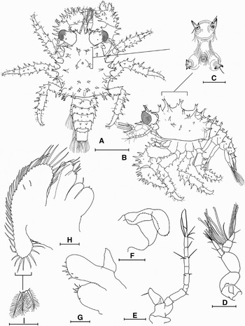
Cephalothorax (Figure 8A–C): the carapace was approximately 1.4 times as long (including the rostrum) as it was wide (not including spines). The branchial regions were inflated and the posterior margin was broadly concave. Beginning with a pair of bifid or trifid spines at the base of the rostrum and moving posteriorly there were 5 pairs of mediodorsal spines. Like all other spines on the glaucothoe, they bore a single seta. The second and third most anterior of these pairs were at the four corners of a distinct gastric lobe. A simple blunt spine was located mid dorsally on the posterior part of this lobe; it lacked an accessory seta. Posterior to the last pair of mediodorsal spines was a short but pronounced midline ridge. At least another 16 pairs of lateral and marginal spines were arranged symmetrically on opposite sides of the carapace; these were simple, bifid or trifid.
Lopholithodes foraminatus: (A) glaucothoe (dorsal); (B) (lateral); (C) mid-dorsal lobe of carapace; (D) antennule (right medial); (E) antenna (left dorsal); (F) mandible (left internal); (G) maxillule (left); (H) maxilla (right); (I) plumose setae of scaphognathite. Scale bars: A, B = 1 mm; C–E = 250 µm; F–I = 100 µm.

In some cases an additional small spine was present on one or both sides near the anteroventral angle of the pterygostomial region. Several additional spines were often present asymmetrically in the branchial region.
Antennule (Figure 8D): the protopod was three segmented with the proximal segment projecting medially and the distal two segments projecting dorsally. Each of these segments was armed with 1–6 minute setae. The proximal segment had a distinct dorsal invagination with a distal opening. The endopod was two segmented with 2 simple setae on the proximal segment and 7–8 simple setae on the distal segment. The exopod was 3 or 4 segmented with 9–11 aesthetascs and 4–5 simple setae on the middle segment(s) and 2–3 subterminal aesthetascs and 3–6 simple setae on the terminal segment.
Antenna (Figure 8E): the peduncle was indistinctly 2 segmented and bore a pronounced ventral spine on its distal margin. The endopod was 9 segmented and progressing distally bore 0, (1–2), 2, 0, (2–4), (0–2), (5–6), (1–4) and (7–8) simple setae. The exopod was a blunt process which in some specimens bore a single seta and/or spine.
Mandible (Figure 8F): the mandibles were uncalcified and proportionally much smaller than in the zoeal stages. The denticulations present in the zoeal stages were also absent. The palp was large and incompletely 2 segmented; it was held against the internal surface of the mandible.
Maxillule (Figure 8G): the coxal endite now bore 4–8 simple and minute marginal setae. The basial endite had 2 simple submarginal setae, 5–6 small simple spines and 2–4 small marginal denticulations. The endopod lacked setae or clear segmentation.
Maxilla (Figure 8H, I): the proximal and distal lobes of the coxal endite bore 0–2 minute marginal setae and 1 simple seta respectively. Both lobes of the basial endite had 3–4 pappose and/or simple setae. The endopod was reduced and in some cases bore 2 minute setae. The scaphognathite now had an elongated posterior lobe and 42–49 marginal plumose setae. There were 1 or 2 simple setae on the internal and external surfaces of the scaphognathite.
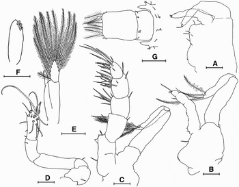
First maxilliped (Figure 9A): the coxa and basis were incompletely delineated with 1 and 6–11 simple and/or pappose setae respectively. The endopod was not clearly segmented and had 3–6 minute terminal processes. In some specimens the exopod was indistinctly two segmented with several minute terminal processes and/or a single terminal plumose seta.
Lopholithodes foraminatus glaucothoe: (A) first maxilliped (left); (B) second maxilliped (left); (C) third maxilliped (left); (D) fifth pereopod (left dorsal); (E) second pleopod (right anterior); (F) endopod of pleopod; (G) telson, abdominal somite 6 and part of 7 (dorsal). Scale bars: A–D = 100 µm; E, G = 250 µm; F = 50 µm.

Second maxilliped (Figure 9B): the protopod was indistinctly delineated into coxa and basis and bore 1 or 2 simple setae. The endopod was indistinctly segmented and had several minute setae and terminal exoskeletal projections. The exopod was two segmented and bore 1 simple seta and 5 plumose setae on the terminal segment.
Third maxilliped (Figure 9C): the segmentation of the coxa and basis was indistinct. The coxa in some cases bore a small simple seta. The endopod was 5 segmented. The proximal segment had 0–2 proximal simple setae, one distal denticulate seta, and 2–5 small denticulations of the cristae dentata along the medial margin. Moving distally the 4 remaining segments of the endopod had 3–5, 7–9, 14–18 and 14–17 setae of various types. The exopod was two segmented with one simple or denticulate seta on the proximal segment, and 1 simple and 5 plumose setae on the terminal segment.
Pereopods: the pereopods now resembled those of the adult in proportion and segmentation. The chelae were large and obviously asymmetrical, with the right being larger in approximately 70% of individuals (a manuscript discussing the 30% incidence of reversed asymmetry is in preparation). The chelae and walking legs were armed with numerous simple spines, each bearing a single seta. The terminal articles of the pereopods bore numerous additional setae. The fifth pereopods (Figure 9D) were folded back on themselves and held in the branchial chamber. The propodus bore several long hooked setae and had small terminal denticulations. These denticulations appeared to interdigitate in a sub-chelate manner with similar structures on the minute dactylus.
Abdomen: somites 1–6 bore 4, 8, 6, 6, 4 and 2 dorsal spines respectively. The spines on the anterior segments were mostly bifid or trifid and became smaller and were mostly simple on the posterior segments; each bore a single seta. Somites 1 and 6 had 2 additional setae while the remaining somites had 6 each. Somites 2–5 were each armed with a pair of simple lateral spines and a pair of marginally bifid posterolateral spines. Somites 2–5 bore pairs of two segmented pleopods, each with an endopod bud projecting from the proximal segment (Figure 9E). The endopod bud was armed with three minute hooked setae mediodistally (Figure 9F). In intact specimens these hooked setae were occasionally observed to be linked with those on the opposite pleopod. The exopod had 11–12 plumose natatory setae. The uropods were more obviously two segmented than in zoea IV but were otherwise unchanged.
Telson (Figure 9G): the telson was now rectangular. Each side had 2 small bulbous processes laterally and 4 plumose setae along the posterior margin. The three most lateral of these setae had distinctly bulbous bases. In many specimens several telsonal processes were missing.
FIRST CRAB INSTAR (FIGURES 10 & 11)
Colour: the exoskeleton of the carapace was either completely white or had small orange patches. There were scattered red chromatosomes in the antennae and mouthparts and in the carapace, primarily under the ventral and lateral margins. The walking legs were distinctly banded. The exoskeleton of the dactyl was orange with a proximal concentration of red chromatosomes forming a distinct dark band. The propodus was bright white distally and red orange proximally.
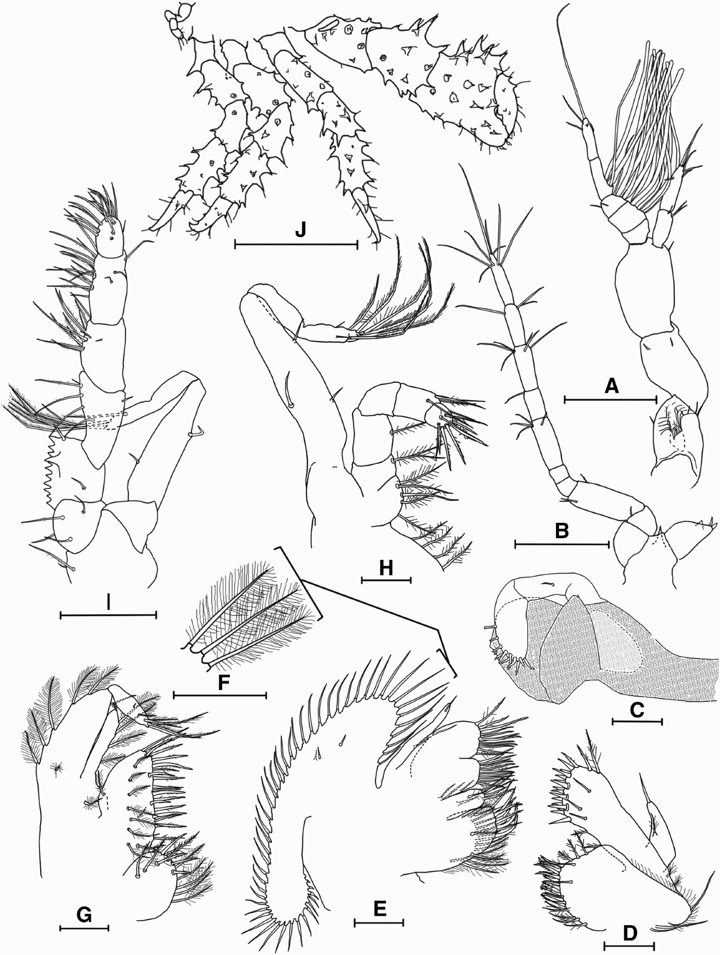
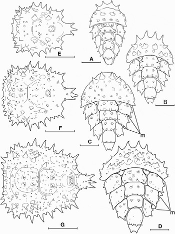
Carapace (Figure 11E): the overall shape of the carapace was unchanged from the glaucothoe. The spines at the base of the rostrum now had marginally bifid tips. The cervical groove was now clearly defined and separated the gastric and cardiac regions of the carapace. The distinct gastric lobe of the glaucothoe was no longer evident. The anterolateral carapace margin was armed with 2 large spines flanking a single small spine. Just posterior to these were three very large marginal spines. The majority of spines on the first crab stage were simple and bore a single seta.
Antennule (Figure 10A): the invagination in the proximal segment of the peduncle now had 5–6 stout setae projecting distally across the opening; at least two of which were plumose. About 6 setae were completely contained in the proximally projecting lumen of the pit. This segment also bore 5–7 pappose and minute setae. In some specimens the endopod was now 3 segmented. When present, the second segment was unarmed. The setation of the endopod was otherwise unchanged. The exopod was 4 or 5 segmented. The first segment was unarmed. The 2nd segment had 6–10 aesthetascs and the 3rd had 2–5 aesthetascs and 5 setae. The 4th segment had 2–4 aesthetascs near the base and was in some cases delineated from the terminal segment which had 3–5 simple setae.
Lopholithodes foraminatus 1st crab instar: (A) antennule (left medial); (B) antenna (right dorsal); (C) mandible (right internal; stippling indicates calcification); (D) maxillule (left); (E) maxilla (right); (F) plumose setae of scaphognathite; (G) first maxilliped (right); (H) second maxilliped (right); (I) third maxilliped (left); (J) pereopods (right dorsal, in situ). Scale bars: A, B, I = 250 µm; C–E, G, H = 100 µm; F = 50 µm; J = 1 mm.
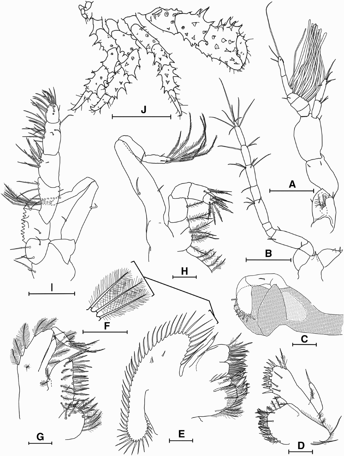
Antenna (Figure 10B): the peduncle had up to 2 simple setae. The endopod was 9 or 10 segmented with the 4th most proximal segment usually subdivided into 2 sections. Progressing distally the endopod bore (0–1), (1–2), (2–3), 0, (2–4), (0–1), 6, (3–4) and (7–8) simple setae. The exopod had 1 or 2 spiniform projections and in some cases a single seta.
Mandible (Figure 10C): the mandible was now proportionally much larger than in the glaucothoe and with the exception of the mandibular palp was completely calcified. The mandibular palp was incompletely 3 segmented with one simple seta on both the 1st and 2nd segments and 11–14 stout denticulate setae on the terminal segment.
Maxillule (Figure 10D): the protopod had 3–4 long, simple or marginally denticulate setae at its base and 1–2 plumose setae laterally, posterior to the base of the endopod. The coxal endite had fine hairs along the anterolateral margin. It was armed with 1 or 2 external, submarginal, simple setae and 20–22 plumodenticulate, pappose, and simple marginal setae. The basial endite had 3 submarginal setae; 2 or 3 pappose setae along the proximal margin; 2 or 3 distal plumodenticulate and denticulate setae; and 16–17 stout marginal spines, some of which were denticulate. The endopod was armed with 2–3 setae.
Maxilla (Figure 10E–F): the proximal lobe of the coxal endite now had 9–10 stout plumodenticulate setae arranged in loose pairs along the margin. One seta in each pair was strongly internally curved while the other was not. There was also a distinct submarginal row of 14–16 internally curved plumose setae. Both the internal and external surfaces of the coxal endite had several additional submarginal setae. The distal lobe of the coxal endite had 1–2 external, submarginal, plumose setae; and 1 denticulate and 3–4 plumose internal submarginal setae. It also had 6 plumodenticulate, denticulate and simple marginal setae. The basial endite had two small simple setae externally near the junction of the proximal and distal lobes. These lobes bore 10–15 and 12–15 marginal and slightly submarginal setae respectively (denticulate, plumodenticulate and simple). The endopod was reduced and bore 1–3 setae. The scaphognathite had 2 minute simple seta on the middle of the external surface, 1 on the internal surface and 49–53 marginal plumose setae.
First maxilliped (Figure 10G): the protopod generally had 2 external plumose setae, one at the base of the endopod and one at the base of the exopod. The coxal endite was armed with 10–13 denticulate, plumodenticulate, and plumose setae. The basial endite had a submarginal row of 7–8 externally projecting, weakly plumose setae; 3–5 internal submarginal simple setae; and 19–21 marginal denticulate and plumodenticulate setae. There was often one plumose seta at the base of the lateral margin. The endopod was unsegmented and bore 5–6 plumose setae along its medial margin. In some specimens an apical seta and/or 1–3 additional setae were present. The exopod was 2 segmented with 3 to 5 plumose setae along the lateral margin of the proximal segment and 1 or 2 simple setae on the medial margin. The distal segment bore 4–7 plumose, and sometimes 1–2 simple setae.
Second maxilliped (Figure 10H): the coxa was armed with 3–4 weakly plumose marginal setae and 1 small external plumose seta. The basis and ischium were incompletely delineated. The basis bore 5 pappose setae and a small simple seta. The ischium had 1–2 pappose setae. Progressing distal to proximal the remaining segments of the endopod bore 1–3, 1, 5–6 and 7–9 setae. The exopod was 2 segmented with the proximal segment bearing 1–3 simple setae and the distal segment generally bearing 1–2 simple setae and 6 plumose setae.
Third maxilliped (Figure 10I): the basal articles of the third maxilliped were now well calcified. The coxa bore 1–3 pappose and several weakly plumose setae. The basis of the endopod had 4–6 setae and the ischium had 2–3 subterminal setae and one denticulate seta at the mediodistal angle. The cristae dentata consisted of 7–11 teeth along the medial margin of the ischium and one larger external tooth. The merus of the endopod had 5–7 setae and the distal 3 articles all bore dense clumps of long, primarily denticulate setae. The first segment of the exopod was armed with 2–4 simple setae while the terminal segment generally had 6 plumose and 2 simple setae.
Pereopods: (Figure 10J): the pereopods, in particular the chelae, were somewhat more bulky than in the glaucothoe. However, they were otherwise largely unchanged.
Abdomen (Figure 11A): the abdomen was now flexed against the underside of the cephalothorax with the dorsal surface directed ventrally. The first and second somites were covered by calcified tergal plates which were partially fused to each other. The tergite of the second somite was considerably larger. The 4th and in most cases the 5th abdominal tergites were divided into a pair of lateral plates and a medial plate. On the third somite the lateral plates were frequently subdivided on one or both sides, with the anteromedial spines forming small separate plates. Similarly the portions of the medial plate bearing the anterior 2 spines were usually partially or completely separated. The 6th abdominal tergite and telson consisted of single plates and the telson lacked spines. The exoskeleton between tergites was thin and membranous. Pairs of degenerate pleopods lacking setae were often present on the ventral surface of somites 2 through to 5.
Lopholithodes foraminatus: (A) abdomen (1st crab instar); (B) abdomen (2nd crab instar); (C) abdomen (female 3rd crab instar), [m]arginal plates; (D) abdomen (female 4th crab instar), [m]arginal plates; (E) carapace (1st crab instar); (F) carapace (2nd crab instar); (G) carapace (3rd crab instar). Scale bars: A–D = 500 µm; E–G = 1 mm.

SUBSEQUENT INSTARS (FIGURE 11)
Colour: several distinct colour morphs were observed in juvenile crabs. By the third crab instar most individuals had developed brilliant orange chelae, with the dactyl and tip of the propodus normally remaining white. In some individuals the carapace remained white while in others it became dull orange, brilliant orange, dull red, or in several cases white anteriorly and brilliant orange posteriorly. Once a colour pattern had been established it was maintained through subsequent moults. The banding pattern of the walking legs was maintained in all individuals. A distinctly lighter distal band on the propodus is evident even in adult L. foraminatus.
Cephalothorax: the carapace of the second crab instar (Figure 11F) was somewhat less elongate than that of the first instar: approximately 1.3 times as long (including the rostrum) as it was wide (not including spines). This ratio was maintained through the subsequent 2 stages. The anterolateral margin of the carapace was now armed with 2 small spines flanked by two large spines. Posterior to this was an alternating sequence of 3 small and 3 large spines. This basic pattern was maintained through subsequent instars, but generally became less regular and more complex. The carapace of the third crab instar (Figure 11G) was more dorsally peaked than in previous stages. There were two distinct peaks in the gastric region and one in the cardiac region. The pair of spines at the base of the rostrum now bore small, medial, accessory projections. In subsequent instars the carapace became broader relative to its length (in adult specimens it is wider than it is long).
Pereopods: the distinct interdigitating invaginations of the carpi of the chelae and first walking legs, characteristic of adult L. foraminatus, were not evident by the fifth crab instar. However, these structures were almost completely developed in 1 year old juveniles, likely in the 7th or 8th instar.
Abdomen: the abdomen of the 2nd crab instar (Figure 11B) was broader and more triangular than that of the 1st instar. The tergites of the 1st and 2nd segments were now completely fused. In most individuals the medial tergite of the third segment now consisted of a single fused plate. In some specimens the telson bore small terminal spines. Pleopods were totally absent. In the third crab instar (Figure 11C) minute marginal plates were present on the right hand side of the 3rd through to 5th somites in some females and on both sides in males. Minute, membranous pleopod buds also appeared on the left side of the ventral surface of the abdomen of some females. By the 4th instar (Figure 11D) marginal plates were always present and sex could be clearly determined. In females with a larger left cheliped, abdominal asymmetry was also reversed.
DISCUSSION
Behaviour
Lopholithodes foraminatus zoeae were apparently positively phototactic, although this was not confirmed experimentally. Positive phototaxis at high light intensities, and negative phototaxis at low light intensities, has been demonstrated for Paralithodes camtschaticus (Shirley & Shirley, Reference Shirley and Shirley1988). This response likely facilitates the reverse diel vertical migration of P. camtschaticus, which consists of rising to the surface at sunrise and descending below 30 m during the night (Shirley & Shirley, Reference Shirley and Shirley1987).
It is difficult to determine whether the observed decline in swimming activity by L. foraminatus zoeae over the course of development reflects a naturally occurring ontogenetic shift in behaviour or is an artefact of laboratory culture. If reduced swimming indeed reflects a natural ontogenetic change in behaviour, it could indicate transition to a demersal habit late in development. An almost complete lack of swimming activity in the lecithotrophic zoeae of Lithodes aequispinus was interpreted by Shirley & Zhou (Reference Shirley and Zhou1997) as evidence of a benthic habit. However, there is also a tendency for laboratory cultured decapod larvae to be less active and robust than those observed in the field (G. Jensen, personal communication, 2006). Given the planktotrophic development of L. foraminatus zoeae and their apparent positive phototaxis, it seems likely that they spend considerable time in the photic zone. Behavioural experiments in the laboratory combined with plankton sampling in the field will be necessary to obtain a complete understanding of the movements of L. foraminatus zoeae in the water column.
Flaring of the antennae by L. foraminatus zoeae in response to chemical or physical trauma is likely a defence mechanism against gape-limited predators. Flaring of the antennal spines of the zoeae of the brachyuran Rhithropanopeus harrisii is part of a spine based defence that is effective against predation by small fish (Morgan, Reference Morgan1989). Morgan (Reference Morgan1987) suggested that the degree of spination among brachyuran zoeae may be correlated with the zoeal habitat. Species that undergo larval development in estuarine environments heavily populated by fish exhibit more pronounced spines than those from non-estuarine coastal waters. Some lithodid crabs have much smaller antennal scaphocerites than L. foraminatus, e.g. Cryptolithodes expansus (Kim & Hong, Reference Kim and Hong2000). It would be interesting to know if species such as C. expansus also exhibit a spine flaring defence, or if some aspect of their life history provides an alternative protection against small gape-limited predators.
The almost complete absence of cannibalistic behaviour by zoeae in this study is surprising given the reports of frequent cannibalism by Paralithodes camtschaticus (Epelbaum et al., Reference Epelbaum, Borisov and Kovatcheva2006) and the very similar size (see below: Growth) and mouthpart morphology of these larvae. It is possible that this behavioural difference is related to differences in trophic ecology between these two species.
Lecithotrophy and secondary lecithotrophy
Lopholithodes foraminatus passed through 4 planktotrophic zoeal stages and died without moulting to the second zoeal stage when not provided with food. Development through 4 feeding zoeal stages is likely a primitive character in the Lithodidae (Konishi, Reference Konishi1986; Zaklan, Reference Zaklan, Paul, Dawe, Elner, Jamieson, Kruse, Otto, Sainte-Marie, Shirley and Woodby2002).
The term ‘secondary lecithotrophy’ was suggested by Anger (Reference Anger1989) to describe the situation where a larval stage does not feed and instead relies upon energy reserves accumulated by preceding stages. Lopholithodes foraminatus glaucothoe were not observed to feed; they moulted to the first crab stage after the same period and with the same survivorship whether or not provided with food; and they exhibited reduced mouthparts (Figures 8 & 9) that were less well armed than those of the 4th zoeal (Figure 7) and 1st juvenile (Figure 10) stages. The stored reserves required for this secondary lecithotrophic development were visible as lipid droplets in the digestive gland of both the late zoeal stages and the early glaucothoe.
Evidence for secondary lecithotrophy in the lithodids Paralithodes camtschaticus, P. brevipes and P. platypus is provided by a reduction in function of the mouthparts and foregut of the glaucothoe relative to both the zoeal and juvenile crab stages (Abrunhosa & Kittaka, Reference Abrunhosa and Kittaka1997). Descriptions of a feeding glaucothoe stage in Paralithodes species by previous authors may have been based on disappearance of food items due to feeding by 4th zoeae or first stage juveniles present in the culture vessels (Epelbaum et al., Reference Epelbaum, Borisov and Kovatcheva2006). The glaucothoe of Hapalogaster mertensii Brandt, 1850 was also described as a non-feeding stage by Miller & Coffin (Reference Miller and Coffin1961) (confirmed by Duguid, personal observation). Descriptions of development for Acantholithodes hispidus (Hong et al., Reference Hong, Perry, Boutillier and Kim2005), Lopholithodes mandtii (Crain & McLaughlin, Reference Crain and McLaughlin2000a), Hapalogaster dentata (Konishi, Reference Konishi1986) and Cryptolithodes expansus (Kim & Hong, Reference Kim and Hong2000) all illustrate a reduction of mouthpart armature in the glaucothoe similar to that described by Abrunhosa & Kittaka (Reference Abrunhosa and Kittaka1997) and observed in the present study. A less dramatic reduction in mouthparts is also evident in illustrations of the glaucothoe of Placetron wosnessenskii (Crain & McLauglin, 2000b). The glaucothoe has also been demonstrated to be a non-feeding stage in lithodids with lecithotrophic zoeal development: Lithodes aequispinus (Shirley & Zhou, Reference Shirley and Zhou1997); Lithodes maja (Anger, Reference Anger1996); Lithodes santolla and Paralomis granulosa (Calcagno et al., Reference Calcagno, Anger, Lovrich, Thatje and Kaffenberger2004); and Paralomis spinosissima (Watts et al., Reference Watts, Thatje, Clarke and Belchier2006). In the absence of experimental evidence of feeding, it seems possible that all lithodid glaucothoe are lecithotrophic.
Secondary lecithotrophy also occurs in other crustaceans, including the cypris larvae of barnacles, the puerulus stage of rock lobsters and the glaucothoe stage of pagurid crabs (reviewed in Anger, Reference Anger and Vonk2001). Secondary lecithotrophy could facilitate location of a suitable substrate for settlement and might be associated with a high degree of juvenile habitat specialization (Anger, Reference Anger1989; Epelbaum et al., Reference Epelbaum, Borisov and Kovatcheva2006). Lithodids occupy diverse habitats and it would be surprising if they were to have settlement requirements more specific than other decapods such as brachyuran crabs. On the other hand, in pagurid hermit crabs, a requirement for gastropod shells at settlement provides a potential explanation for secondary lecithotrophy (Anger, Reference Anger1989). An alternative explanation is the massive reorganization of the digestive system that occurs between the last zoeal and first juvenile stage of pagurids, including the movement of the midgut gland from the cephalothorax to the abdomen (Thompson, Reference Thompson1903). This reorganization may preclude feeding (Harvey & Colasurdo, Reference Harvey and Colasurdo1993).
Regardless of the underlying cause, secondary lecithotrophy in pagurids provides an intriguing alternative explanation for the non-feeding glaucothoe of lithodid crabs. Recent molecular evidence (Cunningham et al., Reference Cunningham, Blackstone and Buss1992; Morrison et al., Reference Morrison, Harvey, Lavery, Tieu, Huang and Cunningham2002; Zaklan, Reference Zaklan, Paul, Dawe, Elner, Jamieson, Kruse, Otto, Sainte-Marie, Shirley and Woodby2002; Tsang et al., Reference Tsang, Ma, Ahyong, Chan and Chu2008) unanimously nests the lithodids within the hermit crab family Paguridae (but see review and criticism of this hypothesis in McLaughlin et al., Reference McLaughlin, Lemaitre and Sorhannus2007). Therefore, if secondary lecithotrophy is indeed a universal character of lithodids, it may be the result of ancestry rather than adaptation. The atavistic reacquisition of structures required for feeding by planktonic larvae is less likely than their original loss (Strathmann, Reference Strathmann1978). This could provide an explanation for why secondary lecithotrophy persists in lithodids despite the lack of an apparent life history advantage. Should future research support universal secondary lecithotrophy in lithodids, this could provide further support for a pagurid ancestry of the Lithodidae.
Growth
The carapace length of Lopholithodes foraminatus larvae and postlarvae was very slightly longer at each stage than that described for both L. mandtii (Crain & McLaughlin, Reference Crain and McLaughlin2000a) and Paralithodes camtschaticus (Epelbaum et al., Reference Epelbaum, Borisov and Kovatcheva2006; carapace length and rostrum length reported separately). Similarly the dry and wet weights of L. foraminatus larvae and postlarvae were higher at each stage than those of Paralithodes camtschaticus, with the exception of the dry weight of the first crab instar and the wet weight of the third zoeal stage (Epelbaum et al., Reference Epelbaum, Borisov and Kovatcheva2006). Overall the larval size of these planktotrophic north-east Pacific lithodids is apparently very similar.
The first crab instars were also only marginally larger than glaucothoe in carapace dimensions and wet and dry weight. These results are not surprising considering the non-feeding habit of the glaucothoe stage. The slight increases in wet and dry weight could reflect the deposition of calcium sequestered from seawater in the shell of the first crab instar.
Stage duration
Mean surface water temperature in the northern Strait of Georgia increases from approximately 8°C in the winter (1 December–28 February) to almost 9.5°C in the spring (1 March–31 May) and 16°C in the summer (1 June–31 August). At 120 dbar (approximately 120 m) the mean temperature ranges between 8°C and 9°C over the same period (Fisheries and Oceans Canada, 2008). Given an extended hatching period in the late winter and spring (Table 1; Duguid, unpublished data) it seems likely that L. foraminatus in this region would reach the glaucothoe stage within 22–39 days of hatching (based on durations of development at 8°C and 12°C calculated using the relationship in Figure 3E). The duration of the glaucothoe stage at temperatures below 11.5°C was not determined. However, based on a requirement of 219 degree days at 11.5°C, and assuming ambient temperatures of 9°C or above, this stage would be unlikely to last more than 25 days in the field.
The 39 days required for zoeal development of L. foraminatus at 8°C agrees well with 39 days for red king crab reared at 7–8°C (Epelbaum et al., Reference Epelbaum, Borisov and Kovatcheva2006) and 44.9 for blue king crab reared at 9°C (Stevens et al., Reference Stevens, Persselin and Matweyou2008). Zoeal development of lithodids with lecithotrophic larvae is much more rapid; for example, 9.6 days at 9°C for Paralomis granulosa (Anger et al., Reference Anger, Thatje, Lovrich and Calcagno2003) and 23.9 days at 9°C for Lithodes aequispinus (Paul & Paul, Reference Paul and Paul1999). This reflects both a reduction in the number of zoeal stages and the provisioning of those stages with maternal reserves for development.
For all larval stages investigated in the present study, an increase in temperature from 8°C to 12°C led to a greater reduction in stage duration than an increase from 12°C to 16°C (Figure 2). A similar levelling out of the relationship between stage duration and temperature has been observed for other lithodids including Lithodes aequispinus (Paul & Paul, Reference Paul and Paul1999), Paralomis granulosa (Anger et al., Reference Anger, Thatje, Lovrich and Calcagno2003) and Paralithodes platypus (Stevens et al., Reference Stevens, Persselin and Matweyou2008). However, in all of these species, decrease in the sensitivity of stage duration to temperature occurred over a lower temperature range than observed in the present study. For example, the duration of the combined zoeal stages of Paralithodes platypus decreased from 71.8 days at 3°C to 44.7 days at 6°C, but was not lower at 9°C (44.9 days) (Stevens et al., Reference Stevens, Persselin and Matweyou2008). The sensitivity of stage duration over a higher temperature range in the present study may reflect adaptation to the relatively warm waters of southern British Columbia. Lopholithodes foraminatus occurs from California to Alaska (Jensen, Reference Jensen1995), a much greater latitudinal range than other large coastal lithodids for which development data are available. It would be interesting to know if the relationship between larval development rate and temperature is constant throughout this range.
Colour
The brilliant orange colour of Lopholithodes foraminatus zoeae is similar to that described for L. mandtii (Crain & McLaughlin, Reference Crain and McLaughlin2000a) and Placetron wossenenski (Crain & McLaughlin, Reference Crain and McLaughlin2000b). However, it differs from the largely transparent appearance of some other lithodid zoeae, such as Hapalogaster mertensii (Duguid, personal observation) and Paralithodes camtschaticus (colour photographs in Kovatcheva et al., Reference Kovatcheva, Epelbaum, Kalinin, Borisov and Levdev2006). It seems likely that this distinct difference in appearance is related to differences in planktonic ecology between lithodid species.
Juvenile colour polymorphism in decapods has been demonstrated to provide protection from small visual predators in the recruitment habitat (Palma & Steneck, Reference Palma and Steneck2001). Early juvenile stages of blue king crab (Paralithodes platypus) occur in multiple colour morphs (Jensen, G., personal communication, 2007). Armstrong et al. (Reference Armstrong, Palacios, Williams, Jensen, Pearson and Melteff1985) found that juvenile P. platypus abundance around the Pribilof Islands was strongly correlated with a heterogeneous substrate composed of broken mollusc shells. The authors concluded that the availability of this shellhash substrate could be a limiting factor in blue king crab distribution. The habitat and ecology of early ontogenetic stages of Lopholithodes foraminatus are completely unknown. The colour polymorphism observed in juveniles suggests a habitat with considerable small scale visual heterogeneity. The lack of such heterogeneity on a mud bottom could explain the tendency of adult L. foraminatus to be distributed on soft substrates in the immediate vicinity of rock features (Zhang et al., Reference Zhang, Workman and Phillips1999).
Identification
To our knowledge, the only prior account of Lopholithodes foraminatus larvae is a series of figures of putative Lopholithodes spp. C, D, and E zoeae and glaucothoe (chelae only) in Lough (Reference Lough1975). Lough's species C is almost certainly Acantholithodes hispidus, development of which is described by Hong et al. (Reference Hong, Perry, Boutillier and Kim2005). The identity of species D is less clear; however, the proportions of the rostrum and posterolateral spines preclude both species of Lopholithodes. The gross morphology of Lough's species E agrees very well with the description of L. foraminatus in the present study. Two specimens of this species (zoea III) were collected on April 22, 1971 in a plankton tow approximately 18 km off the Oregon coast, in water 85 m deep. The peaked mid-dorsal carina observed in Lough's species E and in all zoeal stages in this study is a feature that is not shared with any other north-east Pacific lithodid for which development has been described. We suggest this feature as diagnostic of L. foraminatus zoeae.
Lopholithodes foraminatus glaucothoe are very similar in morphology to L. mandtii glaucothoe as described by Crain & McLaughlin (Reference Crain and McLaughlin2000a). Abdominal somites 2–5 of L. foraminatus bear 8, 6, 6 and 4 dorsal spines whereas those of L. mandtii bear 8 dorsal spines each. The carapace spination of L. foraminatus glaucothoe is apparently different from that illustrated for L. mandtii, but this may be due to differing levels of detail between illustrations. The distinct mid-dorsal lobe of L. foraminatus (Figure 8C) is unique among described lithodid glaucothoe and could be useful for identification.
There are number of differences between the juvenile crab stages of Lopholithodes foraminatus and those of L. mandtii as described by Crain & McLaughlin (Reference Crain and McLaughlin2000a). The lateral carapace spination of the first 2 crab stages of Lopholithodes foraminatus (Figure 11E, F) is distinctive and differs from that illustrated for L. mandtii and from other described lithodid juveniles. Also, unlike in L. mandtii, the lateral and medial plates of the third abdominal somite are generally not entire in the first crab instar of L. foraminatus. The overall white appearance of the first crab stage is different from the bright orange carapace of L. mandtti. However, subsequent crab stages sometimes develop into a bright orange colour morph, and the pigmentation of L. mandtii juveniles past the first crab stage has not been documented. The distinct red proximal bands on the propodi of the walking legs also differ from the solid white propodi described for L. mandtii. The distinctive smooth invaginations in the carpi of the chelae and first walking legs apparently cannot be relied on as a diagnostic feature until crabs are in their second year.
ACKNOWLEDGEMENTS
We thank Dr Greg Jensen (School of Aquatic and Fishery Science, University of Washington, USA), Dr Anna Epelbaum (Pacific Biological Station (PBS), Department of Fisheries and Oceans (DFO), Canada), Antan Phillips (PBS, DFO), and Dr Stefanie Duff (Fisheries and Aquaculture Department, Vancouver Island University) who answered numerous questions in the early stages of this work. Helpful comments were also provided by Dr Akash Sastri (Institute of Oceanography, National Taiwan University) and two anonymous referees. We are grateful to the staff of the Aquatics Unit of the Animal Care Facility at the University of Victoria, in particular Brian Ringwood, who provided invaluable assistance with animal husbandry. Finally, this project would never have been initiated without Joe Watson of Campbell River BC who donated boat time and expertise to capture adult box crabs. This work was funded by a Natural Sciences and Engineering Research Council of Canada (NSERC) Discovery Grant to Dr Louise Page.