


Highlights
-
• COVID-19 associated Guillain-Barré syndrome (C + GBS) exhibited higher prevalence of the demyelinating subtype.
-
• Severity of disease did not differ between C + GBS and pre-pandemic patients.
-
• C+G BS is not associated with anti-ganglioside antibodies.
Introduction
Guillain-Barré syndrome (GBS) is an acute inflammatory polyradiculopathy of dysimmune origin, which is the most common cause of acute neuromuscular paralysis. Reference Willison, Jacobs and van Doorn1 GBS is divided into two main subtypes, demyelinating and axonal, according to electrodiagnostic (EDX) features. Approximately two-thirds of patients have a history of gastroenteritis or upper respiratory tract infection (URTI). Reference Jacobs, Rothbarth and van der Meche2 The International Guillain-Barré Syndrome Outcome Study reported URTI as the most common antecedent event in Europe, North America and East and Southeast Asia, whereas gastroenteritis was the most common in Bangladesh. Reference Doets, Verboon and van den Berg3 Many infectious agents such as Campylobacter jejuni, Epstein-Barr virus, cytomegalovirus, influenza A, Mycoplasma pneumoniae, Haemophilus influenzae, hepatitis E, Zika virus and severe acute respiratory syndrome coronavirus 2 (SARS-CoV-2) trigger GBS. Reference Jacobs, Rothbarth and van der Meche2,Reference Hadden, Karch and Hartung4–Reference Zhao, Shen, Zhou, Liu and Chen10
In 2020, SARS-CoV-2 caused a pandemic and spread to Turkey in March 2020. SARS-CoV-2 was thought to cause pneumonia and many neurological diseases, including musculoskeletal disorders, encephalitis, cerebrovascular disease, myelitis, cognitive dysfunction, headache and GBS. Reference Mao, Jin and Wang11–Reference Ozdag Acarli, Samanci and Ekizoglu13 COVID-19-associated-GBS (C + GBS) has attracted attention because it is unclear whether it presents with any characteristic clinical and laboratory features. Reference Gigli, Bax and Marini14–Reference Filosto, Piccinelli and Gazzina19 Some previous studies reported clinical and EDX differences between patients with C + GBS and those having GBS related to other conditions. The most common features of patients with C + GBS are suggested to present with sensorimotor deficits, facial palsy, greater weakness, a higher incidence of intensive care unit (ICU) admission, poorer outcomes and a demyelinating subtype. Reference Uncini, Vallat and Jacobs18, Reference Filosto, Cotti Piccinelli and Gazzina20–Reference Luijten, Leonhard and Van Der Eijk23 However, it has been observed in other studies that similar features were present in C + GBS and the others. Reference Keddie, Pakpoor and Mousele24,Reference Umapathi, Er, Koh, Goh and Chua25
A multicenter prospective study was started in April 2019 to determine the EDX and clinical features of GBS subtypes in Istanbul, and it was planned to include GBS cases consecutively for 3 years. Reference Tasdemir, Sirin and Cakar26 The SARS-CoV-2 pandemic coincided with the study from March 2020, when the first patient was seen in Turkey, till March 2022, when the multicenter study was ended. In the present study, we aimed to analyze the clinical, EDX and serologic features of patients with C + GBS included in this multicenter study. The control group consisted of patients with GBS who were admitted to the same multicenter study prior to the onset of the pandemic (between April 2019 and February 2020).
Materials and Methods
Patients
Patients aged 18 years and older who had acute-subacute onset progressive muscle weakness and/or sensory symptoms and were admitted to 13 tertiary centers between April 2019 and March 2022. The exclusion criteria were the diagnosis of GBS mimicking conditions such as acute-onset transverse myelitis, myopathy or neuromuscular junction disorder, critical illness neuromyopathy and any acute neuropathy with another etiology. A total of 177 patients were included in the previous multicenter study in Istanbul. Reference Tasdemir, Sirin and Cakar26 Two groups (C + GBS and control group) were extracted from these patients: (i) the pre-pandemic (PP) group, patients diagnosed with GBS during the era before March 2020 (April 2019–February 2020), when the first COVID-19 infection was diagnosed in Turkey, and (ii) C + GBS group, patients with a positive COVID-19 polymerase chain reaction (PCR) swab test performed in the 6 weeks before the onset of GBS symptoms between March 2020 and March 2022. The decision to perform a PCR test for SARS-CoV-2 was taken in response to symptoms consistent with a URTI. The PCR tests were not included in the method of the multicenter study, nor did they constitute a component of an epidemiological study conducted in Turkey. One patient who had two episodes of URTI 1 month apart, one of which was COVID-19 (the first one), and another patient whose PCR was negative at the time of GBS symptoms but whose thorax CT was compatible with COVID-19 pneumonia were excluded from the C + GBS group. Patients during the pandemic period who did not meet the criteria for C + GBS were excluded from the control group because COVID-19 infection as an antecedent event could not be excluded by PCR analysis.
Patients who received a COVID-19 vaccination within the 6 weeks preceding the symptom onset and who did not have any other possible antecedent events were also analyzed separately.
The patients were subjected to two clinical evaluations: first in the 21 days after the onset of symptoms and the second in the 4th–6th weeks. The sensory, motor, autonomic and respiratory, cranial neuropathy and ataxia symptom and sign categories were labeled as positive if the patient had ≥1 symptoms and/or signs that fell into any of these categories. Reference Tasdemir, Sirin and Cakar26 CSF cell counts and protein levels were noted when available. The clinical conditions of the patients were rated using the Medical Research Council (MRC) sum score, the Hughes functional scale and the Erasmus GBS score. Reference Hughes, Newsom-Davis, Perkin and Pierce27–Reference van Koningsveld, Steyerberg, Hughes, Swan, van Doorn and Jacobs29
Ethics
The local ethics committee approved the study (Istanbul Faculty of Medicine, 02.08.2019, number 2019/179), and all participants or their relatives provided written informed consent.
Electrodiagnostic tests
Following a previously described stringent methodology, sensory/motor nerve conduction studies (NCS) and F-waves were performed on upper and lower limbs on the same day as the clinical evaluations. Reference Tasdemir, Sirin and Cakar26 In all subjects, median and ulnar sensory and motor NCS were performed in the upper extremity. Tibial and peroneal motor, superficial peroneal and sural sensory NCS were conducted in the lower extremity. Twenty F-waves were also recorded from the relevant muscles by stimulating the ulnar, median and tibial nerves at the wrist/ankle levels. Values outside the normal reference limits of each laboratory for a specific examination were considered abnormal.
The data were then printed out by the clinical neurophysiologist who performed the study, and the printouts were evaluated by the same investigator (NGS). In each patient, GBS subtypes (demyelinating, axonal, equivocal and inexcitable) were classified according to the criteria by Uncini et al. Reference Uncini, Ippoliti, Shahrizaila, Sekiguchi and Kuwabara30 Accordingly, the patients who could not be examined in a second EDX study were not evaluated using those criteria, except for those diagnosed as having the demyelinating subtype or an inexcitable/equivocal study in the first examination. Reversible conduction failure (RCF) and length-dependent conduction failure (LDCF) were also defined in the axonal group according to the criteria of Uncini et al. Reference Uncini, Ippoliti, Shahrizaila, Sekiguchi and Kuwabara30
Anti-ganglioside antibody tests
Sera were collected venously before initiating immune treatment, then transferred to the laboratory (Istanbul University, Aziz Sancar Experimental Medicine Research Institute) and stored in a −80 °C freezer until use. IgG antibodies against GM1, GM2, GM3, GD1a, GD1b, GT1b and GQ1b gangliosides in the sera were determined using an immunoblotting kit according to the manufacturer’s recommendations (Euroimmun, Anti-Gangliosides Profile 2 (IgG), DL1130-1601-2G). All positive results were confirmed using homemade Enzyme-Linked Immunosorbent Assay.
Statistical analysis
Statistical analyses were performed using the SPSS version 21.0 software package (IBM, Armonk, New York). The Mann–Whitney U test was used to compare continuous variables, and the chi-square (χ2 ) test was used to compare categorical variables between the PP and C + GBS groups. A binary logistic regression analysis was performed to determine the validity of the significant parameters found in the univariate analysis. P ≤ 0.05 was accepted as the limit of significance.
Results
Patients and clinical features
Between March 2020 and 2022 (pandemic period), 108 consecutive patients with GBS were included in the multicenter study, and among these 108 patients, 22 had a positive COVID-19 PCR test performed 6 weeks before the onset of the symptoms related to GBS. In addition to these 22 patients in C + GBS, 69 consecutive patients were recruited during the PP period of the study, resulting in a total of 91 patients.
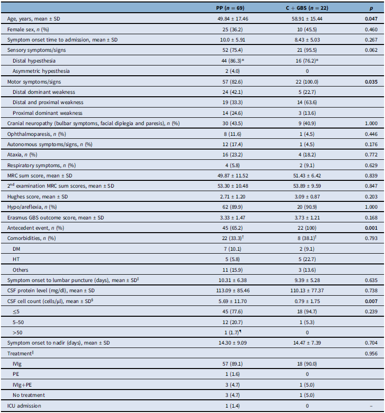
In the univariate analysis, the patients with C + GBS were found to be older than those in the PP period (Table 1). Among the PP patients, 75.4% had sensory symptoms/signs, and 82.6% had weakness. Motor signs/symptoms were significantly more common in the C + GBS group than in the PP group, even though their frequency was also higher in the PP group (100% vs. 83%, Table 1). Although the times from symptom onset to lumbar puncture and CSF protein levels were not different, the mean CSF cell count was higher in the PP group than in the C + GBS group (5.69 ± 11.70 cells/μL vs. 0.79 ± 1.77 cells/μL, p = 0.007). Other clinical features did not differ between the groups (Table 1).
Comparison of clinical features between the PP and C + GBS groups

Table 1. Long description
A table comparing clinical features between the PP and C + GBS groups. The table has 25 rows and 5 columns. Column headers are PP (n = 69), C + GBS (n = 22), and p. Row labels include Age, Female sex, Symptom onset time to admission, Sensory symptoms/signs, Distal hypesthesia, Asymmetric hypesthesia, Motor symptoms/signs, Distal dominant weakness, Distal and proximal weakness, Proximal dominant weakness, Cranial neuropathy, Ophthalmoparesis, Autonomous symptoms/signs, Ataxia, Respiratory symptoms, MRC sum score, 2nd examination MRC sum scores, Hughes score, Hypo/areflexia, Erasmus GBS outcome score, Antecedent event, Comorbidities, DM, HT, Others, Symptom onset to lumbar puncture, CSF protein level, CSF cell count, 5-50, >50, Symptom onset to nadir, Treatment, IVIg, PE, IVIg+PE, No treatment, ICU admission. Each row provides data for the PP group, C + GBS group, and p-value.
C + GBS, COVID-19-associated Guillain-Barré syndrome; DM, diabetes mellitus; GBS, Guillain-Barré syndrome; HT, hypertension; ICU, intensive care unit; IVIg, intravenous immunoglobulin; MRC, Medical Research Council; PE, plasmapheresis; PP, pre-pandemic; SD; standard deviation.
* The data of six patients in the PP group and six patients in the C + GBS group were missing.
† The data of four patients in the PP group and one patient in the C + GBS group were missing.
‡ Sixty-two patients in the PP group and 19 patients in the C + GBS group had lumbar puncture.
§ The data of 11 patients in the PP group and 2 in the C + GBS were missing.
∥ The data of five patients in the PP group and two patients in the C + GBS group were missing.
¶ The patient’s CSF cell count was 58 cell/μl. Distal dominant weakness and hypesthesia were found on his examination. The symptoms lasted less than 4 weeks, and electrophysiological subtype was acute inflammatory demyelinating polyneuropathy (AIDP). The significant p-values are indicated by bold font.
Electrodiagnostic tests
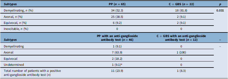
Four patients’ EDX subtypes could not be determined in the PP group because a second test could not be performed. Among patients with C + GBS, 81.8% were classified as having the demyelinating subtype, 9.1% as the axonal subtype and 9.1% as equivocal (Table 2). The frequency of the demyelinating subtype was significantly higher in patients with C + GBS than in those in the PP period (81.8% vs. 52.3%, p = 0.031).
EDX subtypes in the patients in the PP and C + GBS groups and in patients with a positive anti-ganglioside antibody test

Table 2. Long description
A table comparing EDX subtypes in patients with PP and C + GBS groups and those with a positive anti-ganglioside antibody test. The table has two main sections. The first section compares subtypes between PP (n = 65) and C + GBS (n = 22) groups. Row 1: Demyelinating, 34 (52.3 percent) for PP, 18 (81.8 percent) for C + GBS, p value 0.031. Row 2: Axonal, 25 (38.5 percent) for PP, 2 (9.1 percent) for C + GBS. Row 3: Equivocal, 6 (9.2 percent) for PP, 2 (9.1 percent) for C + GBS. Row 4: Inexplicable, 0 for both. The second section compares subtypes with an anti-ganglioside antibody test between PP (n = 46) and C + GBS (n = 12). Row 1: Demyelinating, 1 (9.1 percent) for PP, 0 for C + GBS. Row 2: Axonal, 7 (63.9 percent) for PP, 1 (100 percent) for C + GBS. Row 3: Equivocal, 2 (18.2 percent) for PP, 0 for C + GBS. Row 4: Undetermined, 1 (9.1 percent) for PP, 0 for C + GBS. The total number of patients with a positive anti-ganglioside antibody test is 11 (23.9 percent) for PP and 1 (8.3 percent) for C + GBS.
C + GBS, COVID-19-associated Guillain-Barré syndrome; EDX, electrodiagnostic; PP, pre-pandemic.
* This patient’s electrodiagnostic subtype could not be determined because of the single electrodiagnostic test.
Only two patients with C + GBS (9.1%) had EDX features of acute motor axonal neuropathy (AMAN). In these two patients with axonal subtype, one had both LDCF and RCF, and the other had only RCF. In the PP group, 19 of 25 patients with axonal GBS had RCF (76%), and 4 had LDCF (16%).
Distal latencies, base-to-peak amplitudes and negative peak durations of compound muscle action potentials (CMAPs), motor conduction velocities, minimal F-wave latencies and F-wave persistences in the first EDX test showed no statistically significant difference between the patients with GBS having a demyelinating subtype in the PP and C + GBS groups (Supplementary Table 1). The significant p-values are indicated by bold font.
Anti-ganglioside antibodies
Among the patients with C + GBS, anti-ganglioside antibodies were tested in 12 patients (54.5%), and only one patient had both anti-GM1 and anti-GD1b antibodies. The EDX subtype of this patient was axonal, presenting with both RCF and LDCF. In the PP group, among the 46 patients who had anti-ganglioside antibody tests (66.7%), 11 gave positive results, and 7 were classified as having axonal subtype (Table 2). Three of these patients were positive for anti-GM1 (all axonal), five for anti-GM1 and anti-GD1b (axonal n = 3, demyelinating n = 1 and equivocal n = 1), one for anti-GM1 and anti-GM2 (axonal) and two for anti-GQ1b (equivocal n = 1 and undetermined n = 1).
Multivariate analysis
A binomial logistic regression was performed to ascertain the effects of the significant parameters in the univariate analysis (age, the presence of motor symptoms, CSF cell count, EDX subtype) on the difference between the C + GBS and PP groups. The logistic regression model was statistically significant, χ2(5) = 23.551, p ≤ 0.001. The model explained 40.1% (Nagelkerke R 2) of the variance in C + GBS and correctly classified 74.3% of cases. The only statistically significant parameter was demyelinating subtype (p = 0.031). However, the presence of motor symptoms (p = 0.999), age (p = 0.072) and CSF cell count (p = 0.059) were not statistically significant.
COVID-19 vaccination as an antecedent event
Nine patients had COVID-19 vaccination within the 6 weeks before the onset of GBS-related symptoms. Eight of these patients had Pfizer–BioNTech vaccines, and one had the Sinovac vaccine. Their clinical and EDX features are presented in Supplementary Table 2. All patients had sensory signs, six had motor signs, one had autonomic symptoms/signs, four had cranial neuropathy and two had ataxia. All but one had hypo/areflexia, and one patient had hyperreflexia. Six of these patients had demyelinating, and two had axonal subtypes, and the other was categorized as undetermined because of a single EDX test. Six patients had anti-ganglioside antibody tests with negative results.
Discussion
Beginning early in the pandemic, several reports have been published to reveal the relationship between COVID-19 and GBS, indicating an increase, decrease or no change in the cases of GBS. Reference Fragiel, Miro and Llorens15–Reference Tatu, Nono, Gracio and Kocer17,Reference Filosto, Piccinelli and Gazzina19,Reference Filosto, Cotti Piccinelli and Gazzina22–Reference Umapathi, Er, Koh, Goh and Chua25,Reference Hafsteinsdottir, Dalemo, Eliasdottir, Olafsson and Axelsson31 A possible explanation for this discrepancy between these reports is the use of different methods of recruitment. Reference Foschi, D’Anna and Abdelhak32–Reference Lunn, Carr, Keddie, Pakpoor, Pipis and Willison34 Although the causal link between COVID-19 and GBS remains controversial, classic sensorimotor presentation and demyelinating EDX subtype were found to be the most frequent in GBS with antecedent COVID-19 infection. Reference Gigli, Bax and Marini14,Reference Martinelli-Boneschi, Colombo and Bresolin16,Reference Uncini, Vallat and Jacobs18,Reference Filosto, Piccinelli and Gazzina19,Reference Filosto, Cotti Piccinelli and Gazzina22,Reference Luijten, Leonhard and Van Der Eijk23,Reference Hafsteinsdottir, Dalemo, Eliasdottir, Olafsson and Axelsson31,Reference Caress, Castoro and Simmons35–Reference Pimentel, Luchsinger and Carvalho38 Similar to those, all patients in our study had motor signs, and all but one had sensory symptoms, and the demyelinating subtype was more frequent in the C + GBS group when compared with the PP group. Some studies also reported that patients with C + GBS frequently had facial palsy; Reference Martinelli-Boneschi, Colombo and Bresolin16,Reference Uncini, Vallat and Jacobs18,Reference Luijten, Leonhard and Van Der Eijk23,Reference Palaiodimou, Stefanou and Katsanos37 however, this was not confirmed by others. Reference Filosto, Piccinelli and Gazzina19,Reference Filosto, Cotti Piccinelli and Gazzina22,Reference Keddie, Pakpoor and Mousele24 We also found no relationship between the presence of cranial neuropathy including facial palsy and C + GBS.
In our cohort, patients with C + GBS were found to be older than the PP group in the univariate analysis. However, this finding was not confirmed in the multivariate analysis. Previous reports comparing patients with the C + GBS and PP groups also found no such difference related to age. Reference Fragiel, Miro and Llorens15,Reference Uncini, Vallat and Jacobs18,Reference Filosto, Piccinelli and Gazzina19,Reference Uncini, Foresti and Frigeni21,Reference Filosto, Cotti Piccinelli and Gazzina22,Reference Ahmad, Businaro and Regalbuto39,Reference Sanchez-Boluarte, Aguirre-Quispe, Tacunan-Cuellar, Sanchez-Boluarte and Segura-Chavez40
The majority of previous reports showed that no significant difference was found in GBS severity between the C + GBS and non-COVID-19 groups, similar to our findings. Reference Luijten, Leonhard and Van Der Eijk23,Reference Keddie, Pakpoor and Mousele24,Reference Hafsteinsdottir, Dalemo, Eliasdottir, Olafsson and Axelsson31,Reference Palaiodimou, Stefanou and Katsanos37 However, some studies revealed lower MRC scores, higher rates of mechanical ventilation need, ICU admission and disability scores. Reference Uncini, Vallat and Jacobs18,Reference Filosto, Piccinelli and Gazzina19,Reference Filosto, Cotti Piccinelli and Gazzina22,Reference Caress, Castoro and Simmons35 In these studies during the early pandemic period, patients with C + GBS mainly presented with severe symptomatic COVID-19 infection, which might contribute to the poor outcome measures. Reference Uncini, Vallat and Jacobs18,Reference Filosto, Piccinelli and Gazzina19,Reference Filosto, Cotti Piccinelli and Gazzina22,Reference Caress, Castoro and Simmons35 On the other hand, all our patients with C + GBS – except two who had mild COVID-19 symptoms – did not have active COVID-19 infections at the onset of GBS symptoms. Our low ICU admission rate – a study limitation – likely reflects the direct referral of severe cases from secondary centers to ICUs, resulting in fewer admissions to tertiary neurology departments.
Among the C + GBS group, we only found anti-ganglioside antibodies in one patient with axonal GBS (both anti-GM1 and anti-GD1b antibodies). This patient had a positive PCR test for COVID-19 infection with symptoms of URTI. The 6-week period before GBS was unremarkable for any other antecedent event. His EDX study revealed RCF and LDCF, compatible with the anti-ganglioside antibody status. The lumbar puncture performed on the 4th day of his symptoms revealed normal protein levels with no cells. Previously, only a few C + GBS patients reported anti-ganglioside antibodies (GD1b, GM2 and asialo GM1) alongside cranial neuropathy (Supplementary Table 3). Reference Lantos, Strauss and Lin41–Reference Guilmot, Maldonado Slootjes and Sellimi44 No EDX studies were performed in any patients. The other studies screening for anti-ganglioside antibodies found no antibodies in patients with C + GBS. Reference Toscano, Palmerini and Ravaglia8,Reference Gigli, Bax and Marini14,Reference Martinelli-Boneschi, Colombo and Bresolin16,Reference Uncini, Vallat and Jacobs18,Reference Caress, Castoro and Simmons35–Reference Palaiodimou, Stefanou and Katsanos37,Reference Garnero, Del Sette and Assini45–Reference Manganotti, Bellavita and D’Acunto47 This is the first study to consecutively analyze anti-ganglioside antibodies in patients with C + GBS, and our results mostly imply that there is no relationship between anti-ganglioside antibodies and C + GBS.
Uncini et al. reported increased CMAP duration in the EDX tests of patients with COVID-19-associated acute inflammatory demyelinating polyneuropathy (AIDP) because of the additional muscle fiber conduction slowing due to the hyperinflammatory stage. Reference Uncini, Foresti and Frigeni21 We did not record similar findings in our group. The patients of Uncini et al. had a considerably higher rate of respiratory failure and ICU admissions than those of our patients. It is known that critical illness can cause myopathy and prolonged CMAP duration may serve as a diagnostic marker of this myopathy despite its low sensitivity. Reference Allen, Arunachalam and Mills48–Reference Z’Graggen and Tankisi50 Therefore, the prolonged CMAP duration in Uncini et al.’s group may be more closely related to the presence of critical illness than COVID-19-associated GBS.
In our multicenter study, the methodology for CSF cell count did not change during the pandemic period in all centers. Knowing that, univariate analysis revealed that the cell count in CSF was lower in the C + GBS group when compared with the PP group in the present study, without any difference in the CSF protein levels and the time of lumbar puncture. However, the CSF cell count was not statistically significant in the multivariate analysis. In previous reports, albuminocytologic dissociation was present in nearly two-thirds of patients with C + GBS. Reference Martinelli-Boneschi, Colombo and Bresolin16,Reference Luijten, Leonhard and Van Der Eijk23,Reference Caress, Castoro and Simmons35–Reference Pimentel, Luchsinger and Carvalho38,Reference Sanchez-Boluarte, Aguirre-Quispe, Tacunan-Cuellar, Sanchez-Boluarte and Segura-Chavez40,Reference Ergin Beton, Ozturk Tan and Bilen51 Similar to our results, the comparison between patients with C + GBS and non-COVID-19-associated GBS showed no difference in the protein levels in most previous studies. Reference Filosto, Piccinelli and Gazzina19,Reference Luijten, Leonhard and Van Der Eijk23,Reference Palaiodimou, Stefanou and Katsanos37,Reference Ahmad, Businaro and Regalbuto39,Reference Sanchez-Boluarte, Aguirre-Quispe, Tacunan-Cuellar, Sanchez-Boluarte and Segura-Chavez40 Among two previous studies reporting CSF findings, one showed no difference between the C + GBS and non-COVID-19-associated GBS groups, Reference Luijten, Leonhard and Van Der Eijk23 while another reported lower CSF cell count and higher CSF protein levels in the C + GBS group. Reference Sanchez-Boluarte, Aguirre-Quispe, Tacunan-Cuellar, Sanchez-Boluarte and Segura-Chavez40 As a marker of COVID-19, serum lymphopenia might result in low CSF counts. Reference Ahmad, Businaro and Regalbuto39,Reference Ergin Beton, Ozturk Tan and Bilen51–Reference Jafarzadeh, Jafarzadeh, Nozari, Mokhtari and Nemati56 Unfortunately, we did not collect data on serum lymphocyte counts in our multicenter study and could not perform analyses for a possible relationship between serum and CSF lymphocyte levels, which is a limitation of the study, although the multivariate analysis did not confirm this finding.
There are several limitations of this study. First, the PCR tests were not obtained in all of the patients because our study was not designed for this purpose at the beginning. The patients with an URTI and a positive PCR test at the time of the URTI might represent only a part of C + GBS at the same time. Second, the decision to perform a PCR test was taken in response to the symptoms of URTI by the responsible physician at the time of the antecedent event, and the symptoms of the URTI and their clinical conditions were not handed over to the present study. Furthermore, it should be noted that the patients were not analyzed for SARS-CoV-2 PCR at the time of GBS symptoms. Although the method might contain a selection bias, the physicians recruited the patients from their clinics and checked for all PCR tests that had been performed at the time of the antecedent infections.
As a result of the selection of the patients in this study, the 86 patients during the pandemic period (the remaining patients after 22 patients with C + GBS were subtracted from the total of 108 patients during the pandemic period) were excluded. As these remaining patients were not analyzed for SARS-CoV-2 PCR at the time of both the antecedent event and the onset of the symptoms for GBS, there was an uncertainty surrounding their potential infection with SARS-CoV-2. Therefore, it was determined that these patients were not a suitable group for either a disease control or for the study itself.
Conclusion
Although a causal relationship between COVID-19 infection and C + GBS remains controversial, the results of our study indicate that C + GBS is related more to the demyelinating subtype. Despite the presence of significant correlations in the univariate analysis, such as older age and the presence of motor symptoms more frequently, the multivariate analysis failed to demonstrate their validity.
Supplementary material
The supplementary material for this article can be found at https://doi.org/10.1017/cjn.2026.10659.
Data availability statement
The data that support the findings of this study are available from the corresponding author upon reasonable request.
Author contributions
NGS: Conceptualization, Methodology, Investigation, Resources, Visualization, Writing – original draft, Project administration, Funding acquisition. VT: Investigation, Visualization, Writing – original draft. ArC: Investigation, Writing – review & editing. AC: Investigation, Writing – review & editing. AyS: Conceptualization, Investigation, Supervision, Writing – review & editing. ADE: Investigation, Writing – review & editing. AG: Conceptualization, Methodology, Investigation, Writing – review & editing. BA: Investigation, Writing – review & editing. Destina Yalcin: Investigation, Supervision, Writing – review & editing. DA: Investigation, Supervision, Writing – review & editing. EKO: Conceptualization, Methodology, Investigation, Supervision, Writing – review & editing. ES: Investigation, Visualization, Writing – review & editing. Erdem Tuzun: Investigation, Supervision, Writing – review & editing. ErG: Investigation, Supervision, Writing – review & editing. EsG: Investigation, Supervision, Writing – review & editing. FKS: Investigation, Supervision, Writing – review & editing. Ferda Ilgen Uslu: Investigation, Writing – review & editing. FA: Conceptualization, Investigation, Methodology, Supervision, Writing – review & editing. HD: Investigation, Supervision, Writing – review & editing. HB: Investigation, Writing – review & editing. FIE: Investigation, Writing – review & editing. KU: Conceptualization, Investigation, Supervision, Writing – review & editing. KT: Conceptualization, Investigation, Supervision, Writing – review & editing. LB: Investigation, Writing – review & editing. Mehmet Baris Baslo: Conceptualization, Methodology, Investigation, Supervision, Writing – review & editing. MK: Conceptualization, Investigation, Supervision, Writing – review & editing. MM: Investigation, Writing – review & editing. NP: Investigation, Writing – review & editing. NU: Investigation, Supervision, Writing – review & editing. Onur Akan: Investigation, Writing – review & editing. OC: Investigation, Supervision, Writing – review & editing. PKK: Investigation, Writing – review & editing. RS: Investigation, Writing – review & editing. SG: Investigation, Writing – review & editing. SA: Investigation, Writing – review & editing. SAB: Conceptualization, Investigation, Writing – review & editing. ViY: Investigation, Supervision, Writing – review & editing. VuT: Conceptualization, Methodology, Investigation, Supervision, Writing – review & editing. YP: Conceptualization, Investigation, Supervision, Writing – review & editing. ZM: Investigation, Supervision, Writing – review & editing. ZUA: Investigation, Writing – review & editing. AEO: Conceptualization, Methodology, Investigation, Supervision, Writing – review & editing, Project administration.
Funding statement
This study was funded by the Scientific Research Projects Coordination Unit of Istanbul University (project numbers: TTU-2020-36952 and TSA-2020-36889).
Competing interests
The authors have no conflicts of interest to declare.


 Open access

