Introduction
Following recognition of larvae of Toxocara canis as the causative agent of visceral larva migrans (VLM) in children (Beaver et al., Reference Beaver, Snyder, Carrera, Dent and Lafferty1952), many studies have been carried out to highlight this property in various abnormal hosts (Beaver, Reference Beaver1952, Reference Beaver1962; Tomimura et al., Reference Tomimura, Yokota and Takiguchi1976; Pahari & Sasmal, Reference Pahari and Sasmal1990a). To date, the majority of these reports have been restricted mainly to the migratory behaviour of T. canis larvae in laboratory animals such as rodents, apart a few invertebrates, birds and other mammals (Galvin, Reference Galvin1964; Sharma & Bhatia, Reference Sharma and Bhatia1983; Pahari & Sasmal, Reference Pahari and Sasmal1991; Cho et al., Reference Cho, Egami, Ohnuki, Saito, Chinone, Shichinohe, Suganuma and Akao2007). There have also been a few reports in food animals, such as pigs and piglets (Done et al., Reference Done, Richardson and Gibson1960; Helwigh et al., Reference Helwigh, Lind and Nansen1999; Acharya et al., Reference Acharya, Sasmal, Jana and Roy2002). Recently, the acceptance of pork as human food has increased throughout the world, and support for the zoonotic risk of T. canis infection through consumption of pig or poultry viscera has been reported by Taira et al. (Reference Taira, Saeed, Lind, Murrel and Kapel2004). Thus the cultural dietary preference of many people for eating raw or undercooked animal tissues has enhanced the risk of infection with T. canis larvae through paratenism. Therefore, further investigations on migration and the pathology caused by these migratory larvae in pig tissues or organs and, subsequently, the migratory potential of these porcine tissue adapted larvae in the second host, are needed. With this aim, the present study was conducted to investigate (1) the migratory involvement of different tissues; (2) the extent of pathology caused in vital organs of pigs by these larvae; and (3) the capacity of these somatically migrated larvae to migrate and adapt further in the second paratenic host, the mouse. Thus the zoonotic potential of T. canis larvae when consumed alive through infected meat could be ascertained.
Materials and methods
Infective embryonated eggs were prepared following the method described by Pahari & Sasmal (Reference Pahari and Sasmal1991) in their studies with Japanese quail. The doses of embryonated eggs in water suspension used as inoculum were standardized by the dilution method of Oshima (Reference Oshima1961).
Experimental infection of piglets
Seven healthy 3-day-old piglets of one Large White Yorkshire, procured from the university farm, were isolated and maintained under strict hygienic measures. They were divided into two groups: group I (infected), consisting of five piglets, three females and two males; and group II (uninfected control), consisting of two piglets, one male and one female. Each piglet of group I was infected with 5 × 10⁴ embryonated eggs of T. canis in 5 ml suspension, administered directly with a 10 ml glass syringe deep into the buccal cavity of each piglet. Following infection, the syringe was flushed with 5 ml of distilled water and thus remaining eggs were introduced into the mouth. These piglets were allowed no food and water for 6 h before and 2 h after infection, to facilitate better intraintestinal penetration.
All piglets of both these groups were maintained under identical conditions and fed mother's milk five times a day and water ad libitum. Beginning from 1 day post-infection (DPI), one piglet from group I was sacrificed by electrical stunning and necropsied on 1, 3, 7, 15 and 30 DPI. Identically, the two piglets of group II were also necropsied, on 7 and 15 DPI. Following necropsy, intestine, mesenteric glands, liver, lungs, heart, kidney, spleen, tongue, brain and muscles of breast, thigh and eyes were collected aseptically, kept separate and divided into three parts. The first part of the tissues/organs was examined for recovery of larvae by tissue digestion with artificial gastric juice (pepsin, 0.5 g; HCl, 0.7 ml; 0.9% saline, 99.3 ml; pH adjusted to 1.0 with HCl) following the method described by Mitra & Sasmal (Reference Mitra and Sasmal1985). The percent inoculum was calculated following the formula of Pahari & Sasmal (Reference Pahari and Sasmal1990b): percent inoculum = (number of larvae recovered/number of eggs inoculated) × 100.
The second part of each of these tissues was preserved in 10% neutral formalin. Following the standard method of Culling (Reference Culling1974), serial sections were prepared, stained with haematoxylin and eosin or periodic acid–Schiff haematoxylin and studied histopathologically under both low ( × 100) and high ( × 450) power of a light microscope. Third part of livers collected from piglets of group I, necropsied on 15 DPI, was used for feeding the mice for producing subsequent experimental infections.
Experimental infection of mice
Seven healthy outbred adult Swiss albino mice, four males and three females, of equal body weight were obtained from the animal breeding unit, Veterinary Biologicals, Government of West Bengal. They were maintained under strict hygienic measures, with identical feeding and management. They were divided randomly into two groups: group I (infected), consisting of five mice, three females and two males; and group II (uninfected control), consisting of two mice, one female and one male; and kept in separate iron cages. Each mouse of group I was fed immediately with 5 g liver (last third part) of infected piglets of group I, necropsied on 15 DPI when larval concentration was highest in the piglet liver. All mice consumed the liver without any loss and they were not allowed any food and water for 6 h before and 2 h after infection. The two mice of control group II were kept isolated without any infection, under identical feeding and management. All female and male mice of groups I and II were killed by cervical dislocation and necropsied on 10 DPI and their liver, lungs, heart, brain and muscles were collected separately and searched for larvae by the artificial gastric juice digestion technique, following the method described above for recovery of larvae from the tissues of piglets. Larvae were counted separately for each organ and mean values of five mice were expressed.
The differences in migratory efficiency of T. canis larvae due to sex variation of both the infected piglets and mice have been considered separately.
Results
Experimental infection of piglets
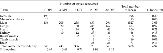
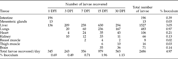
Results of the migration study have been summarized in table 1. Symptoms such as cough, anorexia, loose stool, anaemia, loss of growth and progressive debility were noticed in all piglets of group I. No such symptoms were present in piglets of group II (control). During 30 days of this experiment only one piglet of group I died, on 30 DPI. Larvae were recovered from 1 to 30 DPI, with total recovery of 345 larvae (0.69%) on 1 DPI, 243 (0.49%) on 3 DPI, 356 (0.71%) on 7 DPI, 979 (1.96%) on 15 DPI and with 563 (1.13%) larvae on 30 DPI. Within the first 24 h a total of 345 (0.69%) larvae were recovered from intestinal walls, mesenteric glands and liver. Thereafter, larvae began to appear in other organs (by day 3) and then in skeletal muscles and the brain. The highest total numbers of larvae were recovered on 15 DPI (979 (1.96%)) and from the liver (1527 (3.05%)). This suggests the migratory nature of larvae from intestine to other organs which commenced through the liver. A small percentage (0.49–1.96%) of larvae in comparison to inoculum (5 × 10⁴) were recovered from various organs. No larvae were recovered from the spleen and eye muscles.
Recovery of Toxocara canis larvae on different days post-infection (DPI) from various tissues of piglets inoculated with 5×10⁴ infective eggs.
Pathological changes
On postmortem examination, major pathological changes were observed in the liver, lung and brain. In all organs, the pathological lesions were massive and dependent on the level of larval migration. This was highest up to 15 DPI in the liver and lungs, and then gradually organized and subsided. But in brain tissue this change continued beyond 15 DPI. The observations in the brain tissue of piglets on 14 DPI were of congestion, oedema, shrinkage of nerve cells, vacuolization with invaded larvae deep in the brain tissues. There were gliosis, satellitosis, neurophagia with arrested larvae within the microglial cells causing complete destruction by phagocytosis and liquefactive necrosis.
Experimental infection of mice
No abnormal clinical signs were observed in any of the five infected mice of group I. They appeared as normal and healthy as the two control mice of group II. On 10 DPI, all mice of groups I and II were necropsied and searched for recovery of larvae. One gram of liver tissue was examined by the squash method for estimation of the total number of larvae present in each mouse liver. Five such examinations were done and an average was taken. The mean larval burden in the five mice of group I were: 20 ± 3.62, 17 ± 5.10, 3 ± 1.26, 12 ± 3.92 and 30 ± 5.69 in liver, lungs, heart, brain and muscles, respectively. No larvae were recovered from any other tissues of infected mice on 10 DPI. The degree of pathological lesions in these organs was also insignificant in comparison to pigs. This was directly related with the level of infection present in 5 g of infected piglet liver. No larvae were recovered from any tissues of the two mice of uninfected control group II.
Data obtained from both female and male piglets and mice were analysed statistically for significant variation in migratory efficiency of T. canis larvae in both these hosts. But with these limited number of animals, no such significant variation was obtained.
Discussion
Observations up to 30 DPI indicated that infective second-stage larvae of T. canis can persist for a relatively long period in the tissues of piglets. Within this period the levels of larval recovery suggest that migration of larvae from intestine to other organs has occurred mainly through the liver. This is in agreement with the observations of Beaver (Reference Beaver1956), Galvin (Reference Galvin1964), Sosipatrova (Reference Sosipatrova1973), Pahari & Sasmal (Reference Pahari and Sasmal1990a) and Acharya et al. (Reference Acharya, Sasmal, Jana and Roy2002). The duration and severity of liver involvement were greater in comparison with other organs and gradually extended to lungs, heart, kidney, brain and muscles. No larvae were recovered from spleen and eye muscles, which contradicts the results of Olson (Reference Olson1976) and Mahapatra et al. (Reference Mahapatra, Pramanik and Sasmal1996) where T. canis larvae were recovered from both these organs. The character of histopathological lesions as observed in the present study are identical to the earlier reports of Mehl et al. (Reference Mehl, Boch, Heine, Weiland and Henkel1983) and Lohmann (Reference Lohmann1985).
A total of 2486 (4.97%) larvae were recovered from these organs, indicating the suitability of pig tissues for migration of T. canis larvae, like many other abnormal hosts. This agrees with the previous reports of Done et al. (Reference Done, Richardson and Gibson1960), Galvin (Reference Galvin1964), Tomimura et al. (Reference Tomimura, Yokota and Takiguchi1976) and Acharya et al. (Reference Acharya, Sasmal, Jana and Roy2002). From the present observation it can also be concluded that T. canis larvae follow the identical migratory pattern in White Yorkshire piglets as in other paratenic hosts so far tested (Sinha, Reference Sinha1970; Tomimura et al., Reference Tomimura, Yokota and Takiguchi1976; Sharma & Bhatia, Reference Sharma and Bhatia1983; Pahari & Sasmal, Reference Pahari and Sasmal1991; Acharya et al., Reference Acharya, Sasmal, Jana and Roy2002).
The same picture has also been reflected in a pathogenicity study of different organs. In all these organs, severe to moderate pathological lesions with mortality of one piglet are also suggestive of the pig as a suitable transport host for T. canis larvae. Severe pathological lesions are created by these larvae in different organs and the mortality occurred due to the use of the massive dose of experimental infection. The pathogenic potential in the transport host, the pig, shown in this study was also observed by Tomimura et al. (Reference Tomimura, Yokota and Takiguchi1976) and Cho et al. (Reference Cho, Egami, Ohnuki, Saito, Chinone, Shichinohe, Suganuma and Akao2007) in their studies with monkeys and gerbils, respectively. The observations of Sinha (Reference Sinha1970) in his study with goats, Pahari & Sasmal (Reference Pahari and Sasmal1990b, Reference Pahari and Sasmal1991) in their studies with Japanese quails and earthworms are in agreement with the present study.
Another important observation noted here is that among most of the abnormal hosts the level of pathogenicity is always severe in nature, whereas it is absent in the definitive, carnivorous host. On the other hand, in other mammals, having similar anatomy and physiology to pigs, the reported migratory pathology was identical. It has been reported by Beaver (Reference Beaver1956) that in the Far East when raw liver was prescribed for treatment of pernicious anaemia in humans, a high and persistent eosinophilia appeared within a short period in nearly all of those treated. This eosinophilia associated with the raw liver diet might have been caused by larval Toxocara harboured in the liver, as suspected by Beaver (Reference Beaver1956) and Pahari & Sasmal (Reference Pahari and Sasmal1990a). The danger of these larvae to humans as a paratenic host must be considered, particularly in those places where the rearing and eating of raw or undercooked pork is a common practice. This zoonotic risk has been reaffirmed by Taira et al. (Reference Taira, Saeed, Permin and Kapel2003, Reference Taira, Saeed, Lind, Murrel and Kapel2004).
In the present experiment subsequent infection of mice by somatically migrated larvae was examined. A comparatively small number of larvae were recovered from liver, lungs, heart, brain and muscles, related to the infective dose of larvae present in 5 g of liver, but from this finding, the potentiality of these T. canis larvae to adapt and migrate in the second paratenic host has been shown. This observation agrees with previous reports by Pahari & Sasmal (Reference Pahari and Sasmal1990b) and Tuzer et al. (Reference Tuzer, Toparlak, Gargili, Gulanber, Keles, Efil and Ulutas Esatgil2002), where they experimentally infected mice with T. canis larvae obtained from Japanese quails and chicks, respectively.
No influence in larval migration due to sex variation of the hosts has been found. This might be due to the use of very young piglets and the limited number of animals used in this experiment. In separate studies by the present authors with adult Rhesus monkeys and adult dogs, significant variations have been obtained with the different sexes of both these hosts. This variation has also been found to be further affected by pregnancy of the female animals (unpublished).
Overall, the control animals, both piglets and mice, showed no larval burden at necropsy, confirming the infection-free status of the experimental animals. From these findings, it can be concluded that, pigs may acquire T. canis infection from the natural source by their voracious coprophagus feeding habits. Danger of these migrated T. canis larvae to human beings as a paratenic host, either directly or indirectly infected, is to be considered seriously, as shown in the mouse model used here.
Acknowledgements
The authors are grateful to the West Bengal University of Animal and Fishery Sciences, Kolkata-37, West Bengal, India, for providing financial assistance and facilities.