Isolated congenital complete atrioventricular block in the absence of structural heart disease affects ~1 in 20,000 newborns. The prenatal incidence is likely higher due to the increased rate of in-utero fetal demise with this condition, which may occur as early as week 11 of gestation.Reference Brucato, Cimaz and Caporali 1 Isolated congenital complete atrioventricular block is usually associated with the in-utero exposure of the fetus to maternal antibodies directed against Ro antigens. These antibodies may trigger inflammation, necrosis, and fibrosis of the atrioventricular node and/or the myocardium, and typically manifests as congenital complete atrioventricular block and/or endocardial fibroelastosis – cardiac neonatal lupus. It is estimated that 2% of pregnant women carry anti-Ro antibodies and that 1–2% of their fetuses will develop cardiac neonatal lupus, mainly between 18 and 24 weeks of gestation.Reference Gladman, Silverman and Yuk 2 Maternal treatment with a corticosteroids and, if the fetal heart rate is <50 beats per minute, with a β-mimetic agent has reportedly improved the outcome in antibody-mediated congenital complete atrioventricular block.Reference Jaeggi, Fouron and Silverman 3 We present an unusual case of heart block with a favourable evolution despite profound fetal bradycardia and the parental decline of prenatal medical treatment.
Case report
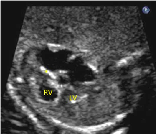
A 30-year-old gravida1 woman of Afghani origin was referred at 24 weeks for fetal bradycardia. She had no symptoms or history of an underlying autoimmune disease. Her initial fetal echocardiogram revealed third-degree heart block with an atrial rate of 110 to 115 beats per minute, a ventricular rate of 55–60 beats per minute, and occasional premature ventricular contractions. This was associated with increased endocardial echogenicity of the right ventricle and the region of the atrioventricular node suggestive of antibody-mediated inflammation and tissue damage (Fig 1, Supplementary video 1). Given the above findings, transplacental treatment with maternal oral dexamethasone and intravenous immunoglobulin was recommended as per our institutional guidelines to prevent more severe cardiac damage and to improve the odds of survival of this fetus.Reference Hutter, Silverman and Jaeggi 4 The parents were also counselled that β-sympathomimetic treatment during the pregnancy and a permanent pacemaker after birth would be required if fetal heart rate decreased further during the remainder of the gestation. Despite the serological confirmation of very high maternal anti-Ro antibody titres (>100 U/ml) by enzyme-linked immunosorbent assay, the family declined prenatal treatment and follow-up visits at our centre and considered no active postnatal care should the baby survive to birth and be significantly compromised.Reference Jaeggi, Laskin and Hamilton 5
Two-dimensional fetal echocardiogram of the right ventricle (RV) and left ventricle (LV) showing endocardiofibroelastosis of the RV. * indicates where the endocardiofibroelasosis is seen in the atrioventricular groove.

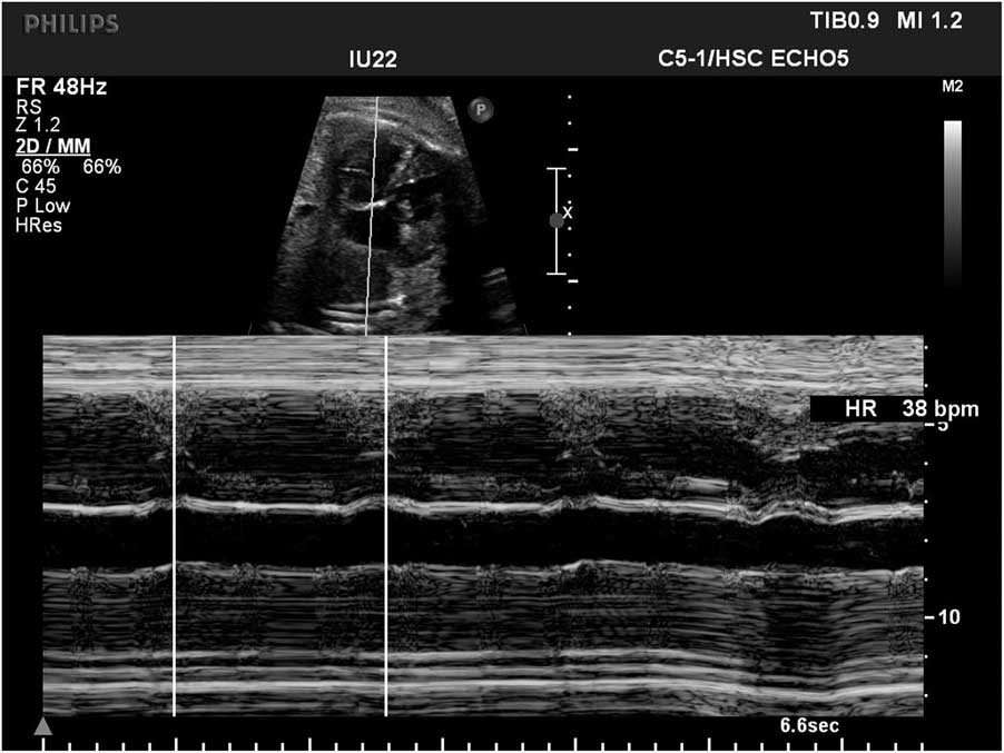
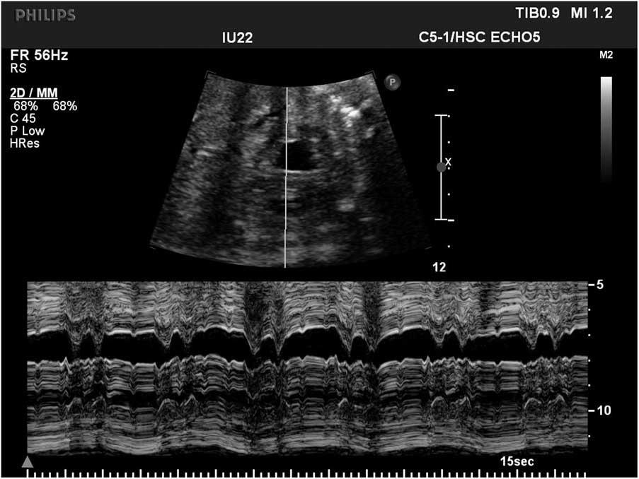
Her obstetrician continued to serially follow the pregnancy from 24 to 34 weeks of gestation. At 28 weeks, the fetal heart rate was between 40 and 50 beats per minute and regular, while the ventricular rate was 35–38 beats per minute and the atrial rate 100–110 beats per minute at 34 weeks, which triggered the patient to return to our centre for ongoing care. The findings were confirmed at our institution a week later (Fig 2) but we also noted fetal hydrops, mild ascites, skin oedema, decreased ventricular function, and the presence of frequent premature ventricular contractions. Because of the slow heart rate and fetal hydrops, the family was informed about the high likelihood of in-utero demise. The family declined premature delivery by caesarean section for neonatal pacing. At 36 weeks the fetal findings had deteriorated further, showing severely decreased right ventricular and moderately reduced left ventricular contractility, ascites, skin oedema, and frequent premature ventricular contractions. On her final prenatal assessment at 37 weeks the ventricular ectopy was significantly more frequent and present throughout the echocardiogram. Premature ventricular contractions were now present every second beat, ventricular bigeminy, which resulted in an averaged ventricular rate of 58 beats per minute (Fig 3). Peak flow velocities of the “normal” and the premature ventricular beats were 120 cm/second and 80–100 cm/second, respectively, suggestive of adequate ventricular stroke volumes with both the normal and the premature ventricular beats. With an average heart rate of 58 beats per minute, including the ventricular ectopy in bigeminy, there likely was an improved cardiac output in comparison to when the ventricular escape rate was between 35 and 38 beats per minute, and there was no significant ectopy seen on the previous assessment. Moreover, the ascites had resolved, the skin oedema was minimal, the left ventricular function had improved, and the fetus appeared appropriately grown and normally active. Following vaginal delivery at term, the boy was initially vigorous and did not require neonatal resuscitation. Isoproterenol infusion was started due to neonatal bradycardia of 40 beats per minute, which increased the ventricular rate to 50 beats per minute. A few hours later, a permanent epicardial pacemaker was implanted following parental consent as the baby had developed signs of low cardiac output including poor peripheral pulses, cold extremities, and progressively increasing lactate levels. The postoperative course was uneventful and at 8 months of age the baby is thriving and developing normally.
M-Mode showing a ventricular rate of 38 beats per minute.

Ventricular bigeminy demonstrated by M-Mode.

Discussion
Fetal hydrops is known to be associated with a high risk of fetal demise unless a treatable cause is identified and treatment rapidly initiated.Reference Jaeggi, Hamilton and Silverman 6 The emergence of hydrops in association with immune-mediated congenital complete atrioventricular block has been attributed to low cardiac output due to bradycardia and myocardial inflammation or damage. Once fetal hydrops is present the mortality risk of a fetus with congenital complete atrioventricular block is as high as 83–100%.Reference Groves, Allan and Rosenthal 7 Other factors significantly associated with perinatal death include an early congenital complete atrioventricular block diagnosis before 20 weeks of gestation, a ventricular rate less than 50 beats per minute, ventricular dysfunction, the presence of endocardial fibroelastosis, and premature delivery before 32 weeks of gestation.Reference Jaeggi, Hamilton and Silverman 6 In the presence of one or more of these factors, the odds of fetal and neonatal demise are increased by 16-fold.Reference Eliasson, Sonesson and Sharland 8 Most fetal survivors with congenital complete atrioventricular block will require permanent pacing in the first years of life.Reference Jaeggi, Hamilton and Silverman 6
Our case presented with several predictors associated with death, including developing a very low ventricular rate, a low atrial rate, ventricular dysfunction, carditis, and evolving hydrops.Reference Chockalingam, Jaeggi and Rammeloo 9 A possible explanation of the unexpected spontaneous resolution of hydrops and survival of our case is the progressive development of ventricular ectopy which resulted in an increase in ventricular rate above 50 beats per minute and, concomitant with the higher ventricular rate, an improved cardiac output in late gestation. The appearance of premature ventricular contractions may be secondary to spontaneous impulse generation within the ventricular myocardium due to underlying myocardial inflammation or due to after depolarisations in the setting of profound fetal bradycardia.Reference Wakisaka, Niwano and Niwano 10 In our case, the premature ventricular contractions progressively increased in frequency as the fetal bradycardia worsened, eventually leading to ventricular bigeminy, which almost doubled the average heart rate. Although the amount of cardiac output provided by extrasystolic beats may vary, the ventricular extra-systole in this particular case provided a flow velocity reaching up to 100 cm/second, which was most likely sufficient to provide adequate cardiac output and survival to birth. In our own experience, previous fetuses with heart rates below 45 beats per minute became or were hydropic and eventually died in-utero despite anti-inflammatory and β-sympathomimetic treatment. None of these cases presented with significant premature ventricular contractions, which raises the possibility that steroids, in the setting of an intrinsically slow ventricular escape rhythm and frequent ventricular extra beats, may not be helpful but possibly detrimental if the treatment abolishes the inflammation-mediated ventricular ectopy.
Our previously published institutional experience showed that a standardised protocol using dexamethasone in addition to intravenous immunoglobulin and β-sympathomimetics had significantly improved the mortality of fetuses with congenital complete atrioventricular block, presumably by reducing the severity of antibody-mediated cardiac damage before birth.Reference Jaeggi, Fouron and Silverman 3 OthersReference Eliasson, Sonesson and Sharland 8 , Reference Lopes, Tavares and Damiano 11 have failed to demonstrate a difference in fetal and neonatal survival between treated and untreated congenital complete atrioventricular block. The disease course of this baby with steroid treatment from the time of diagnosis to birth is speculative. Nonetheless, with standardised treatment we have reliably seen the stabilisation of the ventricular escape rate above 50 beats per minute. Without transplacental anti-inflammatory medication, the condition of this fetus including the heart rate had severely deteriorated by 34 weeks to a stage that precluded survival based on all known outcome predictors. The unexpected survival to birth of this child is best explained with the equally unexpected increase in fetal heart rate secondary to the development of significant ventricular ectopy.
Conclusion
We present a unique case of a prenatally untreated fetus with profound bradycardia and hydrops that survived probably due to the development of frequent premature ventricular contractions in late gestation.
Acknowledgements
The authors thank Nathalie Dutil for her help in this case report.
Financial Support
This research received no specific grant from any funding agency, commercial or not-for-profit sectors.
Conflicts of Interest
None.
Ethical Standards
Informed consent was obtained from all individual participants included in this case.
Supplementary material
To view supplementary material for this article, please visit https://doi.org/10.1017/S1047951117001937