Coronary anomalies are frequent in patients with pulmonary atresia and intact ventricular septum, especially when right ventricular hypoplasia is severe.Reference Freedom, Yoo, Dyamenahalli, Freedom, Yoo, Mikailian and Williams1–Reference Calder, Peebles and Occleshaw3 They may impact on early and late clinical management and worsen prognosis.Reference L’Ecuyer, Poulik and Vincent4,Reference Elias, Poh and du Plessis5
Case presentation
An 8-year-old girl was referred with chest pain, troponin rise, and presumed diagnosis of myocarditis. Neonatal diagnosis consisted of pulmonary atresia and the intact ventricular septum, with severely hypoplastic unipartite right ventricle and quasi-atresia of the tricuspid valve, requiring uni-ventricular management. Neonatal Blalock-Taussig shunt insertion, superior cavo-pulmonary connection, and finally fenestrated Fontan procedure (at the age of 7 years) had been successfully performed in another hospital. No selective coronarography had been performed at any stage but pre-Fontan CT-scan had failed to show coronary anomalies. Anticoagulation therapy had been switched to anti-aggregation, following spontaneous fenestration closure, 18 months before the current event.
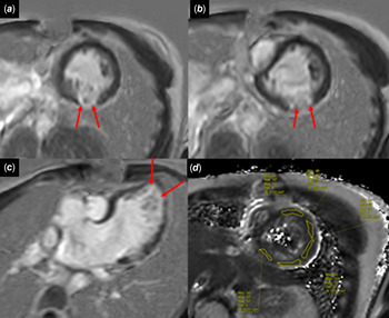
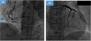
Upon admission, the child described intense chest pain during normal activity, radiating to the left arm. No clinical signs of congestive heart failure were reported. Heart sounds and vital signs were normal. Twelve-lead ECG showed repolarisation anomalies with negative T waves in V4 to V6, AVF, and DII. Ultra-sensitive troponin level, positive on admission (330 ng/L), rose to 1200 ng/L 6 hours later (standards: 0–14 ng/L). Left ventricular function was preserved on echocardiography. The rudimentary right ventricular cavity was unchanged compared to previous investigations, with no intra-ventricular thrombus nor tricuspid regurgitation. Cardiac MRI showed no myocarditis but ischaemic sequelae in three segments corresponding to the territory of the right coronary or the circonflex artery (apical inferior, apical lateral, basal inferolateral, and mid-inferolateral segments of left ventricle) (Fig 1, and video in the supplementary material). Late gadolinium enhancement (LGE) revealed a myocardial scar with enhanced T2 mapping in left basal, mid-inferolateral, and mid-inferior segments in favour of myocardial oedema and recent ischaemia. No enhancement was seen in the hypoplastic right ventricle. On selective coronarography, the proximal left and right coronary arteries were normal but distal right coronary branches seemed rather slender, with the abnormal diffuse collateral network, mainly with regards to the right ventricular cavity (Fig 2). Myocardial ischaemia was considered secondary to peripheral stenosis with an abnormal coronary microvasculature. Low dose aspirin was continued; ACE inhibitors and beta-blockers (bisoprolol) were begun. Further clinical evolution was uneventful with no arrhythmias. Ultra-sensitive troponin level decreased after one week (194 ng/L) and subsequently normalised. During the next months, the child remained asymptomatic. Twelve-lead ECG remained abnormal with inverted T waves in V4 to V6 and inferior leads. Strenuous exercise was withheld up to an uneventful, maximal exercise test 8 months later. Repeat cardiac MRI performed 7 months later was unchanged, but perfusion MRI was not performed.
Cardiac MRI with LGE in left ventricular basal inferolateral, mid-inferolateral, and apical lateral segments (red arrows); label D shows an elevated T2 mapping signal (72 mseconds) in basal inferior, basal inferolateral, and mid-inferolateral segments, confirming the recent character of myocardial ischaemia.

Coronarography. ( a ) non-stenotic right coronary artery with diffuse collateral network (blue arrow). ( b ) non-stenotic left coronary artery.

Discussion
Pulmonary atresia with intact ventricular septum is rare congenital heart disease with an estimated prevalence of 4.5/100.000 births.Reference Freedom, Yoo, Dyamenahalli, Freedom, Yoo, Mikailian and Williams1 The presence of coronary anomalies and the degree of right ventricular hypoplasia affect therapeutic management and prognosis.
The coronary anomalies are described in up to 75% of the patients, and more frequently in the presence of marked right ventricular hypoplasia (unipartite right ventricle).Reference Freedom, Yoo, Dyamenahalli, Freedom, Yoo, Mikailian and Williams1–Reference Calder, Peebles and Occleshaw3 They include: 1) myocardial sinusoids, connecting coronary capillaries to the right ventricular inter-trabeculated spaces; 2) ventriculo-arterial communications or fistulas, connecting coronary main trunks to inter-trabeculated spaces and sometimes associated with stenosis or interruptions of the coronaries; 3) the anomalous origin of the left or right coronary artery. Histologically, one-third of the patients has normal coronary artery walls; one-third shows thickening of the media and intima; the last third has very thickened walls with the destruction of the wall architecture resulting in stenosis or obstructions.Reference Calder and Sage2,Reference Calder, Peebles and Occleshaw3 Significant fistulas and/or coronary stenosis or atresia may result in a right ventricle-dependant coronary circulation, contra-indicating right ventricular decompression. Indeed, the sudden drop of right-ventricle pressure may reduce coronary blood flow and cause ischaemia.
When the right ventricle is tripartite with adequate tricuspid valve z-score, decompression of the right ventricle is considered after birth, usually by percutaneous radiofrequency perforation, in order to achieve a bi-ventricular circulation. Although coronary anomalies are less frequently associated with a well-developed right ventricle, imaging of the coronary perfusion remains highly recommended. Myocardial ischaemia may occur during initial management, as a result of misappraisal of those coronary anomalies.Reference Popoiu, Eicken, Genz, Schreiber and Hess6
In the presence of a small, unipartite right ventricular cavity, right ventricle decompression is not an option and the child is oriented towards uni-ventricular palliation. Right ventriculography and aortic root opacification, are recommended in all such patients before initial surgery, to document the frequently associated coronary anomalies.Reference Calder and Sage2 Our clinical case reminds us that ischaemic events may happen during remote follow-up, even late after uneventful surgical completion.Reference Calder and Sage2,Reference Calder, Peebles and Occleshaw3 The natural history of the coronary lesions remains poorly described and understood, but it is possible that late ischaemic events relate either to the persistence of macroscopic fistulas, to evolving main coronary artery stenosis, or to distal anomalies of microvasculature, as suggested in our patient by the important collateral network.
In everyday practice, it remains difficult to investigate evolving coronary lesions and the risk of myocardial ischaemia in those children. No clear recommendations exist for follow-up and long-term treatment. The high incidence of coronary anomalies must encourage us to systematically perform selective coronarography and/or perfusion MRI before the Fontan operation, which was not performed in our patient. Events such as the appearance of tricuspid valve regurgitation, or right ventricular cavity thrombosis must be looked for as they may induce ischaemia in the right ventricle-dependant coronary circulation.Reference Freedom, Yoo, Dyamenahalli, Freedom, Yoo, Mikailian and Williams1
Regular exercise testing and imaging, including MRI with perfusion myocardial sequences and stress cardiac MRI, must be considered in order to detect ventricular ischaemia, dysfunction, and arrhythmias. Anti-aggregant therapy, beta-blockade, and strenuous exercise avoidance seem reasonable in the presence of coronary lesions with ischaemic events but no scientific data exist to support this. Cardiac transplantation or implantable cardioverter-defibrillator (ICD) may be considered in case of respectively terminal heart failure or arrhythmic ventricular events.Reference Calder, Peebles and Occleshaw3
Conclusion
Myocardial ischaemia after Fontan completion in patients with pulmonary atresia and the intact ventricular septum is a severe complication. Coronary lesions are frequent in the presence of unipartite right ventricles and may be evolving. Early recognition and regular re-evaluation are important to allow proper management.
Video
Cardiac MRI 4 cavities with myocardial ischaemia in apical lateral, basal inferolateral, and mid-inferolateral; right ventricle is very hypoplasic and left ventricle has preserved systolic function.
Acknowledgements
The authors certify that they have no affiliations with or involvement in any organisation or entity with any financial or non-financial interest in the subject matter or materials discussed in this manuscript.
It was approved by the ethical committee of the Timone Hospital. Informed consent was obtained from the parents of the child.
This research received no specific grant from any funding agency, commercial or not-for-profit sectors.
None.
Parental consent was obtained for anonymised publication, as required by French Ethics laws.
Supplementary material
To view supplementary material for this article, please visit https://doi.org/10.1017/S104795112100144X