Critical pulmonary valve stenosis in the neonate is characterised by ductus-dependent pulmonary circulation. In this CHD, cyanosis is related to a right–left interatrial shunt, secondary to a restrictive physiopathology of the right ventricle.
In 1982, Kan et alReference Kan, White, Mitchell and Garder 1 performed the first percutaneous balloon valvuloplasty, which is now the treatment of choice for pulmonary valve stenosis in the paediatric population.
Several studiesReference Kan, White, Mitchell and Garder 1 – Reference Lee, Peng, Tu, Chen, Lee and Chang 19 have shown the immediate success of percutaneous balloon valvuloplasty, the low immediate re-intervention requirement, and qthe adequate growth of the right ventricle and of the pulmonary valve in infants and children with critical pulmonary valve stenosis. A few studies, however, have described the long-term results of this technique, frequently associated with a significant percutaneous or surgical re-intervention rate. According to LatsonReference Saad, Roushdy and Elsayed 20 , some patients need an additional source of pulmonary flow after percutaneous balloon valvuloplasty, and around 5–10% undergo surgery to relieve any residual valvular or subvalvular stenosis. In addition, his research indicates that in long-term follow-up about 30% of the patients need a percutaneous re-intervention. Similarly, Karagoz et alReference Latson 2 , studying a cohort where 56% of the patients had critical pulmonary valve stenosis, demonstrated that freedom from re-intervention was 76% at long-term follow-up.
The aim of this study was to assess the immediate and long-term results of percutaneous balloon valvuloplasty in neonates with critical pulmonary valve stenosis followed-up over 20 years in a single, tertiary-care centre.
Materials and methods
Population
We performed a retrospective analysis of all consecutive neonates who underwent percutaneous balloon valvuloplasty for critical pulmonary valve stenosis from January, 1994 to December, 2014. We identified 24 patients who fulfilled the following criteria: presence of critical pulmonary valve stenosis with supra-systemic right ventricle pressures, hypoplasia of the right ventricle, and ductus-dependent pulmonary circulation as well as those who have undergone percutaneous balloon valvuloplasty within 28 days of age. Patients with pulmonary atresia, severe tricuspid annulus hypoplasia (<−4 Z scoreReference Rao 21 ), coronary circulation dependent on the right ventricle, and patients with any other CHD that needed treatment in the neonatal period were excluded from the study.
Variables assessed
We retrospectively analysed the clinical records of the selected patients, and we grouped the data into three distinct periods: pre-intervention, intervention, and follow-up.
In the pre-intervention period, the following variables were selected: sex, age, weight, prenatal diagnosis, symptoms, oxygen saturation in room air, and period of treatment with prostaglandin E1 before percutaneous balloon valvuloplasty. From the transthoracic echocardiographic assessment, we retained the following data: existence of an atrial septal defect/patent foramen ovale, right ventricle morphology, classified as unipartite, bipartite, or tripartite according to the number of portions that constitute the right ventricle, that is, inlet, outlet, and apical trabecular region, presence of dysplastic pulmonary valve, defined as a thick leaflet valve with fusion of the commissures and opening in doming during systole, larger pulmonary annulus diameter measured at systole, and pulmonary transvalvular peak gradient.
During intervention, we assessed the following parameters: age, haemodynamic pre- and post-percutaneous balloon valvuloplasty pulmonary variables such as transvalvular peak gradient, right ventricle pressure, and right ventricle/systemic pressure ratio, procedural details such as diameter of the largest balloon and ratio of the largest balloon/diameter of the pulmonary annulus, and complications during cardiac catheterisation.
In the follow-up period, we analysed the following data: treatment duration with prostaglandin E1 after percutaneous balloon valvuloplasty, percutaneous re-intervention, surgical intervention, and medical outpatient treatment. For transthoracic echocardiographic variables, we selected the short-term (3–6 months and 1 year), mid-term (3–5 years), and long-term (5–20 years) pulmonary transvalvular peak gradient and, in the most recent transthoracic echocardiographic assessment, the presence of tricuspid valve regurgitation, as well as the degree of pulmonary valve regurgitation, which is classified as none, light, moderate, and severe. We chose not to present other echocardiographic data such as right ventricular dimensions and interventricular septum position and movement due to the retrospective nature of the study and inconsistencies over the study period.
Cardiac catheterisation
All procedures were performed under general anaesthesia according to the previously described technique.Reference Burzynski, Kveselis, Byrum, Kavey, Smith and Gaum 22 Venous and arterial accesses were placed by cannulation of the femoral vessels. After cannulation of the femoral vein, right cardiac catheterisation was performed. Pressure gradients were measured, followed by right ventricular angiography in lateral projection and/or left anterior oblique, to measure the diameter of the pulmonary valve annulus in systole. The valvuloplasty balloon was centred on the annulus valve and inflated until complete resolution of the waist. Finally, pressure measurements were obtained, and a final angiography was performed.
Definitions
In this study, the safety of the procedure was assessed by mortality and major complications; the success of the procedure by the reduction in pulmonary transvalvular peak gradient after percutaneous balloon valvuloplasty for values ≤40 mmHg and by the need for percutaneous re-intervention or surgical intervention before hospital discharge; and the long-term results were evaluated by the evolution of the pulmonary transvalvular peak gradient at long-term follow-up and by the need for long-term re-intervention.
Cardiac catheterisation complications were classified according to CHARM’s – Catheterization for CHD Adjustment for Risk MethodReference Colli, Perry, Lock and Keane 23 – severity scale.
Statistics
Continuous variables are expressed as averages and standard deviations. Qualitative variables are expressed as percentages. The univariate analysis for categorical variables was performed using χ2 and Fisher’s precise tests, and for the continuous variables we used Wilcoxon’s test. The multifactorial analysis was carried out using simple and multiple linear regression and the Kaplan–-Meier survival curve. In both tests, the re-intervention variables, surgical intervention, time until re-intervention, and time until surgical intervention were used as dependent variables and as interest variables. For this analysis, a p value <0.05 was considered statistically significant. All statistical analyses were performed using SPSS® (v 20.0; IBM SPSS Software for Predictive Analytics; SPSS, Chicago, IL, United States of America).
Results
Over the two decades of review, 24 neonates with critical pulmonary valve stenosis underwent 33 transcatheter percutaneous balloon valvuloplasties during a mean follow-up time of 8.36±6.45 years (with a range from 0.02 to 18.37 years).
Pre-intervention
From January, 1994 to December, 2014, percutaneous balloon valvuloplasty was attempted in 24 consecutive neonates with the diagnosis of critical pulmonary valve stenosis.
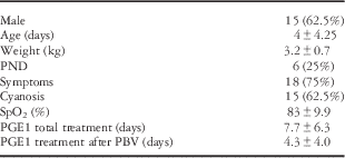
In our sample, 15 (62.5%) patients were male, and the median weight was 3.2±0.7 kg.
Prenatal diagnosis was made in six (25%) patients, and in the others the diagnosis was made, on average, on their 1st day of life. At the time of diagnosis, 18 neonates (75%) had signs of respiratory distress, and 15 neonates (62.5%) had cyanosis, with a median oxygen saturation of 83±10% (Table 1).
Baseline demographics and clinical profile (n=24).

PBV=percutaneous balloon valvuloplasty; PGE1=prostaglandin E1; PND=prenatal diagnosis; SpO2=oxygen saturation in room air
Data are presented as mean±SD or percentages of patients
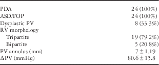
The initial echocardiographic assessment showed that all patients presented patent ductus arteriosus and atrial septal defect/patent foramen ovale. The pulmonary valve was dysplastic in eight neonates (33.3%), and none of them had Noonan syndrome.
The right ventricle was tripartite in 19 (79.2%) and bipartite in five (20.8%) patients; none of the patients had a unipartite right ventricle. The median pulmonary transvalvular peak gradient was 80.6±15.8 mmHg, and the median pulmonary valve annulus diameter was 7 mm (with a range from 5 to 10 mm) (Table 2).
Echocardiographic findings before percutaneous balloon valvuloplasty.

ASD=atrial septal defect; FOP=patent foramen ovale; PDA=patent ductus arteriosus; ∆PV=pulmonary transvalve peak gradient; PV=pulmonary valve; RV=right ventricle
Data are presented as mean±standard deviation or percentages of patients
Almost all neonates (91.7%) required prostaglandin E1 therapy before the procedure for an average of 7.67±6.3 days.
Intervention
The median age at cardiac catheterisation was 4.0±4.3 days. Percutaneous balloon valvuloplasty was performed in 24 patients (100%), but it was unsuccessful in two because of persistent valvular obstruction. Only one of these neonates required percutaneous re-dilation 13 days after the first cardiac catheterisation, and the patient underwent surgical reconstruction of the right ventricular outflow tract, atrial septal defect closure, and patent ductus arteriosus ligation within 1 month of age. The other neonate remained prostaglandin E1 dependent for 14 days after percutaneous balloon valvuloplasty, necessitating a subsequent Blalock–Taussing shunt. At 14 months of age, this patient underwent surgical reconstruction of the right ventricular outflow tract and Blalock–Taussing shunt ligation. Of these two patients, only the first one had a dysplastic pulmonary valve.
There was one death during the entire study period (4.17%). After a successful percutaneous balloon valvuloplasty (pulmonary transvalvular peak gradient of 20 mmHg), the patient remained prostaglandin E1 dependent and showed signs of progressive dysfunction of the right ventricle; the patient died 6 days after cardiac catheterisation because of septicaemia.
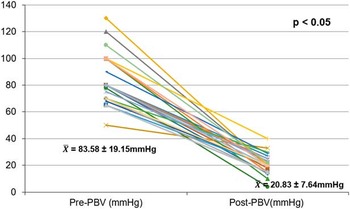
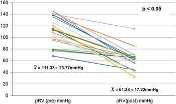
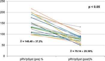
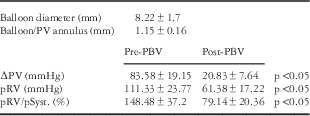
After percutaneous balloon valvuloplasty, the pulmonary transvalvular peak gradient decreased immediately from 83.58±19.6 to 20.83±7.6 mmHg (p<0.05) (Fig 1); the right ventricular pressure decreased from 111.33±23.8 to 61.38±17.3 mmHg (p<0.05) (Fig 2); and the right ventricle/systemic pressure ratio decreased from 148.5±37.2 to 79.1±20.36% (p<0.05) (Fig 3). The diameter of the largest balloon/diameter of the pulmonary annulus ratio was 1.15±0.2 mm (with a range from 0.9 to 1.43 mm) (Table 3).
Comparison of transvalvular systolic gradients (mean value±SD) before and immediately after percutaneous balloon valvuloplasty (PBV).

Comparison of right ventricle (RV) pressure (mean±SD) before and immediately after percutaneous balloon valvuloplasty.

Comparison of right ventricle (RV)/systemic pressure ratio (mean±SD) before and immediately after percutaneous balloon valvuloplasty.

Catheterisation findings before and after percutaneous balloon valvuloplasty (PBV).

∆PV=pulmonary transvalve peak gradient; PV=pulmonary valve; RV=right ventricle; pRV/pSyst=RV/systemic pressure ratio
Data are presented as mean±SD or percentages of patients
No statistically significant differences were found in the results of percutaneous balloon valvuloplasty regarding patients’ sex, age, weight, right ventricle morphology, or pulmonary valve dysplasia.
Prostaglandin E1 therapy was maintained, on average, for an additional 4.3±4.0 days after percutaneous balloon valvuloplasty, and only two patients could not wean off this treatment: the neonate that died and the neonate that underwent Blalock–Taussing shunt. The longest period of treatment with prostaglandin E1 was 27 days in a 2.5-kg premature infant.
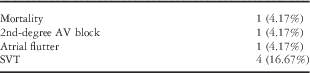
Of the 24 patients who underwent percutaneous balloon valvuloplasty, one died (4.17%), as mentioned above, and six (29.2%) had complications classified as moderate according to the CHARMReference Colli, Perry, Lock and Keane 23 scale.
Of the six neonates, five had tachyarrhythmia – supraventricular tachycardia and atrial flutter – and one had a second-degree atrioventricular block (Table 4).
Complications.

AV=atrioventricular; SVT=supraventricular tachycardia
Data are presented as percentages of patients
In all, 11 patients (45.8%) were discharged home with medications such as furosemide, digoxin, or propranolol that was eventually stopped in all.
Follow-up
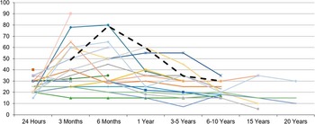
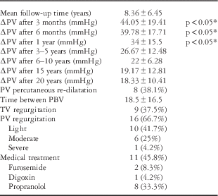
On short-term follow-up, we observed a decrease in the pulmonary transvalvular peak over the 1st year of life (p<0.05), with an average of 44.05±19.41, 39.78±17.71, and 34±15.5 mmHg at 3 months, 6 months, and 1 year after percutaneous balloon valvuloplasty, respectively (Table 5 and Fig 4).
Comparison of transvalvular systolic gradients after percutaneous balloon valvuloplasty over time. The dashed line represents the central tendency curve.

Clinical and echocardiographic findings over time.

PBV=percutaneous balloon valvuloplasty; ∆PV=pulmonary transvalvular peak gradient; PV=pulmonary valve; TV=tricuspid valve
Data are presented as mean±SD or percentages of patients
*Relationship with the pulmonary transvalve peak gradient after PBV
Moreover, during the remainder of the study, we confirmed the maintenance of a low pulmonary transvalvular peak gradient, with an average of 26.67±12.48, 22±6.28, 19.17±12.81, and 18.33±10.41 mmHg at 3–5, 6–10, 15, and 20 years after percutaneous balloon valvuloplasty, respectively (Table 5).
Long-term echocardiographic assessment showed that 37.5% (nine) of the patients had light-to-moderate tricuspid valve regurgitation, and 71% (16) had pulmonary valve regurgitation, classified as severe in one patient (4.2%) and as moderate in six patients (25%) (Table 5). We found no correlation between the ratio of the largest balloon diameter/pulmonary annulus diameter and the degree of pulmonary valve regurgitation.
Re-intervention
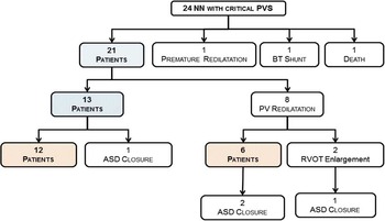
Of the 21 patients in whom percutaneous balloon valvuloplasty was attempted, nine (42.85%) required a second percutaneous or surgical procedure – eight (38.1%) of them underwent percutaneous pulmonary valve re-dilation and one patient underwent percutaneous atrial septal defect closure.
Patients who underwent pulmonary valve re-dilation had higher pulmonary transvalvular residual gradients after the first percutaneous balloon valvuloplasty (p<0.05), and in all of them the right ventricle was tripartite. In 62% of the cases, a second percutaneous balloon valvuloplasty was attempted within 11 months of the index procedure, and in average within 18.5±16.5 months. Later, two of these patients underwent cardiac surgery to enlarge the right ventricular outflow tract, on average, at 9.5 months of age.
Pulmonary valve re-dilation was successful in the remaining six patients; two of them underwent atrial septal defect closure – one by surgery and the other by percutaneous treatment. Atrial septal defect closure was performed, on average, at the age of 9 years (Fig 5).
Follow-up results of 24 newborns with critical pulmonary valve stenosis (PVS) with attempted percutaneous balloon valvuloplasty. ASD=atrial septal defect; BT=Blalock–Taussing; PV=pulmonary valve; RVOT=right ventricle outflow tract.

In the univariate analysis, none of the factors significantly influenced pulmonary valve re-dilation; however, we verified that five (62.5%) of the neonates who underwent pulmonary valve re-dilation had dysplastic pulmonary valves.
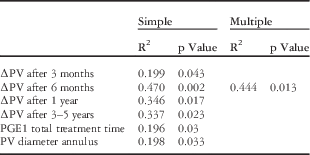
In the simple linear regression analysis, the factors associated with time for re-intervention were the pulmonary annulus size, the increase in pulmonary transvalvular gradient between 3 months and 3–5 years after percutaneous balloon valvuloplasty, and the total prostaglandin E1 treatment time (Table 6). In the multiple linear regression analysis, however, the pulmonary transvalvular gradient 6 months after percutaneous balloon valvuloplasty was the only significant variable, explaining 44.4% of time of re-intervention variance.
Factors associated with time to re-intervention.

PGE1=prostaglandin E1; ∆PV=pulmonary transvalve peak gradient; PV=pulmonary valve
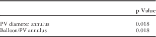
Through a univariate analysis, we also verified that the increase in the pulmonary transvalvular gradient (≥50 mmHg) 6 months after percutaneous balloon valvuloplasty was significantly related to a smaller pulmonary annulus size and with a smaller diameter of the largest balloon/diameter of the pulmonary annulus ratio (Table 7).
Factors associated with a pulmonary transvalve peak gradient≥50 mmHg 6 months after percutaneous balloon valvuloplasty.

PV=pulmonary valve
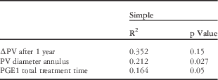
Analysing time to first surgery, by simple linear regression, we found a significant relation with the pulmonary annulus size, with an increase in the pulmonary transvalvular gradient 1 year after percutaneous balloon valvuloplasty, and with the total prostaglandin E1 treatment time (Table 8).
Factors associated with time to first surgical intervention.

PGE1=prostaglandin E1; ∆PV=pulmonary transvalve peak gradient; PV=pulmonary valve
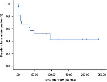
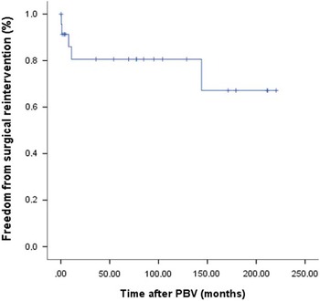
In this study, freedom from re-intervention was 75% at 7 months, 50% at 8 years, and 43% at 10, 15, and 18 years (Fig 6), and freedom from surgical intervention was 80.6% at 1 and 10 years and 67.1% at 15 and 18 years (Fig 7).
Freedom from re-intervention after balloon dilatation for neonatal critical pulmonary stenosis. PBV=percutaneous balloon valvuloplasty.

Freedom from surgical re-intervention after balloon dilatation for neonatal critical pulmonary stenosis. PBV=percutaneous balloon valvuloplasty.

Discussion
Percutaneous balloon valvuloplasty is a technique that, since 1982,Reference Kan, White, Mitchell and Garder 1 has been extensively used for pulmonary valve stenosis treatment. Several investigationsReference Karagoz, Asoh and Hickey 3 – Reference Kaku, Pinto, Agualusa and Sampayo 5 , Reference Zeevi, Keane, Fellows and Lock 24 , Reference Gildein, Kleinert, Goh and Wilkinson 25 have supported the safety and immediate success of this technique.
In 1984, Tynan et alReference Bergersen, Gauvreau and Foerster 6 , described the results of percutaneous balloon valvuloplasty in neonates with critical pulmonary valve stenosis, and since then many other studies have shown its efficacy and success as first-line treatment for this age group.Reference Latson 2 , Reference Chubb and Simpson 4 – Reference Lee, Peng, Tu, Chen, Lee and Chang 19 , Reference Zeevi, Keane, Fellows and Lock 24 , Reference Gildein, Kleinert, Goh and Wilkinson 25 Long-term results of percutaneous balloon valvuloplasty in the neonatal period, however, were less studied;Reference Latson 2 furthermore, studies with medium-term outcomes of percutaneous balloon valvuloplasty for critical pulmonary valve stenosis usually include infants above 28 days of age and without ductus-dependent pulmonary blood flow.Reference Chubb and Simpson 4 , Reference Tabatabaei, Boutin, Nykanen, Freedom and Benson 8 – Reference Holzer, Gauvreau and Kreutzer 10
To the best of our knowledge, this is the only study with the longest follow-up period on neonates with critical pulmonary valve stenosis and ductus-dependent pulmonary blood flow, who underwent percutaneous balloon valvuloplasty before 28 days of life at a single, tertiary-care centre. Half of the patients were followed-up for more than 7.5 years and ¼ of them for more than 14.8 years.
Immediate results of percutaneous balloon valvuloplasty
In this study, percutaneous balloon valvuloplasty was performed in all patients (100%) with immediate success in 91.7%, with no mortality associated with the procedure. These results overlap with those presented in previous studies that have shown a procedure performance rate of 93–98% with an immediate success of 87.5–100%.Reference Latson 2 , Reference Chubb and Simpson 4 , Reference Rao 7 , Reference Tabatabaei, Boutin, Nykanen, Freedom and Benson 8 , Reference Jarrar, Betbout and Farhat 11 – Reference Gildein, Kleinert, Goh and Wilkinson 13
As demonstrated by others,Reference Latson 2 – Reference Lee, Peng, Tu, Chen, Lee and Chang 19 , Reference Gildein, Kleinert, Goh and Wilkinson 25 there was a significant reduction in the pulmonary transvalvular peak gradient, in right ventricular pressure, and in the right ventricle/systemic pressure ratio after percutaneous balloon valvuloplasty.
After percutaneous balloon valvuloplasty, 79% of the patients remained under prostaglandin E1 therapy, and we verified that 50% needed a longer period of treatment (>3 days), which was significantly higher than what has been reported in previous studies (17–27%).Reference Latson 2 , Reference Chubb and Simpson 4 , Reference Gournay, Piéchaud, Delogu, Sidi and Kachaner 14 , Reference Santoro, Formigari, Carlo, Pasquini and Ballerini 15 This was probably related to the characteristics of the population selected for this study, as we included only newborns with ductus-dependent critical pulmonary valve stenosis.
Re-intervention
In this study, the re-intervention rate for patients who underwent successful percutaneous balloon valvuloplasty was 42.8%, which is significantly higher compared with other paediatric age groups (8–10%).Reference Holzer, Gauvreau and Kreutzer 10 , Reference Tynan, Jones, Joseph, Deverall and Yates 12 , Reference Gildein, Kleinert, Goh and Wilkinson 25 This ratio was also higher compared with other studies (17–36%) performed in neonates with critical pulmonary valve stenosis.Reference Latson 2 , Reference Chubb and Simpson 4 , Reference Tabatabaei, Boutin, Nykanen, Freedom and Benson 8 , Reference Holzer, Gauvreau and Kreutzer 10 , Reference Tynan, Jones, Joseph, Deverall and Yates 12 , Reference Gildein, Kleinert, Goh and Wilkinson 13 This disparity may be explained by the following motives. First, this study only included neonates with critical pulmonary valve stenosis and ductus-dependent pulmonary blood flow. In fact, Tabatabaei et alReference Chubb and Simpson 4 showed no differences in the immediate haemodynamic results between neonates dependent and non-dependent on patent ductus arteriosus; however, no study so far has compared the evolution and re-intervention rate in these two groups of patients. Second, our study had the longest follow-up period for neonates with critical pulmonary valve stenosis, with ¼ of the patients followed-up for longer than 14.8 years. Finally, we assumed as re-interventions all the procedures that patients underwent after percutaneous balloon valvuloplasty including percutaneous atrial septal defect closure.
In this study, freedom from re-intervention was 50% at 8 years and 43% at 10 and 18 years, which is lower than that reported in previous studies (84% at 8 years and 64–87% at 10 years).Reference Latson 2 , Reference Chubb and Simpson 4 , Reference Tabatabaei, Boutin, Nykanen, Freedom and Benson 8 , Reference Holzer, Gauvreau and Kreutzer 10 , Reference Gildein, Kleinert, Goh and Wilkinson 25 On the other hand, freedom from surgical intervention (80.6% at 1 and 10 years and 67.1% at 15 and 18 years) was higher than that described by Karagoz et alReference Latson 2 , which represents, to the best of our knowledge, the study with the longest follow-up period of neonates on whom percutaneous balloon valvuloplasty was attempted; however, in their study, only 56% of infants had critical pulmonary valve stenosis.
Despite the high re-intervention rate, 12/24 patients (50%) underwent only one percutaneous intervention, and 18/24 (75%) patients were successfully treated exclusively by percutaneous balloon valvuloplasty, which is in line with what was previously reported by several authors (74–86%).Reference Latson 2 – Reference Kaku, Pinto, Agualusa and Sampayo 5 , Reference Rao 7 , Reference Tabatabaei, Boutin, Nykanen, Freedom and Benson 8 , Reference Holzer, Gauvreau and Kreutzer 10 , Reference Tynan, Jones, Joseph, Deverall and Yates 12 , Reference Zeevi, Keane, Fellows and Lock 24 , Reference Gildein, Kleinert, Goh and Wilkinson 25
Previous studiesReference Latson 2 have shown a correlation between right ventricular morphology and re-intervention. In our study, one of the five patients with bipartite right ventricle had an unsuccessful percutaneous balloon valvuloplasty for persistent valvular obstruction. The remaining four patients had successful percutaneous balloon valvuloplasties and did not require further interventions. All the patients who underwent pulmonary valve re-dilation had a tripartite right ventricle; therefore, bipartite right ventricle was not a significant predictor of re-intervention as found in other studies.
In the present study, we also verified that the factors associated with time for re-intervention were pulmonary annulus size, increase in pulmonary transvalvular gradient between 3 months and 3–5 years after percutaneous balloon valvuloplasty, and total prostaglandin E1 treatment time. Among these, the most significant factor was the increase in pulmonary transvalvular peak gradient 6 months after percutaneous balloon valvuloplasty, which was related to a smaller pulmonary annulus size and to a smaller diameter of the largest balloon/diameter of the pulmonary annulus ratio. Given the size of our sample, however, these data do not allow us to infer conclusions applicable to other populations.
Complications
Percutaneous balloon valvuloplasty is a more complex and lengthy technique in neonates than in other paediatric age groups, particularly with regard to critical pulmonary valve stenosis, because of the morphology and hypoplasia of the right ventricle and the tricuspid annulus size.
This reflects on a high intra-catheterisation major complications rate (29.2%) and mortality (4.2%), which are similar to what has been reported in several studies between 14 and 31%Reference Latson 2 , Reference Chubb and Simpson 4 , Reference Tabatabaei, Boutin, Nykanen, Freedom and Benson 8 – Reference Holzer, Gauvreau and Kreutzer 10 , Reference Fedderly, Lloyd, Mendelsohn and Beekman 26 and 3 to 25%,Reference Latson 2 , Reference Chubb and Simpson 4 , Reference Tabatabaei, Boutin, Nykanen, Freedom and Benson 8 , Reference Jarrar, Betbout and Farhat 11 , Reference Gildein, Kleinert, Goh and Wilkinson 25 , Reference Fedderly, Lloyd, Mendelsohn and Beekman 26 respectively.
It is noteworthy that, after percutaneous balloon valvuloplasty, patients show an increase in pulmonary valve regurgitation over time, which can be moderate to severe.Reference Latson 2 – Reference Kaku, Pinto, Agualusa and Sampayo 5 , Reference Tabatabaei, Boutin, Nykanen, Freedom and Benson 8 , Reference Holzer, Gauvreau and Kreutzer 10 , Reference Tynan, Jones, Joseph, Deverall and Yates 12 , Reference Gournay, Piéchaud, Delogu, Sidi and Kachaner 14 , Reference Gildein, Kleinert, Goh and Wilkinson 25 , Reference Ladusans, Qureshi, Parsons, Arab, Baker and Tynan 28 In our study, pulmonary valve regurgitation was light in 10 neonates (41.7%), moderate in six (25%), and severe in only one patient (4.2%). Serious pulmonary valve regurgitation was noted in a neonate who underwent cardiac surgery to enlarge the right ventricular outflow tract, and at the age of 10 years he needed a surgical pulmonary valvuloplasty to reduce the size of the pulmonary annulus.
Pulmonary regurgitation was analysed primarily with color flow Döppler and by the presence of reverse diastolic flow in the distal pulmonary arteries. Echocardiography is not the best technique for quantification of pulmonary regurgitation, and therefore these data should be analysed by taking into account the method used.
Study limitations
This study presents some limitations that are primarily related to its retrospective nature and sample size. In fact, small samples hamper the determination and the statistical evaluation of the predictors of success and efficiency of percutaneous balloon valvuloplasty in neonates with ductus-dependent pulmonary valve stenosis, compromising its extrapolation tor the general population.
On the other hand, the retrospective nature of the study did not allow an evaluation of right ventricular function and dimensions after percutaneous balloon valvuloplasty over time, and neither the consequences of pulmonary valve regurgitation on the right ventricle.
Conclusions
Percutaneous balloon valvuloplasty is a safe and effective technique in neonates with ductus-dependent pulmonary valve stenosis; however, in this age group, the complexity inherent to the execution of this technique is associated with significant morbidity and mortality.
During a mean follow-up time of 8.36 years, this study showed maintenance of low pulmonary transvalvular peak gradient and low incidence of severe pulmonary valve regurgitation, with a large proportion of patients remaining free from surgery (80.6% at 1 and 10 years and 67.1% at 15 and 18 years) and from percutaneous re-intervention (50% at 8 years and 43% at 10 and 18 years).
Despite the high re-intervention rate, 75% of the neonates were exclusively treated by percutaneous balloon valvuloplasty.
Acknowledgements
None.
Financial Support
This research received no specific grant from any funding agency or from commercial or not-for-profit sectors.
Conflicts of Interest
None.
Ethical Standards
The authors assert that all referenced work contributing to this review complies with the ethical standards of biomedical or medicolegal investigation.