Tetralogy of Fallot is the most common cyanotic CHD, with a prevalence of approximately 0.5 per 1000 live births. Reference Hoffman and Kaplan1 Associated anomalies are frequently seen. Reference Bailliard and Anderson2 Here, we present a neonate diagnosed as having tetralogy of Fallot, a double aortic arch, and an aortopulmonary window by echocardiographic imaging. To our knowledge, this is the first case report of this triad of congenital heart anomalies in the literature. This case also illustrates that complex and very rare cardiac defects may be asymptomatic in the early neonatal period and progressively have severe congestive heart failure symptoms in a few weeks.
Case report
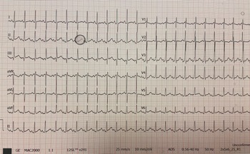
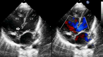
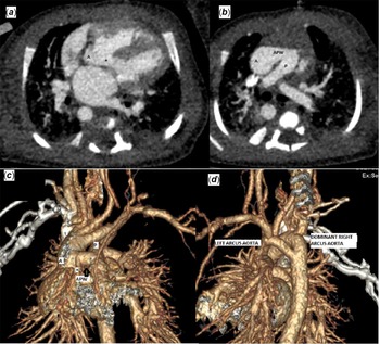
An 8-day-old boy baby weighing 3750 grams who was investigated for microcephaly was referred for assessment of possible concomitant cardiac pathology. He was breastfed totally with weight gain, and there was no tachypnea and tachycardia. A 1/6 soft systolic murmur was heard in the left upper sternal border. Peripheral pulses were palpable. His oxygen saturation of both upper and lower extremities in room air was 96%. Although he was a newborn, a left inferior cardiac axis was noticeable on the electrocardiogram with sinus rhythm (Fig 1). He had a sibling with microcephaly and epilepsy in his family history. Transthoracic echocardiography was performed. A subaortic malaligned ventricular septal defect with overriding aorta were seen in the apical five-chamber view (Fig 2). Although obstructed outflow of the right ventricle (mild) and pulmonary annular hypoplasia (z score: -3.52) was conspicuous, the main pulmonary artery and the branches were well-developed with insignificant gradient on continuous wave Doppler and a McGoon index of 2.8. On the parasternal long-axis view with anterior angulation and on a high parasternal short-axis view, a large and tubular aortopulmonary window was seen between the ascending aorta and the main pulmonary artery (Supplementary Video S1 and S2). Also, a double aortic arch with hypoplasia of the left arch was demonstrated. On the parasternal short-axis view, the right coronary artery was thought to originate from the aortopulmonary window, not from the right coronary sinus (Supplementary Video S2). To support our diagnosis, thorax CT angiography was performed (Fig 3). In addition to echocardiographic findings, CT angiography showed that the right coronary artery originated from the aortic side of the aortopulmonary window.
Electrocardiogram of the infant on 8th day of life demonstrating left axis.

Apical five-chamber view of transthoracic echocardiogram demonstrating malaligned ventricular septal defect and overriding aorta. LV left ventricle, RV right ventricle, bold arrow ventricular septal defect, asterisk aorta.

( a ) CT angiography demonstrating malaligned ventricular septal defect with an asterisk (*) ( b ) CT angiography image demonstrating aortopulmonary window. A aorta, P pulmonary artery. ( c ) CT angiography with 3D demonstration on anteroposterior position showing large tubular aortopulmonary window (bold arrow), right coronary artery originating from aortopulmonary window (asterisk *) ( d ). On posteroanterior position showing the vascular ring of the double aortic arch. A aorta, P pulmonary artery.

Respiratory distress, tachycardia, and cardiomegaly with pulmonary congestion on X-ray developed on the 20th day of baby. Despite anti-congestive therapy, signs of overflow were evident. Although early surgery is recommended for wide aortopulmonary windows, the surgery was delayed due to intervening late neonatal severe septicaemia and requiring intubation on 28th day. On his 42nd day of life, single-stage surgery was performed. During the surgical procedure with median sternotomy, anterior division of the double aortic arch was performed after cardiopulmonary bypass and aorto-bicaval cannulation. After cooling the patient to 32°C and cardioplegia with del Nido solution, closure of the subaortic malaligned ventricular septal defect with a double velour patch via right atriotomy was performed. The aortopulmonary window was closed via a pulmonary arteriotomy because of the coronary anomaly. The incision extended to the right ventricular outflow tract because of hypoplasia of the pulmonary valve and abnormal muscle bands were resected. The right ventricular outflow tract was enlarged with a bovine pericardial transannular patch and a monocusp pulmonary valve created in view of pulmonary valvar hypoplasia. After cardiopulmonary bypass termination, extra-corporeal membrane oxygenation was indicated for refractory desaturation in the operating room and the sternum was left open with the Bogota technique. He was weaned gradually from extra-corporeal membrane oxygenation during 9 days of intensive care, then requiring inotropic agents, diuretics, and sildenafil therapy for persistent pulmonary hypertension. He was then discharged from hospital uneventfully in the following days with normal cardiac function, no residual aortopulmonary window, and minimal leakage of the ventricular septal defect patch with normal pulmonary pressure.
Discussion
Tetralogy of Fallot is the most common cyanotic cardiac defect, constituting 7–10% of all CHDs. Reference Diaz-Frias and Guillaume3 A 2002 meta-analysis of the incidence of CHD, which included 41 studies about tetralogy of Fallot, suggested that the best estimate of incidence would be 577 cases of tetralogy of Fallot per million live births. Reference Hoffman and Kaplan1 Associated anomalies such as right aortic arch, anomalous origins of coronary arteries, atrial septal defects, additional ventricular septal defects, straddling, and overriding of the tricuspid valve are seen at a substantial level. The most associated anomaly was right aortic arch, which presents in 25% of patients with tetralogy of Fallot. Reference Bailliard and Anderson2
Aortopulmonary window is a communication between the ascending aorta and the pulmonary artery. It is a rare defect accounting for 0.15% of all congenital cardiac defects. Reference Jacobs, Quintessenza, Gaynor, Burke and Mavroudis4 Only 19 cases of aortopulmonary windows associated with tetralogy of Fallot were reported until 2001. Reference Alborino, Guccione, Di Donato and Marino5 Some case series have been published since then. Reference Talwar, Agarwal and Choudhary6,Reference Prabhu, Kasturi and Mehra7 Prabhu et al. found that 0.3% of 2684 patients with tetralogy of Fallot had a double aortic arch, and only one patient had an aortopulmonary window concurrent with tetralogy of Fallot. Reference Prabhu, Kasturi and Mehra7 Talwar et al. reported that only 3 of 62 patients with aortopulmonary windows who underwent surgery had accompanying tetralogy of Fallot. Reference Talwar, Agarwal and Choudhary6 Aortopulmonary window may be isolated or in combination with other cardiac defects such as coarctation or interruption of the aorta, very rarely with tetralogy of Fallot, ventricular septal defect, pulmonary atresia, and coronary anomalies originating from the aortopulmonary window or pulmonary trunk, and these situations, called complex aortopulmonary windows, are known to have high mortality–morbidity before or after surgery. Reference Alakhfash, Tagelden, Almesned and Alqwaiee8,Reference Atalay, Soran Turkcan, Kulahcioglu, Eris, Ece and Aydın9 Pre-operative septicaemia, complex aortopulmonary windows with anomalous coronary artery origins, and longer cross-clamp times during surgery were considered risk factors for increased ICU stay, as in our patient. Reference Vural, Aykan and Aydin10 Pulmonary hypertension is one of the complicating risk factors for surgery and post-operative outcomes. Although our patient was in the neonatal period, he had severe congestive heart failure symptoms after third week of life despite anti-congestive therapy. This situation complicated the early post-operative period with pulmonary hypertensive crises and resolved with extracorporeal membrane oxygenation and specific pulmonary vasodilation therapy.
Conclusion
Tetralogy of Fallot with an aortopulmonary window and a double aortic arch with coronary anomalies is very rare and may be asymptomatic with no physical examination findings in the early neonatal period. Meticulous attention to all structures during transthoracic echocardiography identifies such rare and complex congenital heart malformations.
Supplementary material
To view supplementary material for this article, please visit https://doi.org/10.1017/S1047951122003328
Acknowledgements
None.
Financial support
This research received no specific grant from any funding agency, commercial, or not-for-profit sectors.
Conflicts of interest
None.
Ethical standards
The case report did not require the approval of the local ethics committee according to current legislation. The patient’s parents consented to the publication of this report, and no identifiable information is included.