Introduction
The number of people of very advanced age is burgeoning worldwide, especially in developing countries. The United Nations has estimated that the global population aged 80 years and over will grow from 125 million people in 2015 to 202 million by 2030 (United Nations, 2015). These demographic changes have created a population of oldest-old individuals who may be prone to neuropsychiatric disorders.
Late-life depression (LLD) is one of the most common psychiatric disorders in the elderly and is currently diagnosed based on the occurrence of a major depressive episode in adults aged 65 years or older, as defined by the DSM-IV criteria (American Psychiatric Association, 1994). The prevalence of this condition differs depending on study setting, population, diagnostic instrument and age ranges. One meta-analysis suggested that the prevalence of LLD in those aged 75 years and older was between 4.6% and 9.3% (Luppa et al., Reference Luppa2012). LLD has been reported to be associated with increased rates of morbidity, health service use, and mortality (Wilkins et al., Reference Wilkins, Mathews and Sheline2009), thus making this disorder an important public health issue.
Cognitive symptoms have been found to present in approximately 50% of subjects with LLD (Butters et al., Reference Butters2004). These symptoms have been linked to poorer treatment responses and may further impair functionality (Koenig et al., Reference Koenig, Bhalla and Butters2014). Most studies have reported impairments in processing speed and executive function, while divergent results have been identified regarding the presence of deficits in attention, visuospatial function, and memory (Dybedal et al., Reference Dybedal, Tanum, Sundet, Gaarden and Bjolseth2013).
Despite the prevalence of LLD, the growing oldest-old population and the impact of depressive syndromes on public health, few studies have specifically addressed the issue of cognitive dysfunction in oldest-old individuals with LLD (Xavier et al., Reference Xavier, Ferraz, Bertolucci, Poyares and Moriguchi2001; Chang et al., Reference Chang2013).
Although neuropsychological evaluation remains the gold standard for cognitive function assessment (Harvey, Reference Harvey2012), these evaluations are not widely available in clinical practice, especially in low-income countries such as Brazil and in public health settings. Hence, the development of time-constrained tests to evaluate, quantify and follow such deficits in clinical practice would be of great value.
We sought to investigate whether a brief cognitive-functional battery would capture the cognitive and functional deficits frequently observed in LLD in a community-dwelling oldest-old population with low levels of education.
Methods
Study design and target population
This was a cross-sectional investigation of a subsample of subjects who participated in the Pietà study, an epidemiological survey on successful brain aging conducted in Caeté, Minas Gerais (MG), Brazil, in 2007 (Caramelli et al., Reference Caramelli2011). A total of 1,251 individuals aged 75+ years who were living in the city were invited to participate in the study. Community agents from the family health program and municipality health department were contacted, and announcements were aired on local radio stations and printed in newspapers. The research team visited the two existing institutions in Caeté to identify eligible institutionalized elderly individuals. Six hundred and thirty-nine individuals (51.1% of the target population within this age group) agreed to participate in the study and were subjected to a comprehensive medical, neurological, and psychiatric evaluation. Subjects who refused or who could not sign the informed consent for and had no legally accepted representative were not included. Additionally, individuals with a severe sensory impairment that precluded appropriately answering the questionnaires were excluded. Those who, after the extensive cognitive and functional evaluation, were diagnosed as having dementia based on the DSM-IV criteria (American Psychiatric Association, 1994) were also excluded from the present analysis.
Neuropsychiatric assessment and cognitive evaluations
Subjects were interviewed by board-certified psychiatrists, neurologists, or geriatricians using the Mini International Neuropsychiatric Interview (MINI), a semi-structured psychiatric interview that permits the diagnosis of mental disorders according to DSM-IV criteria – (Sheehan et al., Reference Sheehan1998; Amorim, Reference Amorim2000), which permitted a definite diagnosis of major depressive disorder to be made. Individuals were further evaluated using the following instruments: (1) the Mini-Mental State Examination (MMSE) (Folstein et al., Reference Folstein, Folstein and McHugh1975), which consists of a 30-point test that is used to assess spatial and temporal orientation, attention and calculation abilities, episodic memory, language skills, and visuospatial function, with different cut-off scores applied depending on age and schooling and lower scores signifying higher degrees of cognitive dysfunction; (2) the Geriatric Depression Scale 15-item version (GDS-15) (Yesavage et al., Reference Yesavage1982; Almeida and Almeida, Reference Almeida and Almeida1999), which is an interviewer-administered questionnaire suitable for depression screening in the elderly on which a score equal to or greater than six indicates the presence of significant depressive symptoms; (3) Pfeffer's functional activities questionnaire (FAQ) (Pfeffer et al., Reference Pfeffer, Kurosaki, Harrah, Chance and Filos1982; Assis et al., Reference Assis, de Paula, Assis, de Moraes and Malloy-Diniz2014), a self-rated scale administered to the patient's family member or caregiver on which ten instrumental activities of daily living are rated from zero to three, with higher scores indicating higher degrees of functional impairment; and (4) the Brief Cognitive Battery (BCB) (Nitrini et al., Reference Nitrini1994). The BCB consists of the category fluency test (number of animals per minute), which assesses executive function and processing speed (Caramelli et al., Reference Caramelli, Carthery-Goulart, Porto, Charchat-Fichman and Nitrini2007); the clock-drawing test, which evaluates both executive and visuospatial functions (Sunderland et al., Reference Sunderland1989) and the figure memory test (Nitrini et al., Reference Nitrini2004). In the figure memory test, ten figures are initially shown to a patient to assess recognition and naming ability and then withdrawn. After the initial viewing and without warning, the subject is asked to remember the figures he/she had just seen (incidental memory). Two other opportunities are provided to memorize the figures (immediate and learning memory). After an interference activity (animal category fluency and the clock-drawing test), the subject is once again asked to recall the figures (delayed recall memory). We would like to note that this is a memory test that is designed to be more suitable for the evaluation of individuals with low levels of education, such as the population we evaluated.
Ethics
The study was conducted according to the ethical standards set forth by the Helsinki Declaration (1983) and approved by the Ethics Committee of the Universidade Federal de Minas Gerais. All participants or their legally acceptable representatives signed the written informed consent form.
Statistical analysis
All statistical analyses were conducted using the Statistical Package for Social Sciences, version 20 (SPSS, Chicago, IL, USA). Continuous variables are shown as the mean ± SD or median and interquartile range, when appropriate. The Mann–Whitney test was used to compare medians, and correlation analyses were performed using Spearman's tests. Variables with a p value ≤ 0.2 were entered into the multivariate logistic regression analysis with backward elimination to identify factors independently associated with LLD. A p value <0.05 was regarded as statistically significant.
Results
Sociodemographic
Of the 639 subjects initially evaluated, 174 were diagnosed with dementia and, thus, excluded from the present analysis. Fifty-four of the remaining 465 individuals (11.6%) were diagnosed with LLD; on average, these participants were aged 81.0 ± 4.8 years and had 3.9 ± 3.4 years of schooling, and 77.8% of the subjects with LLD were female. The non-depressed subjects (n = 411) were 59.4% female (p = 0.009) with mean age of 80.1 ± 4.7 years (p = 0.142) and 3.8 ± 2.7 years of schooling (p = 0.399). Apart from their gender ratios, with a female preponderance observed in the LLD group, the groups were basically similar.
Cognitive function analysis
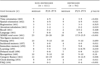
Depressed individuals had lower mean total MMSE and subscale scores, namely, time and spatial orientation, attention/calculation, and language. They also performed worse on the incidental and immediate memory and learning tasks of the ten-figure test. Subjects with depression performed significantly worse on the category fluency test, clock-drawing test and FAQ (Table 1).
Comparison of cognitive tests and subscales between depressed and non-depressed individuals

*MMSE = Mini-Mental State Examination; FAQ = functional activities questionnaire.
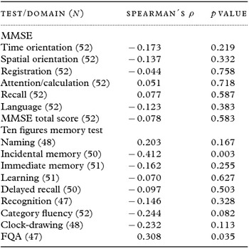
Among the 54 depressed individuals, symptom severity, as measured by GDS-15 score, was correlated negatively with incidental memory (ρ = −0.412; p = 0.003) and positively correlated with FAQ score (ρ = 0.308; p = 0.035), as shown in Table 2.
Correlation between Brief cognitive battery tests’ results and symptom severity measured by GDS-15 among depressed individuals

*MMSE = Mini-Mental State Examination; FAQ = functional activities questionnaire.
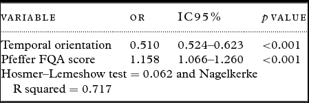
In the multiple logistic regression analysis adjusted for age, gender, years of schooling, marital status, and retirement status, only temporal orientation and Pfeffer's FAQ score remained independently associated with major depression (Table 3).
Multivariate regression analysis final model

Discussion
Our findings suggest that LLD was a prevalent condition in the oldest-old population and that individuals with LLD may present significant cognitive dysfunction. Our data point to the possibility that these cognitive deficits may be identified based on the results of a BCB. Temporal and spatial orientation, attention/calculation, language, immediate, and incidental memory, and executive and visuospatial function were all impaired in depressed individuals. Symptom severity was negatively correlated with incidental memory and functional disability, as measured on the FAQ.
The study sample was representative of the oldest-old Brazilian population, who has been found to exhibit low levels of education and a female preponderance (Instituto Brasileiro de Geografia e Estatística, 2012). In this study, the majority of depressed individuals were women, as has been observed in most LLD studies (Luppa et al., Reference Luppa2012).
The LLD prevalence observed in our sample was between the prevalence rates previously reported by other Brazilian authors. In the study conducted by Xavier and colleagues, 7.5% of the community-dwelling octogenarians sample was diagnosed with LLD (Xavier et al., Reference Xavier, Ferraz, Bertolucci, Poyares and Moriguchi2001), while Costa and coworkers described a prevalence of 15.4% in Bambuí, MG (Costa et al., Reference Costa, Barreto, Uchoa, Firmo, Lima-Costa and Prince2007). Our value was slightly higher than the mean prevalence (7.2%) reported in the meta-analysis conducted by Luppa and colleagues. This discrepancy may be explained, in part, by the use of different instruments used for diagnosis and greater female presence observed in our sample of depressed individuals. In that meta-analysis, the pooled female:male ratio was between 1.4 and 2.2, while in our sample, the gender ratio was 3.5 (Luppa et al., Reference Luppa2012).
In our study, global cognitive functioning was worse in depressed individuals, as indicated by the lower MMSE total scores observed in affected individuals. This finding is in accordance with the results of the study conducted by Butters and colleagues, which showed that individuals with LLD performed poorer on the MMSE (Butters et al., Reference Butters2004). These authors, as well as Woo and colleagues (Woo et al., Reference Woo, Rice, Legendre, Salmon, Jeste and Sewell2004), showed that visuospatial and executive functions were also impaired, as indicated by the results of the clock-drawing test in this study.
Orientation problems could stem from attentional deficits, which is in accordance with the results identified on the attention/calculation subscale of the MMSE. Additionally, it is well known that depressed subjects demonstrate slowed time experience, which could further contribute to disorientation (Bschor et al., Reference Bschor2004). The MMSE language tests, by nature, are highly dependent upon intact attentional function. As such, the observed deficits are in line with the aforementioned attentional dysfunction.
Depressed patients performed worse on the category fluency test, a test that predominantly depends upon executive function and processing speed. Using different instruments, Butters and coworkers demonstrated the presence of highly significant impairments in processing speed and executive function in LLD patients. Additionally, the authors observed overall language impairment in LLD subjects, but between-group differences were only significant in the Boston naming test results and did not reach statistical significance in the results of the animal fluency test (Butters et al., Reference Butters2004).
In a study of men over the age of 75 years, Chang and colleagues could not identify any difference between healthy and depressed subjects in the MMSE recall memory subscale (Chang et al., Reference Chang2013), which is in accordance with findings of our work. In a study conducted by Elderkin-Thompson, the results of the delayed recall memory tests did not differ significantly between depressed and control individuals. These authors proposed that this finding may be explained by the absence of true hippocampal dysfunction and preferential involvement of subcortical-frontal circuits in LLD (Elderkin-Thompson et al., Reference Elderkin-Thompson, Moody, Knowlton, Hellemann and Kumar2011). This alleged subcortical-frontal impairment could underpin the origin of the observed attentional and executive dysfunctions and, thus, impair incidental and immediate memory, as shown by our results.
Symptom severity, as measured by the GDS-15, was negatively correlated with the results of the incidental memory task of the figure memory test, which is in agreement with another Brazilian study (Silva et al., Reference Silva2014). As could be expected, depressed patients, probably due to mood and cognitive symptoms, showed worse functionality, as measured by the FAQ. This finding is in accordance with the psychometric properties of this tool that were reported by another Brazilian group (Assis et al., Reference Assis, de Paula, Assis, de Moraes and Malloy-Diniz2014).
Temporal orientation and FAQ score were the only variables independently associated with LLD in the final multiple regression model. Although the direction of this relationship cannot be inferred due to the cross-sectional nature of the study, it seems reasonable to hypothesize that LLD independently contributed to the development of temporal disorientation and functional impairment. This hypothesis is corroborated by previous research that has shown functionality to be both directly and indirectly impaired in depressed individuals in a process mediated by cognitive impairment (Yen et al., Reference Yen, Rebok, Gallo, Jones and Tennstedt2011).
This is one of the very few studies yet conducted to evaluate cognitive function in oldest-old individuals with LLD. The main strengths of this study reside in the inclusion of a large community-based sample of participants that was representative of the Brazilian oldest-old population and carefully evaluated by experienced physicians. We would also like to note that the research subjects went through a cognitive battery that comprehensively assessed the most important cognitive domains.
The study limitations include the absence of formal neuropsychological confirmation of the observed deficits and lack of depression treatment status data. Additionally, the cognitive battery we used was not specifically designed to address cognition in individuals with depressive syndromes, as no such tests exist at present.
On the other hand, some strengths of the study must be highlighted. The diagnosis of LLD was made via semi-structured interviews performed by experienced physicians; this is different from other studies wherein the diagnosis of LLD relied on lay interviewer application of scales. Furthermore, dementia was diagnosed after the performance of comprehensive cognitive and functional evaluations and established by discussions between study clinicians/researchers until consensus was reached.
Screening elderly individuals with depression using cognitive tools, either to rule out the presence of a neurodegenerative disorder with comorbid depressive symptoms or to detect and/or quantify cognitive symptoms in LLD, is an important step in clinical practice. Moreover, cognitive symptoms may be objectively monitored throughout the course of antidepressant treatment, eventually leading to specific rehabilitative measures. In this scenario, it is of particular interest that physicians can perform cognitive evaluation using time-constrained tools and, therefore, provide better mental healthcare to the oldest-old.
Conclusion
In conclusion, our findings suggest that LLD may occur in conjunction with cognitive dysfunction in the oldest-old and that these deficits may be identified based on the results of a BCB.
Conflicts of interest
None.
Authors’ roles
A. L. Teixeira, M. T. Barbosa, and P. Caramelli conceived and designed the study. A. L. Teixeira, H. Guimarães, M. T. Barbosa, E. Resende, R. Beato, K. Carmona, and P. Caramelli collected the data and revised the article. F. Dias was responsible for carrying out the statistical analysis and wrote the article.
Acknowledgments
We acknowledge the partnership from Instituto Hermes Pardini and Laboratório Geraldo Lustosa. We greatly thank Maria Ângela Dutra Ornellas for the local coordination of the operational arrangements. We are deeply grateful to the City Hall, the Municipal Health Department and the Retired and Pensioner Pavilion from Caeté (MG) for their continuous support to the Pietà study and also to all elderly participants and their families for the generous collaboration and engagement on the research project. The Pietà Study, from which this paper derives, took place in Caeté (MG), Brazil, and is partially supported by the CNPq (Conselho Nacional de Desenvolvimento Científico e Tecnológico - National Counsel of Technological and Scientific Development) and FAPEMIG (Fundação de Amparo à Pesquisa de Minas Gerais – State foundation for research support of Minas Gerais) A. L. Teixeira and P. Caramelli are funded by CNPq, Brazil.