Case report
We experienced two cases of patients with frontotemporal lobe degeneration (FTLD) who showed variants of primary progressive aphasia (PPA) (Gorno-Tempini et al., Reference Gorno-Tempini2011). Patient 1 and 2 showed semantic and logopenic PPA, respectively. Semantic variant PPA is classified as one subtype of FTLD, while logopenic variant PPA is considered Alzheimer's disease or FTLD-associated PPA. Alzheimer's disease was excluded in both patients using Pittsburgh compound B (PiB) – positron emission tomography (PET) amyloid imaging. We aimed to detect atrophy of frontal or temporal lobes using MR images, and performed (18F)-fluoro-D-glucose (FDG)-PET, because hypometabolic activity in the regions where cortical atrophy was observed support the diagnosis of frontotemporal dementia-related PPA (VanDreel et al., Reference VanDreel, Aftab, Sanan, Grant and Lu2016). The major molecular classes of FTLD are FTLD-tau, FTLD-TDP, FTLD-UPS, FTLD-FUS, and FTLD-ni (Mackenzie et al., Reference Mackenzie2010). We hypothesized that the neuropathologic subtypes of these two patients are classified into FTLD-tau; hence we performed 18F-THK-5351 PET. Tau deposits in Alzheimer's disease and corticobasal syndrome have been reported using 18F-THK-5351 PET (Dani et al., Reference Dani, Edison and Brooks2016; Kikuchi et al., Reference Kikuchi2016). To our knowledge, this is the first report showing tau deposits in PPA variants of FTLD using 18F-THK-5351 PET.
Written informed consent was obtained from both patients and their spouses for publishing this report including clinical and imaging data and cognitive estimations.
Patient 1
Patient 1 was a 69-year-old, right-handed woman living with her husband. Her husband claimed that approximately five years ago, she experienced difficulty in recalling words, such as the names of her friends. The patient's duration of education was 12 years. She had visited a neurosurgery clinic, but no particular finding was reported using brain magnetic resonance (MR) imaging. Approximately, two years ago, she started pausing in conversations with her husband. During this time, she still managed to perform household tasks.
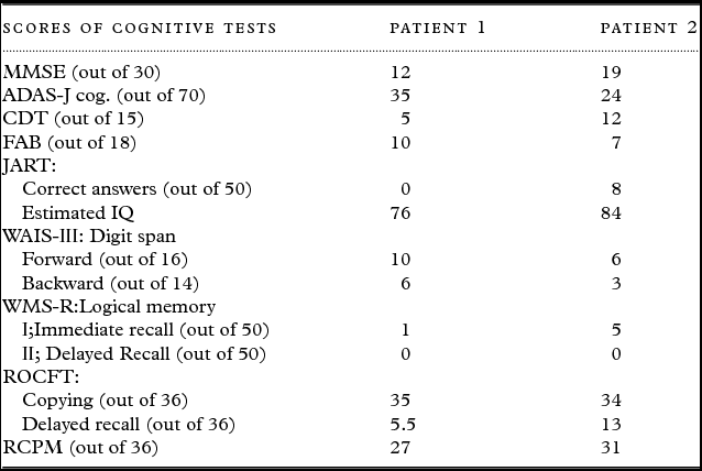
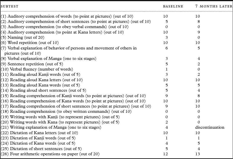
Patient's cognitive ability was assessed using Mini-Mental State Examination (MMSE), cognitive subscale of Alzheimer's Disease Assessment Scale-Japanese version (ADAS-J cog.), Clock-Drawing Test (CDT), Frontal Assessment Battery (FAB), Japanese Adult Reading Test (JART), digit span subtest in the Wechsler Adult Intelligence Scale-Third edition (WAIS-III), logical memory subtest in the Wechsler Memory Scale-Revised (WMS-R), Rey–Osterrieth Complex Figure Test (ROCFT), Raven's Coloured Progressive Matrices (RCPM) (Sugishita and Yamazaki, Reference Sugishita and Yamazaki1993), and Japanese Standard Language Test of Aphasia (SLTA) (Mimura et al., Reference Mimura1998) (Tables 1 and 2). The SLTA comprises 26 subtests. Changes in all SLTA subscale scores, which were repeated after a seven-months interval, are shown in Table 2. The total neuropsychiatric inventory (NPI) score was zero (Cummings et al., Reference Cummings, Mega, Gray, Rosenberg-Thompson, Carusi and Gornbein1994). No parkinsonism was observed.
Cognitive evaluations

MMSE: Mini-Mental State Examination, ADAS-J cog: Alzheimer's Disease Assessment Scale-Japanese version, CDT: Clock Drawing Test, FAB: Frontal Assessment Battery, JART: Japanese Adult Reading Test, WAIS-Ⅲ: Wechsler Adult Intelligence Scale-Third edition, WMS-R: Wechsler Memory Scale-Revised, ROCFT: Rey–Osterrieth Complex Figure Test, and RCPM: Raven's Coloured Progressive Materices
The results of all subtests of SLTA in patient 1

SLTA: Japanese Standard Language Test of Aphasia
She fluently spoke, but all confrontation naming, single-word comprehension, and object knowledge for items were impaired when accessed using lexical processing in aphasia (TLPA) test (Fujita et al., Reference Fujita2000). Semantic aphasia was observed using TLPA at baseline, though it was not clearly observed using SLTA at both baseline and seven months later (Table 2). Surface dyslexia was observed using the JART and SLTA subtest (reading Kanji words aloud) (Table 2). Moreover, dysgraphia of Kanji and Kana was observed using SLTA subtests (Table 2). Word repetition and speech production were spared, which was observed in the results of SLTA (Table 2). Her speech production was spared in grammar and motor speech.
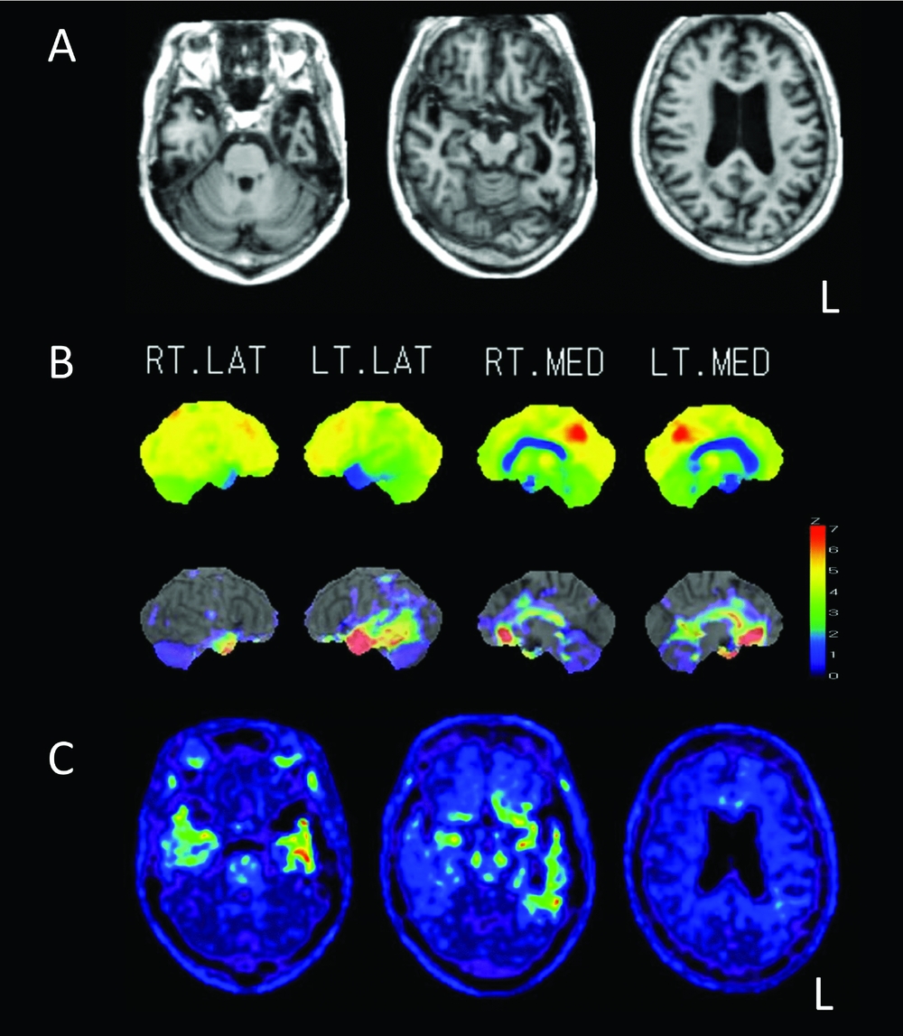
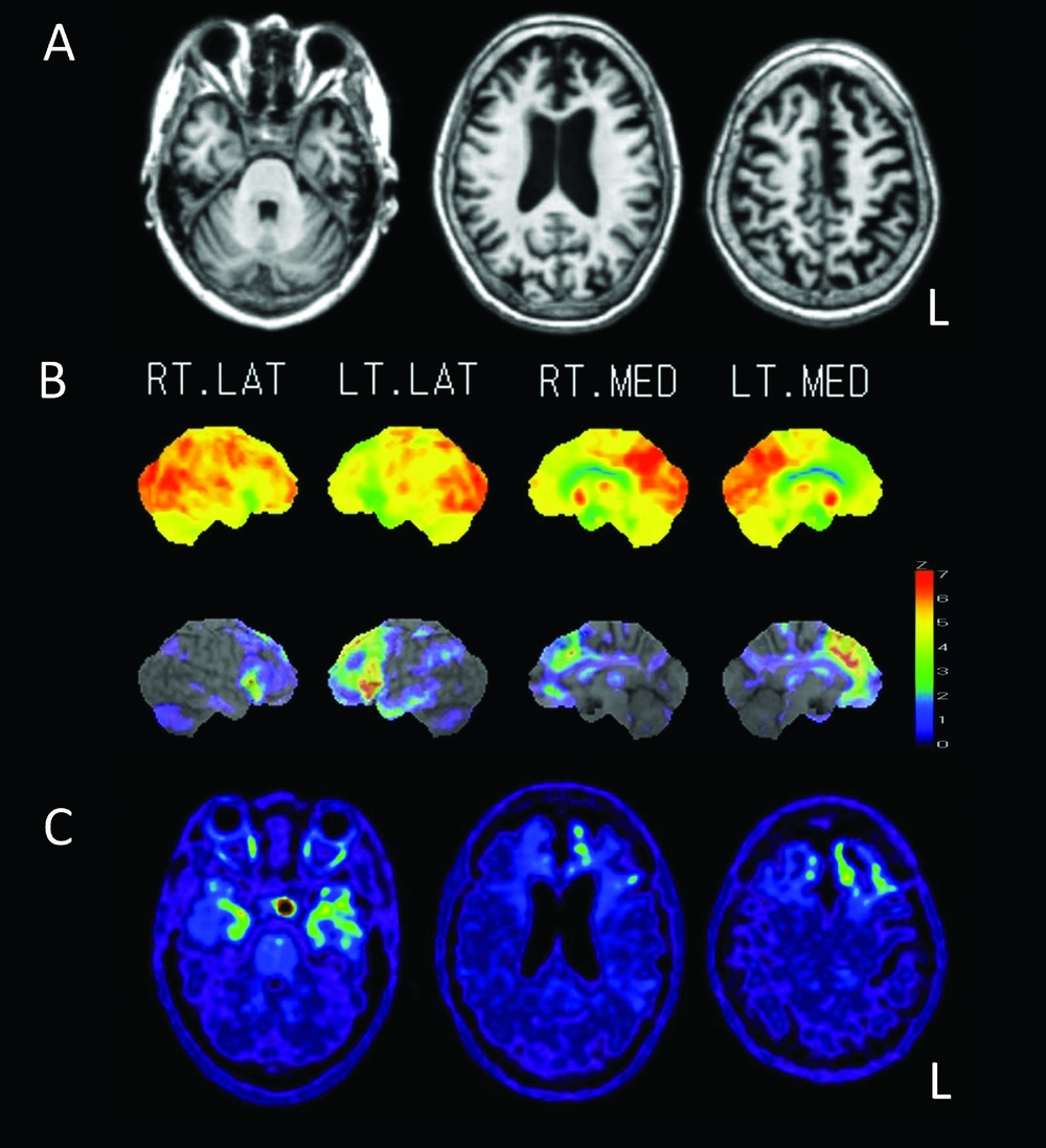
Brain MR images showed diffuse cerebral cortical atrophy. Particularly, atrophy of left anterior inferior and middle temporal gyri was significant (Figure 1(A)). (18F)-FDG-PET images showed reduced glucose metabolism in the temporal and parietal lobes, and in both anterior temporal lobes, with a particular severe reduction in the left anterior temporal lobe. However, glucose metabolism in the posterior cingulate gyrus and precuneus was preserved (Figure 1(B)). PiB-PET amyloid images showed negative amyloid deposition in cerebral cortices. A neuroradiology specialist (KI) read the PiB-PET amyloid images. The criteria are as follow: PiB positivity was evaluated in four cortical regions, including frontal, parietal, and temporal lobes (neocortical association areas) and precuneus/posterior cingulate gyri. According to PiB positivity in each region, each patient was classified as PiB positive, PiB equivocal, or PiB negative. For each region, PiB-positive images were defined as those having a higher accumulation in the cerebral cortex than in the white matter (non-specific accumulation area), and PiB-negative images as those having no cortical accumulation. When cortical accumulation was suspected, but accumulation was not higher in the cortex than in the white matter, an ‘‘equivocal’’ rating was assigned (Hosokawa et al., Reference Hosokawa2015).
Patient 1 (A) MRI T1-weighted axial images, (B) surface map and Z score map of [18F]-fluoro-D-glucose-PET image, (C) 18F-THK-5351 PET images.

Patient 1 was diagnosed with semantic variant PPA according to the criteria (Gorno-Tempini et al., Reference Gorno-Tempini2011), considering the symptoms mentioned above. Using 18F-THK-5351 PET, tau accumulation was observed in bilateral temporal lobes, predominantly in the left anterior temporal lobes. Moreover, non-specific tau accumulation was observed in the medial temporal lobes, indicating positive tau deposition (Figure 1(C)).
Patient 2
Patient 2 was a 64-year-old, right-handed woman. She lived and visited our clinic with her husband. Her duration of education was 12 years. Her husband claimed that she began to have speech difficulties approximately five years ago, which he thought was to be due to his company's financial difficulties. Moreover, he claimed that she sometimes cooked messily or in a wrong way, which was not observed in the past.
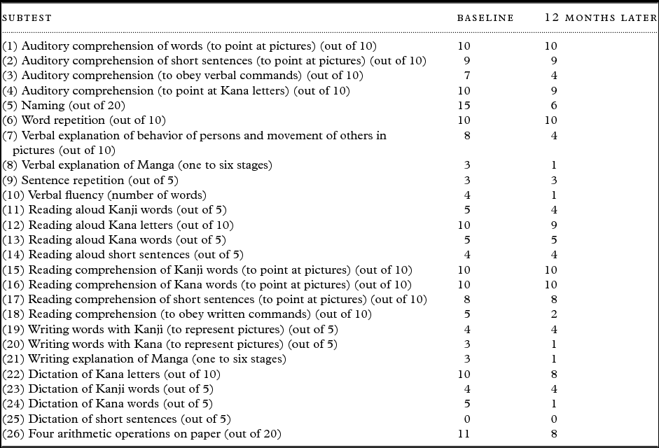
Patient's cognitive ability was assessed using MMSE, ADAS-J cog., CDT, FAB, JART, digit span subtest in the WAIS-III, logical memory subtest in the WMS-R, ROCFT, RCPM, and SLTA (Tables 1 and 3). Changes in the scores of all subtests of SLTA, which was repeated after a 12-month interval, are shown in Table 3. The total NPI score was zero. No parkinsonism was observed.
The results of all subtests of SLTA in patient 2

SLTA: Japanese Standard Language Test of Aphasia.
She had difficulty in recalling words in spontaneous speech and naming. Her speech was fluent but sparse and often stopped after a sentence. Single-word retrieval in spontaneous speech and naming was impaired. Impairment in naming was clearly observed using TLPA and SLTA (Table 3). Moreover, repetition of sentences and phrases was impaired. The patient's score of sentence repetition in SLTA subtest was 3/5, although the word repetition subtest score was 10/10 (Table 3). Her single-word comprehension and object knowledge were spared, which was observed using the TLPA, as was her motor speech. Frank agrammatism, dysarthria, and prosodic disturbance were not observed. Phonemic paraphasia and phonologic speech errors in spontaneous speech and naming were not clearly observed.
Brain MR images demonstrated slight atrophy of the bilateral frontal lobe (Figure 2(A)). FDG-PET images showed left-dominant reduced glucose metabolism in the bilateral frontal lobes. The anterior part of temporal lobes and left parietal area showed reduced glucose metabolism (Figure 2(B)). PiB-PET images showed negative amyloid deposits in cerebral cortices. The method and criteria of reading them are described in part of Patient 1.
Patient 2 (A) MRI T1-weighted axial images, (B) surface map and Z score map of [18F]-fluoro-D-glucose-PET image, (C) 18F-THK-5351 PET images.
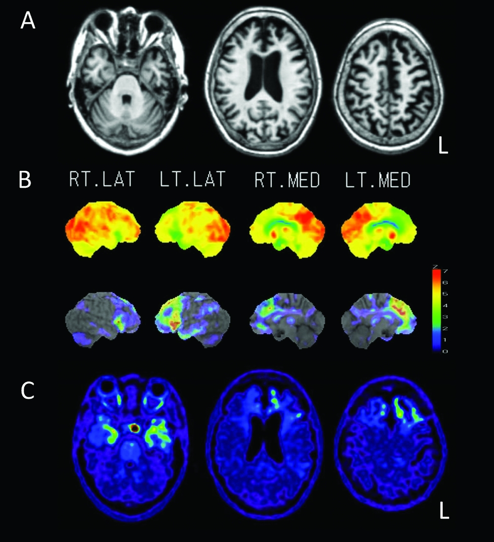
Patient 2 was diagnosed with logopenic variant PPA according to the criteria (Gorno-Tempini et al., Reference Gorno-Tempini2011), considering the symptoms mentioned above. Using 18F-THK-5351 PET, abnormal tau accumulation was demonstrated in the bilateral frontal cortices and left medial frontal cortex. Moreover, increased tau accumulation was observed in the bilateral anterior to medial temporal lobes (Figure 2(C)). Non-specific tau accumulation was observed in brainstem, striatum, thalamus, and hypophysisis.
Conclusion
Our findings suggest an association in the pathology of these three diseases: Alzheimer's disease, corticobasal syndrome, and FTLD. PiB-negative, and Tau-positive images suggested that the patients do not suffer from AD, but perhaps from FTLD-tau. Therefore we can presume that the patients’ FTLD symptoms will continue to progress, allowing for advance planning and better, more effective clinical care.
Conflict of interest
None.
Sources of funding
None.
Description of author's roles
MT formulated the research questions, designed this study and carried it out, analyzed the neurocognitive data, and wrote the case report. KI managed the neuroimaing examinations, read and analyzed the neuroimages, and made the figures. CH carried out the neuroimaging examinations. KS and OS provided useful comments for the case report.