INTRODUCTION
An infection during the antenatal period is able to influence the whole course of pregnancy and the immunological relationship between mother and progeny. The neonatal period is marked by a high susceptibility to infection. Innate immunity represents the first barriers to infection and plays a pivotal role in the induction of adaptive immunity (Kovarik and Siegrist, 1998). Materno-fetal active transfer of IgG is mediated by a specific Fc receptor (FcRn) localized in the murine fetal yolk sac and placenta and the intestinal epithelium of suckling mice, as well as in the human placental syncytiotrophoblast (Simister et al. 1996).
Antibodies, parasitic antigens, immune cells, cytokines and probably other factors may be transferred at the same time during the fetal and/or the post-natal life. Each element can have a potentially passive, stimulating or immunosupressive effect, depending on its concentration, on offspring reactivity and on the time of transfer. Besides protective effects, maternal infection may weaken the offspring's immune response (Carlier and Truyens, 1995). The vertical transmission of a large number of parasites generally leads to congenital infection (Brabin and Brabin, 1992).
Larval toxocariasis belongs to global zoonoses with both transplacental and lactogenic transmission of the causative agent in paratenic hosts, including humans (Glickman and Magnaval, 1993). In the definitive host transplacental transmission is a component part of the parasite life-cycle. The response to larval toxocariasis is an immunopathogenic character with dominating eosinophilia, hypergammaglobulinaemia, enhanced proliferative responses of T and B cells (Kayes, 1997; Takamoto et al. 1998; Dent et al. 1999). The primary function of eosinophils is the protection against parasites, although there is little unequivocal in vivo evidence to prove this (Gon et al. 1997). Eosinophils are also responsible for considerable pathology in mammals because they are inevitably present in large numbers in inflammatory lesions associated with helminth infections or allergic conditions (Behm and Ovington, 2000).
Stimulation or/and suppression of T-cell subpopulations due to T. canis infection during gestation is highly dependent on the period of infection. The elevated production of INF-γ (Th1) correlates with a high rate of miscarriages in infected mouse mothers (Reiterová, Tomasovicová and Dubinský, 2003). Specific features of both innate and acquired early-life immunity have to be carefully dissected. It appears that intrinsic deficiencies of a given lineage may not prevent the induction of effective immune responses if specific activation requirements – distinct from those of later life – are met (Kovarik and Siegrist, 1998).
The aim of the present study was to examine in mice the influence of T. canis infection of mothers during pregnancy on their offspring's resistance towards re-infection in comparison to offspring of non-infected mothers.
MATERIALS AND METHODS
Isolation and maturation of Toxocara canis eggs
The eggs were recovered from the lower part of the uteri of T. canis females which were isolated from naturally infected puppies and incubated in 0·1 M H2SO4 at room temperature (21±3 °C) until infective larvae developed.
Experimental design
In order to determine the influence of maternal infection on offspring resistance to re-infection we monitored cellular and humoral immune responses in the form of serum cytokines IFN-γ and IL-5 and eosinophilia in peripheral blood. We also looked for the presence of migrating larvae in both groups of the offspring.
Experiments were carried out on pathogen-free inbred Balb/c mice kept under routine laboratory conditions under a 12 h light/dark regime (at 19–21 °C, 50–60% relative humidity and fed ad libitum on a commercial food mixture and water according to standard regulations for animal experiments).
Fifteen males and 15 females, 2 months of age (60±3 days), weighing 25–30 g were mated. One male and 1 female were bred in separate cages. Pregnant mice were orally infected with a single dose of 1000 T. canis eggs on day 15±3 of gestation. The females were checked for conception by examination for the presence of a vaginal plug after mating.
The first experimental group of offspring (Group 1) was composed of 25 female mice originating from infected dams (described above). After weaning, mice were coupled from different litters and separated according to sex.
The second group contained 25 female offspring of 10 non-infected healthy mothers of the same age (Group 2). Both experimental groups of offspring were challenged with 500 T. canis eggs per animal at 6 weeks of age.
The control group was composed of 25 healthy 6-week-old non-infected female mice (Group 3).
The samples were collected on days 1, 7, 14, 21, 28, 35, 42 and 49 after infection of offspring. On the stated days, 3 mice from each group were euthanized and their samples of blood, particular organs (brains, spleens) and skeletal muscles were examined separately.
Flow cytometry
Lymphocytes isolated from mouse spleens were depleted of erythrocytes, washed twice with Dulbecco's-PBS at pH 7·2 (Sigma, USA) and once with RPMI 1640 medium (Sigma) and were then resuspended to a final concentration of 1×10⁶ cells in 1 ml. The cell samples (2·5×105) were incubated with FITC-labelled rat anti-mouse CD4 (Sigma cat. No. F-7400) and phycoerythrin-labelled rat anti-mouse CD8a monoclonal antibodies (Sigma cat. no. P-3067) 4 μl/tube at 4 °C for 30 min. Cells were then washed 3 times with PBS (pH 7·2) containing 0·1% sodium azide and analysed by flow cytometry. Data for 104 cells/sample, falling within forward-scatter (FSC) and side-scatter (SSC) gates set specifically for lymphocytes, were collected from each sample with a FACScan flow cytometer (Becton-Dickinson Biosciences, USA). All data files were analysed with CellQuest software.
ELISA for detection of IFN-γ and IL-5
The cytokine concentrations in the sera of the mice were measured by sandwich ELISA on microtitre plates (Nunc, USA). Interferon-γ (IFN-γ) and interleukin-5 (IL-5) were used as marker cytokines for Th1 and Th2 responses, respectively. Antibody pairs for capture and detection (all biotinylated) of the cytokines studied were as follows: R4-6A2 and XMG1.2 for IFN-γ, TRFK5 and TRFK4 for IL-5 (BD Biosciences, PharMingen, USA). The results were expressed in ng/ml using murine recombinant IFN-γ and IL-5 as standards. The detection limit of the assay for both cytokines was 0·02 ng/ml.
Peripheral blood eosinophil count
Blood smears were prepared individually from each animal. After staining by Hemacolor Rapid (Merck, Germany) eosinophils were counted under the light microscope. The numbers of eosinophils were evaluated as a proportion of 100 white blood cells (%).
Enzyme-linked immunosorbent assay (ELISA) for determination of Toxocara-specific antibody production
The production of various specific antibody classes in mouse sera was determined using an ELISA according to the protocol of Havasiova-Reiterova, Tomasovicova and Dubinsky (1995). Anti-mouse IgM, IgG (Sigma) and IgG1, IgG2a (Serotec Ltd, Oxford, UK) peroxidase-labelled conjugates at a dilution of 1[ratio ]5000 and 1[ratio ]1000, respectively and chromogen o-phenylenediamine (Sigma-Aldrich, Steinheim, Germany) with H2O2 were used as substrate. The reaction was stopped with 2 M H2SO4. Optical densities (OD) were recorded spectrophotometrically at 492 nm (Revelation Quicklink, Opsys MR, Dynex Technologies, USA). Cut-off values were determined individually for each antibody type by taking the average OD of non-infected control mice plus 3 standard deviations (S.D.).
Larval recovery
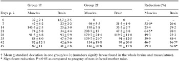
In order to detect the presence of migrating larvae in the infected mice, their brains were collected into Petri dishes, washed with saline and minced. The larvae were isolated by Baermann's method. The samples of muscles were digested according to Velebny, Tomasovicova and Stpiczińska (1992) and larvae were counted under a light microscope. The reduction of larval count was calculated according to the following formula: no. of larvae in second group−no. of larvae in first group/no. of larvae in second group×100 (%).
Data analysis
The experimental results were evaluated statistically in 2 ways: analysis of the reduction in total larval burden was done by χ2-test. Comparison of T-cell subpopulations, cytokines and antibody levels within and among groups was done using paired t-test. The differences among the groups were considered significant at the values of P<0·05 and P<0·001.
RESULTS
CD4+ and CD8+ T cells
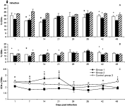
In both experimental groups (Groups 1 and 2) a lower percentage of CD4+ T lymphocytes was detected in comparison to control Group 3, with a significant decrease (P<0·05) at 7, 14 and 35 days p.i. (Group 1), as well as 1, 7, 42 and 49 days p.i. (Group 2). Also their percentage occurrence in Group 1 was not significantly lower from days 7 to 35 p.i., with a successive significant (P<0·05) increase, compared with the second group (Fig. 1A).

Fig. 1(A–C). Occurrence of spleen CD4+ (A), CD8+ (B) and CD4+/CD8+ T lymphocytes in infected offspring (C) originating from Toxocara canis-infected and non-infected mothers and of control healthy Balb/c mice. Results are expressed as the mean percentage of lymphocytes±S.D. (n=3/group). Statistically significant differences (P<0·05) compared between Groups 1 and 2 (+); between Groups 1 and 3 (×); between Groups 2 and 3 (*). □ represents offspring of infected mothers (Group 1); [squf ] represents offspring of non-infected mothers (Group 2); represents healthy mice (Group 3).
In the first group of mice the CD8+ population was gradually increased after an initial decrease from days 7 to 14 p.i., while in the second group the CD8+ population appeared to be suppressed at the beginning of the experiment with a subsequent increase and with a maximum on day 35 p.i. (Fig. 1B). In healthy control mice the concentration of both subpopulations balanced at the mean level of 30·84±3·59% for CD4+ and 13·62±1·69% for CD8+ T cells, respectively without any significant differences (P>0·05) (Fig. 1A,B).
The CD4+/CD8+ ratio, designated as the immunoregulation index, in offspring from infected mothers appeared to be suppressed over the first 3 weeks with significant differences (P<0·05), compared with offspring of the non-infected mothers. However, after this period the immunoregulation index of the second group declined quickly and approached the ratio of the first group. The CD4+/CD8+ ratio in first group of offspring was consistently reduced with significant differences (P<0·05) almost throughout the whole experiment, compared to the healthy control group 3. The exception was on days 28 and 35, when no significant differences were observed (P>0·05) (Fig. 1C).
IFN-γ and IL-5
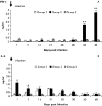
In the offspring of infected mothers (Group 1) the production of serum IFN-γ was not observed throughout the whole experiment. However, in the offspring of non-infected mothers a significant elevation (P<0·001) of this cytokine was detected after day 42 p.i. (Fig. 2A). The production of IL-5 in Group 1 was lower, when compared with Group 2, with significant (P<0·05) differences on days 1 and 7 p.i. (Fig. 2B).

Fig. 2(A–B). Concentration of serum IFN-γ (A) and IL-5 (B) in offspring from Toxocara canis-infected (white and black bars, respectively) or non-infected mothers (grey and shaded bars, respectively) after challenge infection. Data are expressed as the mean concentration of cytokines (ng/ml)±S.D. (n=3/group). Asterisks indicate time-points showing significant differences between both groups of infected offspring (* P<0·05, ** P<0·001).
Peripheral blood eosinophilia
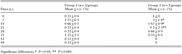
The number of peripheral blood eosinophils in offspring originating from infected mothers appeared to be suppressed in comparison to the second group, with significant differences (P<0·05) from days 7 to 21 p.i. In offspring of non-infected mothers the relative numbers of eosinophils in the bloodstream increased from the beginning to day 28 p.i. with peaks on days 7 and 21. Subsequently the numbers of these cells returned to normal (Table 1).

Parasite-specific IgM, IgG, IgG1 and IgG2 response
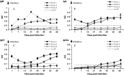
The IgM response of the offspring of infected mothers increased from the first week post-infection and reached its maximum level (P<0·001) on day 21, followed by a gradual decline. In the offspring of non-infected mothers this class of immunoglobulins was detected later, always at a lower level compared with the Group 1 (Fig. 3A).

Fig. 3(A–D). Dynamics of specific IgM (A), IgG (B), IgG1 (C) and IgG2a (D) antibodies in offspring originating from Toxocara canis-infected and non-infected mothers after challenge with 500 T. canis eggs per mouse. Data are arithmetic means of OD values (±S.D.) of 3 mice per infected group. ○ represents infected mothers Group M; [squf ] offspring of infected mothers (Group 1); [utrif ] offspring of non-infected mothers (Group 2).
IgG-specific antibodies in Group 1 slowly increased until the end of the experiment following a temporary decline up to day 14. The seroconversion in the second group occurred later with a gradual increase and reached its maximum level (P>0·05) only on day 49 p.i. (Fig. 3B).
In the offspring of infected mothers the level of specific IgG1 response (passively transmitted from mothers) increased until the end, following a temporary decline. The production of this subclass of specific immunoglobulins came about much later in the second group with a significantly (P<0·05) lower level (Fig. 3C).
The specific IgG2a antibody response was generally weaker. In the offspring of infected mothers it increased after day 28 p.i., whereas in the offspring of the second group it was detectable only after day 49 p.i. (Fig. 3D).
Larval recovery
In the first group of offspring 32±2·1 larvae in the muscles and 12·7±2·5 larvae in the brains, transmitted from the infected mothers, were detected before challenge. A striking reduction of T. canis larvae count was recognized in the first experimental group of offspring (Group 1) compared to the second (Group 2). A significant reduction (P<0·05) of larval numbers was observed in brains on day 42 and 49 p.i. (56·7% and 56·8%, respectively), while in muscles at the same time, a lower reduction, 46·7% and 39%, respectively was detected. The highest reduction (P<0·05) in the offspring's muscles was observed on days 7 and 21 p.i. (Table 2).

DISCUSSION
The present study analysed the influence of maternal Toxocara canis infection on the offspring immune response and later susceptibility to challenge infection. The cellular immune response in T. canis infection depends on two of the most important subpopulations of T cells with the membrane markers of CD4+ and CD8+ (Kayes, 1997). The helper CD4+ T lymphocytes are essential for the activation, differentiation, and isotype regulation of B lymphocytes and are of a significant help in antigen presentation. After antigen stimulation they release a whole range of cytokines, as regulatory mediators that determine the development of the immune response into Th1 or Th2 type (Pichler, 1997). On the other hand, the cytotoxic CD8+ T lymphocytes are responsible for the destruction of structures carrying an antigen on their surface. They also participate in the inhibition of the immune response and regulate eosinophilia (Owhashi, Arita and Niwaq, 1998).
Under physiological conditions the relative proportion of both of these subpopulations (immunoregulation index) in a host organism is relatively balanced (Landers et al. 1991). The changes in their numbers and proportion in the blood and in the organs may influence the production of serum cytokines and therefore disturb the immune balance and usually manifest in malfunctions of immunoregulation. Their determination becomes an important diagnostic tool also in infectious diseases (Levkutova et al. 1998). We observed changes in the proportion of both subpopulations after challenge infection. In both experimental groups a lower percentage of CD4+ T lymphocytes was observed in comparison to the control group. On the other hand, the CD8+ population appeared to be suppressed in the beginning followed by a subsequent increase. In healthy control mice the concentration of both subpopulations was relatively balanced. The CD4+/CD8+ ratio was lower in the offspring of infected mother mice throughout the whole experiment, compared to the young of non-infected mothers as well as to the healthy control group of mice of the same age. In accordance to our results, Aldawek et al. (2002) observed a significant increase of the spleen CD8+ cells and a significant decrease of CD4+ cells after a multiple high-dose infection of mice with T. canis eggs. According to Dvoroznakova, Boroskova and Tomasovicova (2002) immunization of mice with excretory-secretory and somatic antigens of T. canis and subsequent infection induced a predominantly Th2 response within the first 2 weeks of the experiment with a decrease of CD4+ T cells. Later, this type of response switched to Th1.
The present study also focused on the dynamics of 2 inductive cytokines (IFN-γ and IL-5) known as the dominant cytokines in Th1 and Th2 immune responses in larval toxocariasis. We observed a striking decrease in IL-5 concentration, which is closely related to the depletion of CD4+ T splenocytes primarily responsible for the production of this cytokine. Consequently, eosinophil maturation might also be inhibited. It is well known that the accumulation of eosinophils is a key pathogenetic factor in helminthiases. T. canis induces persistent peripheral eosinophilia (Glickman and Schantz, 1981) that markedly increases after infection. It usually reaches its maximum at 10 days p.i. Takamoto et al. (1998) recognized that, in mutant mice deficient in CD4+ T cells and their normal and heterozygous littermates infected with T. canis, the first peak of eosinophilia (on day 10) is CD4 independent and the second peak (on day 21) is CD4 dependent. We observed that eosinophil levels tended not to increase in offspring primarily infected from mothers, in contrast to the second group with a single infection. In those, the eosinophil count increased to peak on day 21, with a subsequent decline. Other investigators observed an earlier peak of eosinophils in peripheral blood in T. canis-infected rats, with a sequential decline (Okada et al. 1996).
Specific antibody production already starts in the early stage of gestation but, due to the sterile surroundings of the fetus its level is very low. The antibodies present in offspring blood originate from the mother and belong only to the IgG class (Buc, 1997). We detected noticeable changes in the production of different antibody classes. However, we observed a rapid IgM secondary immune response in the young from infected mother mice, but its level rapidly declined. Interestingly, the IgG antibody class showed similar patterns in both groups with a non-significant lower level in mice with a single infection. Machnicka-Rowinska et al. (2002) detected a higher level of IgG in sera of mice after multiple infections in comparison with mice after a single infection.
The IgG1 antibody subclass is mainly directed against proteins (somatic antigens of dead larvae) and belongs to the Th2 type of response. IgG1 antibodies in sensitized offspring originated from mothers. After an initial decline, the concentration of these passively acquired antibodies began to increase and remained stable for the remainder of the experiment. Interestingly, in the second group of mice, the seroconversion of this antibody subclass came much later with a gradual elevation.
Secretion of IgG2a isotypes is enhanced by Th1-dependent IFN-γ (Snapper, Finkelman and Paul, 1988). This type of antibody is predominantly produced against polysaccharide components of antigen (Buc, 1997). The sensitization with T. canis larvae, which were lactogenically transmitted from mothers, induced in challenged offspring a more efficient immune response compared to the controls. In the second group of offspring IgG2a started to be produced 3 weeks later. The physiological fetal environment engages the neonatal cells preferentially in a CD4+ Th2-cell response (Bendelac et al. 1992) with increasing production also of IgG1 in mice, involved in the response to helminth infections. There appears to be a Th2-cell-biased offspring response in parasitosis (Carlier and Truyens, 1995). In this high-responder murine Balb/c strain both Th1 and Th2 cells are active, although with a Th2-dominant response during the T. canis infection (Cuellar et al. 2001). Offspring challenged at 6 weeks post-partum, in comparison to offspring after a single infection, had a prompt humoral immune response of Th2 type. These results are associated with the protection resulting in a high reduction of migrating somatic larvae in the challenged group.
The striking immune response is only one side of larval toxocariasis. Another high-risk factor is represented by vertically-transferred migrating larvae. In children, potential paratenic hosts, the most dangerous phenomenon could be the accumulation of larvae in the brain, which may have an unfavourable impact on their mental development (Glickman et al. 1981). Recent studies discovered alterations in the behaviour of Toxocara-infected mice, which were associated with the number of larvae recovered from the brain. Toxocara infection had an impact upon a diverse range of murine behaviours, with little evidence of a specific and hence an adaptive alteration. Many of the effects on murine host behaviour by Toxocara are likely to be pathological side-effects of infection rather than a consequence of adaptive host-manipulation. The offspring of infected mothers were hypo-responsive when mother mice were infected in early pregnancy, but they showed high immune responsiveness when infection was induced in late pregnancy. The changes in murine behaviour may be relevant to human toxocariasis (Holland and Cox, 2001).
Congenital infections may not manifest clinically, however, they could modulate the immune response for a long time after re-infection of offspring by the same parasite. Oteifa et al. (1996) found that exposure to infection during pregnancy, whether early or late, led not only to transmission of larvae into the fetus but also to modulation of its immune response and the course of infection. After infection with different infective doses of T. canis eggs Farjat et al. (1995) observed delayed larval arrival at the brain in an inverse relationship to the inoculums but the percentage of recovery was about 7 and 8%. Abo-Shehada, Al-Zubaidy and Herbert (1991) showed that acquired immunity which developed against T. canis infection in twice-infected mice resulted in a significant reduction (27%) in the number of larvae recovered from the brain compared with once-infected mice. Dvoroznakova, Boroskova and Tomasovicova (2002) observed a higher protective effect of immunization with ES antigen (52·1% in brain and 29·0% in muscles), than with somatic antigen – resulting in a 44·2% reduction of larvae in the brain and 16·0% in muscles.
In the present experiments, the offspring of infected mothers were primarily immunized with antigens from mothers. We expected that offspring would be more susceptible to the challenge infection. In contrast, immunization of the fetus during prenatal development caused a significant protective reaction in offspring with a high reduction in the number of migrating larvae. These results suggest that prenatal immunization accelerated the cell-mediated immune responses and enhanced the ability of immunocompetent cells to reduce the number of Toxocara larvae as early as the first week after infection, with a mean of 40% reduction in the muscles and 38% in the brain. Similarly, Hrckova and Velebny (2001) observed a very high reduction of larval counts only in non-immunized mice with a single infection of T. canis eggs after the treatment with liposomized albendazol in combination with immunomodulator glucan (54% in the brain and 37% in the muscles).
In conclusion, our results revealed a lower percentage of CD4+ T lymphocytes in both experimental groups in comparison to the control group, a higher production of cytotoxic CD8+ T lymphocytes in offspring from infected mothers in comparison with the control group; a suppressed production of both observed cytokines; a synergic effect of maternal and memory antibodies after re-infection and a significant reduction in the number of migrating larvae. However, this protection was only partial and did not allow complete elimination of the parasite.
A better knowledge of this fascinating topic, particularly of the complex mechanisms underlying the immune response to parasites in progeny, could have considerable implications in public health.
This work was supported by the Slovak Science Grant Agency (VEGA), Grant No. 2/4179/04 and partially financed by the State programme ‘Genomics of transmissible diseases for healthier human and animal populations’ No. SP 51/028 08 00/028 08 03. We would like to thank Professor Dr Bruno Gottstein of the University of Bern, Switzerland, for his helpful suggestions and pre-reviewing of this paper.