INTRODUCTION
Purinergic receptors within the central nervous system play an important role in communication between neuronal and glial circuits and also within astrocyte networks (Fields and Burnstock, Reference Fields and Burnstock2006; North and Verkhratsky, Reference North and Verkhratsky2006). Adenosine triphosphate (ATP) is released from astrocytes and acts as a widespread gliotransmitter, triggering and maintaining calcium signalling and calcium oscillations. Purinergic receptors initiate release of Ca2+ from internal stores (Bernstein et al., Reference Bernstein, Behnisch, Balschun, Reymann and Reiser1998).
A variety of purinergic (P2) receptors are expressed on astrocytes that are activated by ATP released from neighbouring astrocytes or neuron terminals during synaptic transmission.
Although there are some studies on the role of purines in mammalian hippocampal long-term potentiation (LTP) as a part of the possible physiological mechanism underlying learning and memory (Yarbrough and McGuffin-Clineschmidt, Reference Yarbrough and McGuffin-Clineschmidt1981; Wieraszko and Seyfried, Reference Wieraszko and Seyfried1989; Pankratov et al., Reference Pankratov, Lalo and Krishtal2002; Yamazaki et al., Reference Yamazaki, Fujii, Nakamura, Miyakawa, Kudo and Kato2002; Almeida et al., Reference Almeida, Rodrigues, de Mendonca, Ribeiro and Cunha2003; Gimenez-Llort et al., Reference Gimenez-Llort, Masino, Diao, Fernandez-Teruel, Tobena and Halldner2005; Rex et al., Reference Rex, Kramar, Colgin, Lin, Gall and Lynch2005; Serrano et al., Reference Serrano, Haddjeri, Lacaille and Robitaille2006), there have been few studies using in vivo animal behaviour models (e.g. Yarbrough and McGuffin-Clineschmidt, Reference Yarbrough and McGuffin-Clineschmidt1981). In the present study we use the post-hatch chick model for investigating the role of ATP released from astrocytes and P2Y1 receptors in the hippocampus in learning. In a recent study using a non-selective inhibitor of purinergic receptors (suramin) and the P2Y receptor antagonist PPADS (pyridoxal phosphate-6-azo-(benzene-2,4-disulphonic acid), we have shown the involvement of purines in memory in the cortical region of the chick brain (intermediate medial mesopallium (IMM)) (Cronin et al., Reference Cronin, Edwards and Gibbs2011).
Over the last 7 years, there have been a number of studies that have shown roles for astrocytes at the level of synapses, circuits and behaviour in organisms ranging from flies to mammals (Pascual et al., Reference Pascual, Casper, Kubera, Zhang, Revilla-Sanchez and Sul2005; Halassa et al., Reference Halassa, Fellin and Haydon2009; Ng et al., Reference Ng, Tangredi and Jackson2011). In the chick, we have shown that astrocytes are intimately involved in memory and learning (Gibbs et al., Reference Gibbs, Hutchinson and Hertz2008a,Reference Gibbs, Bowser, Hutchinson, Loiacono and Summersb; Gibbs and Bowser, Reference Gibbs and Bowser2009, Reference Gibbs and Bowser2010). Astrocytes modulate synapse development, synaptic transmission and neuronal excitability (Haydon, Reference Haydon2001). They express a variety of neurotransmitter receptors and envelop synapses. Neurotransmitters that spill out of the synaptic cleft activate these receptors on the astrocyte cell membrane, activating increases in cytosolic Ca2+ levels and triggering release of vesicles containing neuromodulatory gliotransmitters including l-glutamate, ATP and d-serine (e.g. Haydon, Reference Haydon2001). Although it is clear that astrocytes play essential roles in the function of nervous system, and that gliotransmission modulates synapses, a significant challenge lies in developing an appropriate experimental strategy to allow unequivocal identification of roles for gliotransmission in the function of nervous system, and ultimately their role in learning and memory formation. To date, most research on gliotransmission has focused on observations made in recordings from rodent brain slices or primary neuron-astrocyte cultures. However, the extension of these studies to the level of whole animal behaviour is problematic.
We have used a single-trial bead discrimination learning task in the domestic chick to answer questions regarding the involvement of glial cells in neural plasticity and ultimately in learning and memory formation. Post-hatch chicks are particularly suitable for investigating learning, since learning is clearly and consistently manifested (Gibbs and Summers, Reference Gibbs and Summers2002; Gibbs, Reference Gibbs2008). There has been a major review of avian brain anatomy, which on the basis of molecular genetic studies (Puelles et al., Reference Puelles, Kuwana, Puelles, Bulfone, Shimamura and Keleher2000) and on the connectivity between brain regions (Szekely, Reference Szekely1999; Jarvis et al., Reference Jarvis, Güntürkün, Bruce, Csillag, Karten and Kuenze2005) demonstrates major organizational similarities between avian and mammalian brains (Jarvis et al., Reference Jarvis, Güntürkün, Bruce, Csillag, Karten and Kuenze2005). The IMM receives visual, auditory and somatosensory input from the thalamus, as does the mammalian neocortex. The pallial regions are structurally layered but not in the same manner as in mammals. The morphological organization of the avian hippocampus is also different, but the main areas such as the dentate gyrus and Ammon's horn and subiculum show connectivity to the same brain regions as the mammalian hippocampus (Atoji and Wild, Reference Atoji and Wild2006). Accordingly, the avian brain shows much closer homology to its mammalian counterpart than previously thought, with birds having an impressive behavioural repertoire (e.g. cognitive functions). On the basis of our recent studies and the wider literature, it is obvious that memory processing in the bird is essentially identical to that in mammals (Gibbs, Reference Gibbs2008), emphasizing the usefulness of the chick model to identify the role of astrocytes in memory (Gibbs et al., Reference Gibbs, Hutchinson and Hertz2008b).
OBJECTIVE
In this paper we have examined the role of ATP in the hippocampus in learning and memory formation. We have used a single-trial bead discrimination task in young chicks. Injection of selective P2Y1 receptor agonists and antagonists into the hippocampus at specific times after training demonstrated that when ATP is released endogenously from chick astrocytes it affects memory processing by actions on neighbouring astrocytes and neurons. Challenging studies with astrocytic-specific agents (thrombin, fluoroacetate (FAc) and 1,4-dideoxy-1,4-imino-d-arabinitol (DAB)) were used to determine the likely receptor target and the source of the ATP. The action of the selective ATP agonists on calcium transients measured as intracellular calcium ([Ca2+]i) was determined in astrocytic and neuronal cultures from chick brain.
MATERIALS AND METHODS
Animals and housing
Male domestic chicks from an egg-laying hybrid strain (New Hampshire, Rhode Island Red, White Leghorn and Black Australorp) weighing approx. 35 g, 24–36 h old were obtained from Wagner's Poultry (Coldstream, Australia). The chicks were placed in boxes in pairs on arrival, each pair was differentiated into light and dark by their natural colour variation. The box temperature was maintained between 26 and 30°C by white 15W pilot lamps above each cage, and the humidity was 30–40%. Groups of 14–16 chicks were used for each experimental condition. All experimental procedures are in accordance with the guidelines approved by the Monash University Animal Ethics Committee and comply with the 1997 ‘Australian Code of Practice for the Care and Use of Animals for Scientific Purposes’. All efforts were made to reduce both the suffering and the number of animals used. The chicks were killed at the completion of each experiment by CO2 inhalation.
Discriminative avoidance learning
Before the learning trial and drug treatment, the chicks were familiarized with the new environment, during which time they were presented with beads on the end of a 20 cm stiff wire and encouraged to peck at these. Each pair of chicks received two 10 s presentations of a small shiny bead (2 mm), followed by a 10 s presentation of both a red and a blue glass bead. The beads were dipped in water and pretraining was completed at least 30 min before the training trial.
For training, the chicks were presented for 10 s with an identical red bead dipped in either 100 or 20% methyl anthranilate (Sigma-Aldrich Inc., St. Louis, MO, USA) to induce strongly and weakly reinforced training, respectively. The chicks generally pecked within 1 or 2 s and the bead was kept in sight for 10 s. The chicks registered disgust at the taste by head shaking and bill wiping on the floor of the cage. Memory retention was tested at specified intervals after training by presenting clean, dry, red and blue beads, with an interval of 2.5 min between the beads. The number of pecks at each of the beads in the 10 s presentation, recorded by a hand-held data logger, was used to calculate the retention score, the discrimination ratio for each bird. The discrimination ratio is the number of pecks on the blue bead relative to the pecks at both the red and blue beads.
When a chick remembers, it either avoids or makes less pecks at the red than the blue bead; when it forgets it pecks at both beads equally. Thus, the mean discrimination ratio of a group of chicks approaches 1.0 when they remember and approaches 0.5 when they do not remember. Chicks that either did not train, or did not peck at the blue bead on test were excluded from the data set at the completion of the experiment. These exclusions resulted in the loss of only 1 or 2 chickens leaving group sizes of 14–16 chicks.
Strongly reinforced training resulted in good memory when the chicks were tested at 120 min, whereas weakly reinforced training resulted in chicks remembering for only 30 min. The former paradigm was used to investigate whether a drug prevented memory formation; the latter to determine when a drug enhanced memory. The drugs were injected at times between 5 min before or up to 40 min after the single training trial and memory retention was tested for 120 min after training. Memory following ATP (after weakly reinforced training), MRS2179, PPADS (after strongly reinforced training) treatment was tested in separate groups of chicks at times between 10 and 120 min.
Drugs and injections
All agonists and antagonists were from Sigma-Aldrich (N.S.W., Australia). The minimum effective doses were established by injections into the IMM and expressed as pmol per hemisphere for hippocampal injections. For the behavioural experiments the drugs used were ATP, ADPβS, ATPγS, FAc, PPADS, MRS2179 – all at a dose of 100 pmol per hemisphere. ARL67156 was 50 pmol per hemisphere and thrombin was 3 pmol per hemisphere. Dosages of drugs were chosen on the basis of dose response studies made with injections into chick IMM.
In experiments where we challenged the ability of agonists to facilitate memory, a subcutaneous injection of a low, suboptimal dose of MRS2179 (0.6 nmol per chick) or the glycogen phosphorylase inhibitor DAB (0.1 nmol per chick) were given in 100 µl volume into a fold of skin on the ventral thorax. We have shown that higher subcutaneous doses of MRS2179 (2.0 nmol per chick) and DAB (1.0 nmol per chick; Gibbs et al., 2008a) inhibit memory. In other experiments the inhibitor of astrocytic oxidative metabolism FAc 100 pmol per hemisphere) was injected into the hippocampus at times when it normally had no effect on strongly reinforced memory either 5 min before training or 30 min after weakly reinforced training (see Gibbs and Bowser, Reference Gibbs and Bowser2009).
One microlitre injections of drugs were given by direct injection into the hippocampus of each hemisphere using a 50 µl Hamilton syringe with repeating dispenser. Freehand injections targeted the hippocampus 1–1.15 mm from the midline and 1.0 mm from bregma using these tactile landmarks on the cartilaginous skull. The depth of injection was controlled to 1.5 mm (including the 1.0 mm orifice of the 27 gauge needle) by a plastic sleeve. Accuracy of placement of these injections was monitored at the completion of the experiment and gave mean values of 1.2 mm left and right of midline and 1.03 mm forward of bregma (Gibbs et al., Reference Gibbs, Bowser, Hutchinson, Loiacono and Summers2008a).
Chick forebrain neuronal primary culture
Primary neuronal cultures of E6 telencephalic neurons were prepared according to Pettmann et al. (Reference Pettmann, Louis and Sensenbrenner1979) with some modifications. Briefly, cerebral hemispheres were removed from 6-day-old chick embryos (E6), cleaned of meningeal membranes, digested for 10 min at 37°C with trypsin and mechanically dissociated with a fire-polished Pasteur pipette. Dissociated cells were resuspended in plating media that consisted of neurobasal medium supplemented with 10% B27, 100 U ml−1 penicillin and 0.1 mg ml−1 streptomycin and plated on poly-d-lysine-coated 15 mm coverslips at a density of 1 × 10⁵ cells and incubated at 37°C in a humidified 5% CO2 incubator. After 2 h, media were fully changed, followed by 50% of media change every third day until use after 7 days in culture. Media were changed every day and cultured neurons were used in experiments after 10 days in culture.
Chick forebrain astrocyte primary culture
Astrocyte cultures were prepared from 14-day-old chick embryo telencephic hemispheres (E14) as described above with the following modifications. A density of 0.5 × 10⁵ cells were plated onto uncoated 15 mm coverslips in low glucose Dulbecco's modified Eagle's medium (DMEM) supplemented with 10% fetal bovine serum, 100 U ml−1 penicillin and 0.1 mg ml−1 streptomycin and incubated at 37°C in a humidified 5% CO2 incubator. Astrocyte culture media were changed every 3 days and were used after a week in culture.
Single wavelength calcium imaging
Neuronal or astrocyte cultures grown for 7 days in culture were loaded with 2 µM Fluo-4 acetoxymethyl ester (AM) (Invitrogen, Oregon, USA) in a physiological salt solution (imaging buffer) containing: NaCl 125 mM, KCl 5 mM, CaCl2 2 mM, MgCl2 1 mM, glucose 10 mM, HEPES 10 mM, pH 7.4 as previously described (Bowser and Khakh, Reference Bowser and Khakh2004). The coverslips were placed on an open diamond bath perfusion chamber (Warner Instruments, USA) and mounted onto an Inverted Olympus (IX70) microscope. The cells were perfused with buffer in the presence or absence of the specified drugs at a continuous flow rate of 6 ml min−1. Fluorescence at 516 nm, following excitation at 490 nm using an argon light source was acquired from individual cells using the Metamorph Imaging Software (Molecular devices). These images were then analysed with Image J 1.4 software. The relative change in fluorescent intensity was expressed as an F = F/F0, where F is the change in fluorescent intensity relative to time and F0 is an average baseline value of fluorescence before stimulation.
Western blots from brain and cultures
Briefly, samples were prepared from hippocampi that were dissected from the brain of 1-day-old rats or chicks. Neuronal and astrocyte cell cultures were prepared from rat hippocampi or chick cerebral hemispheres according to the protocol described above. They were homogenized, separated using SDS–PAGE and then transferred to the nitrocellulose membrane. The membrane was then incubated with blocking solution for 1 h followed by incubation with rabbit anti-P2Y1 antibody (abcam) (1/2000 dilution) for 45 min at room temperature. Next, the membrane was washed and incubated with a secondary anti-rabbit antibody (1/10,000 dilution) linked to horseradish peroxidase for 45 min at room temperature. A chemiluminescent method was used for detection and analysis of the samples.
Data analysis
The results for each experiment were analysed using SPSS (Information Analysis Systems SPSS Inc., Chicago, IL, USA) with one- and two-way independent measures ANOVA with either Dunnett's t-test or simple main effect post hoc analyses where appropriate. In the one-way analyses the experimental data were compared with the control. All analyses used unweighted means as the variations in sample size were not due to experimental factors. Two-tailed tests of significance were conducted and a type I error rate of 0.05 was adopted.
RESULTS
Role of ATP in facilitating memory
In chicks, as in mammals, memory storage occurs in three sequentially dependent stages with a characteristic duration peculiar for each learning task – short-, intermediate- and long-term memory and in the bead discrimination task these time durations are 10, 50 and >60 min, respectively (Gibbs and Ng, Reference Gibbs and Ng1979). Each of the stages has different biochemical and physiological characteristics that we, and others, have described extensively (Gibbs and Ng, Reference Gibbs and Ng1977; Gibbs and Summers, Reference Gibbs and Summers2002). As in other animals, memory involves the hippocampal and cortical regions of the brain and interactions between them and other brain regions (Izquierdo et al., Reference Izquierdo, Bevilaqua, Rossato, Bonini, Medina and Cammarota2006; Gibbs, Reference Gibbs2008; Gibbs et al., Reference Gibbs, Bowser, Hutchinson, Loiacono and Summers2008a). Synaptic glutamate receptors (NMDA, AMPA and mGluR) are involved in memory in the chick, as in mammalian memory (Izquierdo et al., Reference Izquierdo, Barros, da Costa, Furini, Zinn and Cammarota2007). Furthermore, memory consolidation from short term to intermediate memory and from labile (short term and intermediate memory) to long-term storage is controlled by neuromodulators such as noradrenaline (Gibbs, Reference Gibbs2008).
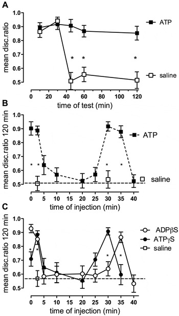
Facilitation of memory by purinergic agonists was examined by injecting ATP into the hippocampus. Memory retention was measured as the mean discrimination ratio (m.d.r.). Chicks were trained with 20% methyl anthranilate, which results in weakly reinforced memory that subsides by 40 min post-training. Injection of ATP (1 nmol) into the hippocampus 2.5 min post-training resulted in facilitation of the weakly reinforced memory (F (1,144) = 8.77, P < 0.001) such that the memory still existed 120 min after training (Fig. 1A) showing that ATP is an important neuromodulator of memory.
Effect of hippocampal administration of ATP, ADPβS and ATPγS on memory. (A) Memory retention tested in separate groups of chicks at different times following training with weak (20%) anthranilate. Chicks were injected with saline or ATP (1 nmol per hemisphere) 2.5 min after training. (B) Memory 120 min after weak anthranilate training and injection of saline or ATP at times shown relative to training. (C) Memory retention 120 min after weakly reinforced training with injection of ADPβS (100 pmol per hemisphere) or ATPγS (100 pmol per hemisphere) at different times relative to training. *P < 0.05.

To find the times at which ATP could be injected to facilitate memory, ATP was injected into the hippocampus of different groups at 0, 2.5, 5, 10, 20, 25, 30 and 35 min post-training, and the m.d.r. determined at 120 min to measure facilitation of long-term memory (Fig. 1B). Interestingly, ATP enhanced long-term memory significantly when injected immediately (0) and 2.5 min after training and again when injected 30 and 35 min post-training (F (10,145) = 13.85, P < 0.001). The two non-hydrolizable ATP agonist analogues ATPγS and ADPβS (Fig. 1C) produced similar but not identical memory profiles. ADPβS enhanced memory when injected immediately, 2.5 and 35 min after training (F (9,148) = 8.15, P < 0.001), whereas ATPγS only enhanced memory when injected 2.5 and 30 min after training (F (8,135) = 5.72, P < 0.001).
Involvement of endogenous ATP in normal memory formation
Activation of purinergic receptors by ADPβS and ATPγS is known to facilitate the consolidation of a weakly reinforced memory to long-term storage. However, by itself this finding does not tell whether endogenous purines are normally involved in memory formation. Astrocytes are known to be a source of ATP in rodent brain and there is no reason to expect that chick astrocytes are any different. To determine whether endogenous ATP is involved in memory processing, we examined the effect of hippocampal P2Y receptor inhibition on learning.
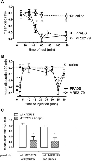
The chicks were trained with 100% methyl anthranilate to produce memory that is normally retained for over 120 min post-training. In this case, injection of the non-specific purinergic receptor antagonist PPADS (100 pmol) or the P2Y1 selective inhibitor MRS2179 (100 pmol) at 2.5 min after training resulted in the loss of memory by 40 min which persisted to at least 120 min after training (Fig. 2A) (F (5,83) = 13.78, P < 0.001; F (6,97) = 11.52, P < 0.001, respectively). For the inhibitors to block memory formation ATP release must occur in the chick hippocampus and the data illustrate the importance of purinergic signalling in chick learning.
Inhibitory effect of the ATP antagonist PPADS and the P2Y1 antagonist MRS2179 on memory and the selectivity of the agonist to the P2Y1 receptor. (A) Memory retention tested in separate groups of chicks at different times following training with strong anthranilate. Saline, PPADS (100 pmol per hemisphere) or MRS2179 (100 pmol per hemisphere) were injected 2.5 min after training. (B) Memory retention tested at 120 min following of injection of saline, PPADS or MRS2179 at different times relative to training on strong (100%) anthranilate. (C) MRS2179 challenge to weak memory facilitation by ADPβS to confirm that ADPβS is acting via P2Y1 receptors. Saline or a low dose of MRS2179 were injected subcutaneously prior to the hippocampal injection of ADPβS (100 pmol per hemisphere) 2.5 or 30 min after training. Memory was tested 120 min after training. *P < 0.05.

Examination of the results from a range of times of injection showed which time points the antagonists could be injected to cause complete loss of memory (Fig. 1E). Importantly, the mirror image of ADPβS facilitation in Fig. 2B was obtained with PPADS and MRS2179, both inhibiting long-term memory formation when injected at 0, 2.5 and 35 min post-training (F (8,104) = 9.44, P < 0.001; F (8,135) = 11.02, P < 0.001, respectively). The selective ectonucleotidase inhibitor (ARL67156) (Gallagher and Salter, Reference Gallagher and Salter2003; Sperlagh et al., Reference Sperlagh, Zsilla, Baranyi, Illes and Vizi2007), which prevents the hydrolysis of ATP to adenosine, when injected into the hippocampus facilitated memory at times similar to those shown by ATP. When injected 2.5 or 30 min after weakly reinforced training ARL67156 led to a significant increase in memory 120 min following training, but at other times of injection (0, 5–25 and 35 min) there was no enhancement of memory (F (9,122) = 7.49, P < 0.001, data not shown).
To show selectivity of ADPβS to P2Y1 receptors, a low sub-optimal subcutaneous dose of MRS2179 was pre-administered (0.6 nmol per chick) prior to weakly reinforced training with injection of ADPβS at 2.5 or 35 min after training. This dose of MRS2179 had only a marginally inhibitory effect on strongly reinforced training (m.d.r. 0.725 ± 0.063, n = 10) when injected subcutaneously 2.5 min after training, whereas a dose of 2.0 nmol per chick inhibited memory (m.d.r. 0.502 ± 0.0056, n = 10). MRS2179 injected 5 min before weakly reinforced training prevented ADPβS at 2.5 min from promoting consolidation, likewise when injected 30 min after training it prevented ADPβS-induced consolidation at 35 min (F (1,56) = 23.41, P < 0.001) (Fig. 2C).
Evidence suggesting that astrocytes are the source of the ATP promoting memory via P2Y1 receptors
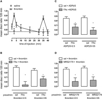
The source of the endogenous ATP that was modulating memory could be either from neurones or from astrocytes. In rodents astrocytes are a major source of the released ATP in the hippocampal slice (Bowser and Khakh, Reference Bowser and Khakh2007). Thrombin selectively activates calcium release in astrocytes (Bowser and Khakh, Reference Bowser and Khakh2004). Thrombin (3 pmol per hemisphere), like the P2Y1 receptor agonist ADPβS, also enhanced long-term memory formation when injected at 2.5 and 35 min after weak training (Fig. 3A) (F (8,122) = 6.43, P < 0.001), raising the possibility that astrocytic stimulation is the source of the ATP facilitating memory formation.
Effect of thrombin on memory and ability of the astrocytic metabolic inhibitor FAc to antagonise thrombin and ADPβS. (A) Memory retention following injection of saline or thrombin at different times relative to training on weak (20%) anthranilate and tested 120 min after training. (B,C) Chicks were preadministered with saline or FAc (100 pmol per hemisphere) 5 min before or 30 min after training, followed by thrombin (3 pmol per hemisphere (B)) or ADPβS (C) 2.5 or 35 min after training. (D) Subcutaneous injection of MRS2179 (0.6 nmol per chick) was administered 5 min before or 30 min after training and before injection of thrombin at 2.5 or 35 min after weak training. *P < 0.05.

When the chicks were dosed with FAc (100 pmol) to inhibit astrocyte oxidative metabolism and activation 5 min before or 30 min after training (Gibbs et al., Reference Gibbs, Hutchinson and Hertz2008Reference Gibbs, Bowser, Hutchinson, Loiacono and Summersb; Gibbs and Bowser, Reference Gibbs and Bowser2009) thrombin was unable to facilitate long-term memory (Fig. 3B) (F (1,55) = 31.92, P < 0.001). We have previously shown that this dose of FAc inhibits memory processing when injected between 0 and 20 min into the hippocampus after strongly reinforced training, but when injected 5 min before or 30 min after training it has no effect on memory processing (Gibbs and Bowser, Reference Gibbs and Bowser2009). This finding supports our interpretation that thrombin is acting in an astrocyte-dependent manner. Facilitation by ADPβS was also prevented by prior administration of FAc (Fig. 3C) (F (1,59) = 22.45, P < 0.001).
To confirm that thrombin was activating astrocytes to release ATP/ADP (adenosine diphosphate), we challenged thrombin facilitation of weakly reinforced learning, by giving a low subcutaneous dose of the P2Y1 receptor antagonist MRS2179 (0.6 nmol per chick). In this experiment, MRS2179 was administered 7.5 or 5 min prior to thrombin injection at 2.5 or 35 min after training, respectively (Fig. 3D). The low dose of MRS2179 prevented thrombin from promoting memory consolidation at both 2.5 and at 35 min after weakly reinforced training (F (1,53) = 23.78, P < 0.001).
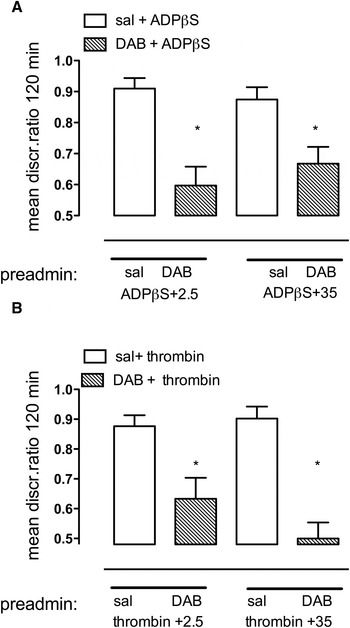
Further support for involvement of astrocytes in the action of ADPβS and thrombin comes from experiments where we challenged the ability of ADPβS and thrombin to facilitate memory consolidation by preventing the breakdown of glycogen in astrocytes. This was achieved by inhibiting glycogen phosphorylase with DAB (Gibbs et al., Reference Gibbs, Anderson and Hertz2006). Administration of a sub-optimal dose of DAB (0.1 nmol per chick) (Gibbs et al., Reference Gibbs, Bowser, Hutchinson, Loiacono and Summers2008a) before the agonist successfully challenged the ability of both ADPβS (Fig. 4A) (F (1,51) = 29.71, P < 0.001) and thrombin to facilitate memory (Fig. 4B) (F (1,53) = 37.27, P < 0.001). In these experiments DAB is being used as a tool to show astrocytic involvement, it implies only an indirect link between the breakdown of glycogen in astrocytes and ATP stimulation. We have shown elsewhere (Gibbs, Reference Gibbs2008) that astrocytic glutamate is dependent on glycogenolyis.
The effect of astrocytic inhibitor of glycogenolysis DAB on the ability of ADPβS and thrombin to facilitate memory. DAB (0.1 nmol per chick (Gibbs et al., Reference Gibbs, Hutchinson and Hertz2008a)) was injected subcutaneously 5 min before training or 5 min before injection of ADPβS (A) or thrombin (B), which were injected into the hippocampus 2.5 or 35 min after weakly reinforced training. *P < 0.05.

Facilitation of memory by ADPβS is prevented by MRS2179, by FAc and by inhibiting glycogenolysis with DAB, the latter two treatments affecting astrocytic metabolism. In addition, we found that facilitation of memory by thrombin which activates astrocytic protease-activated receptor-1 (PAR-1) receptors and increases [Ca2+]i is prevented by MRS2179, FAc and DAB. These results lead us to suggest that the ATP activating P2Y1 receptors comes from astrocytes. However, it is possible that the P2Y1 receptors could be located on astrocytes and/or interneurons.
To see whether we could confirm these findings and clarify the mechanism, we compared the effects of some of these agents in cell cultures of astrocytes and neurones from the chicks.
Activation of chick astrocytes by purinergic agonists
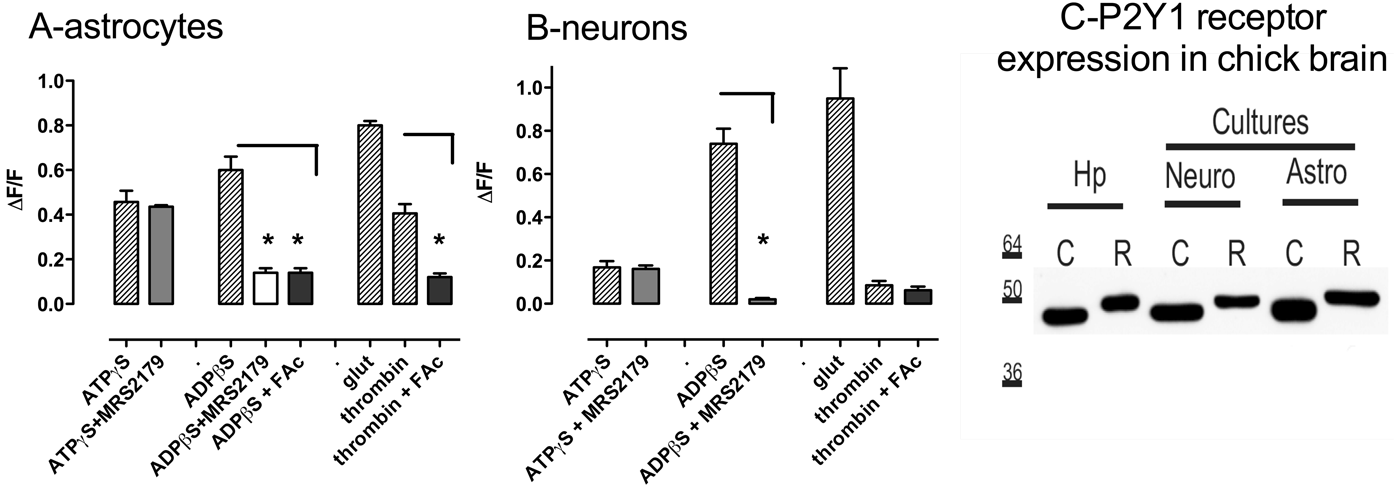
Cultured chick astrocytes were loaded with the calcium indicator dye Fluo-4/AM to monitor changes in their [Ca2+]i levels. They were then superfused with either glutamate (1 µM), ATPγS (100 µM), ADPβS (100 µM) or thrombin (10 U ml−1) for 20 s in order to elicit a calcium response. The collated data are presented in Fig. 5A (data obtained from more than 30 cells in three individual experiments). ATPγS and ADPβS induced calcium transients with similar peak [Ca2+]i. The observation of a similar response to ADPβS and ATPγS suggests that these cells express G-protein coupled P2Y receptors which are unlikely to be ionotropic P2X receptors (Bowser and Khakh, Reference Bowser and Khakh2004). The action of the P2Y1 receptor inhibitor MRS2179 (10 µM) further classified the type of receptor expressed. This antagonist inhibited the response to ADPβS suggesting that at least the P2Y1 purinergic receptor is expressed in astrocytes. Glutamate caused a robust calcium response most likely due to activation of metabotropic glutamate receptors. Thrombin activation of PAR-1 that responds by increasing [Ca2+]i in astrocytes (Junge et al., Reference Junge, Lee, Hubbard, Zhang, Olson and Hepler2004) also induced calcium transients in chick astrocytes. Inhibition of astrocyte metabolism with fluoroacetic acid (Bowser and Khakh, Reference Bowser and Khakh2007) inhibited the response to both ADPβS and to thrombin.
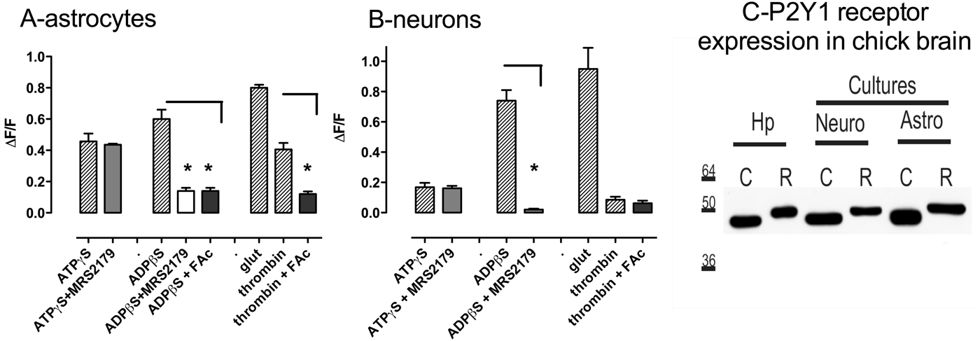
Calcium mobilizing agonists activate astrocytes and neurons in culture. (A) Bar graph summarizing all astrocyte experiments (ATPγS 100 µM, ADPβS 100 µM, glutamate 1 µM, thrombin 10 U ml−1, FAc 1 mM, MRS2179 10 µM). *Indicates that the calcium response to ADPβS in the presence of the MRS2179 or FAc is significantly different (P < 0.05) to the control ADPβS response and also that the response to thrombin in the presence of FAc is significantly different (P < 0.05) than the thrombin control. N is equal to at least 30 cells over three experiments for each experimental group. (B) Bar graph summarizing all neuronal experiments (ATPγS 100 µM, ADPβS 100 µM, glutamate 1 µM, thrombin 10 U ml−1, FAc 1 mM). *Indicates a significantly reduced response to ADPβS in the presence of FAc (P < 0.05). N is equal to at least 30 cells over three experiments for each experimental group. (C) P2Y1 expression analysis in chick (C) compared with rat (R). P2Y1 receptors were expressed in hippocampus, with all cell types, as well as in separately cultured astrocytes and neurons of both chick and rat.
Activation of chick neurons by purinergic agonists
Previous work on purinergic signalling in the rat hippocampus illustrated the importance of astrocyte-derived ATP in modulating neuronal activity (Bowser and Khakh, Reference Bowser and Khakh2004; Shigetomi et al., Reference Shigetomi, Bowser, Sofroniew and Khakh2008). As a surrogate for neuron firing, we used calcium imaging methods identical to that used in the chick astrocytes cultures (Fig. 5B). In this preparation, glutamate (1 µM) caused a rapid increase in [Ca2+]i that rapidly dissipated. This is likely due to the activation of post-synaptic ionotropic glutamate receptors. ADPβS elicited a calcium response peak [Ca2+]i similar to that observed in the astrocytes. However, ATPγS and thrombin elicited only a small and insignificant calcium response. Importantly, the response to ADPβS was inhibited by MRS2179 (10 µM). P2Y1 receptors are the likely purinergic receptors activated in these chick neuronal cultures.
P2Y1 receptors in chick astrocytes and neurons
We have presented pharmacological evidence for the role of P2Y1 receptors in mediating purinergic signalling in astrocytes and neurons in the chick hippocampus. In Fig. 5C we present western blot analysis of P2Y1 protein expression in the cultured chick cells and in the hippocampus of 1-day-old chicks. Using a commercial antibody (ABCam AB74996 diluted 1/500) we compared protein derived from rat neuron and astrocytes cultures and homogenized isolated whole hippocampi. The blot illustrates the expression of the receptor in each preparation. Importantly, chick P2Y1 runs at a lower molecular weight than the rat protein consistent with previous observations (~47 kDa compared to ~50 kDa) (Webb et al., Reference Webb, Simon, Krishek, Bateson, Smart and King1993, Reference Webb, Simon and Barnard1998; Tokuyama et al., Reference Tokuyama, Hara, Jones, Fan and Bell1995). P2Y1 receptors are expressed in both chick astrocytes and neurons, in line with data obtained in the rat (Fig. 5C).
CONCLUSIONS
• Exogenous ATP injected into the hippocampus during short-term memory (0–2.5 min) or at the transition from intermediate- to long-term memory (30–35 min) after weakly reinforced training, facilitated the consolidation of weakly reinforced learning.
• The two ATP analogues, ATPγS and ADPβS, consolidate memory, albeit with slight timing differences and the action of the selective antagonist MRS2179 confirmed that endogenous ATP is involved, as well as showing selectivity for ADPβS to P2Y1 receptors.
• That astrocytes are the source of endogenous ATP release is suggested by the ability of thrombin to promote consolidation at the same time periods as ADPβS, i.e. 2.5 and 35 min after training, and the ability of MRS2179 and the astrocyte-specific toxins FAc and DAB to prevent consolidation by both thrombin and ADPβS.
• In cell cultures ADPβS increased calcium transients in both astrocytes and neurons. Importantly thrombin increased [Ca2+]i in astrocytes but not in neurons, an effect preventable by FAc.
• ATPγS only increased calcium transients in astrocytic cultures, and this effect was not inhibited by MRS2179.
DISCUSSION
The main findings of the present study are: (a) purinergic receptor astrocytes play an important role in the consolidation of short-term memory to long-term memory; and (b) ATP released from chick neurons or astrocytes during the learning process modulates neuronal activity through astrocytic P2Y1 receptors.
As well as being released from astrocytes (Bowser and Khakh, Reference Bowser and Khakh2007; Pryazhnikov and Khiroug, Reference Pryazhnikov and Khiroug2008; Huang et al., Reference Huang, Sudibya, Fu, Xue, Dong and Li2009) ATP is also released from neurons as a co-transmitter with glutamate and other neurotransmitters (Mori et al., Reference Mori, Heuss, Gahwiler and Gerber2001; Fujii et al., Reference Fujii, Sasaki, Mikoshiba, Kuroda, Yamazaki and Mostafa Taufiq2004; Pankratov et al., Reference Pankratov, Lalo, Verkhratsky and North2006; Burnstock, Reference Burnstock2007). In the present study, both astrocytes and neurons from chick forebrain responded to ADPβS and glutamate by a transient increase in [Ca2+]i. The astrocytes also responded by an increase in [Ca2+]i when activated by ATPγS or by the PAR-1 receptor agonist thrombin, whereas the neurons did not. The response to ADPβS, due to activation of P2Y1 receptors, was prevented by the selective antagonist MRS2179. Although neurons express P2Y1 receptors and respond to ADPβS, they do not respond to thrombin, i.e. there is no increase in [Ca2+]i. The suggestion that the thrombin increase in [Ca2+]i was generated by astrocytes is supported by the finding that this increase was prevented by FAc, an inhibitor of astrocytic oxidative metabolism.
ATP injection directly into the hippocampus promoted consolidation of weakly reinforced memory when injected at either of two time periods that correspond to when memory consolidation is vulnerable to facilitation or inhibition. This simple, incidental learning task has a distinct advantage over repetitive or rote learning tasks in that this task reveals the very discrete timing of memory processes. When the actions of the non-hydrolysable purinergic agonists ADPβS and ATPγS in promoting memory consolidation are compared with ATP, it is apparent that the ATP effect is a temporal summation of the individual agonists, suggesting that ADPβS and ATPγS may be activating different purinergic receptors. P2Y1 receptors are activated at 0–2.5 min and again at 35 min after training, whereas activation of ATPγS receptors is also important at 2.5 min, at the later time it is involved a little earlier than ADPβS receptors, at 30 min. The effect of the antagonist PPADS and the selective ADPβS antagonist MRS2179 on consolidation support our interpretation that ATP is released during memory processing. Even though ATP will be broken down by ectonucleotidases to ADP and adenosine, the finding that the effect of MRS2179 inhibits memory occurs at the same timing after training as ADPβS promotes consolidation, strongly suggests that the effects we are seeing here are due to P2Y1 as well as some other P2Y receptor. On the basis of our previous work with these drugs (Bowser and Khakh, Reference Bowser and Khakh2004) we believe that ADPβS and ATPγS do provide sufficient ATP hydrolysis to cause an adenosine receptor response. The selective ectonucleotidase inhibitor (ARL67156) (Gallagher and Salter, Reference Gallagher and Salter2003; Sperlagh et al., Reference Sperlagh, Zsilla, Baranyi, Illes and Vizi2007), which prevents the hydrolysis of ATP to adenosine, when injected into the hippocampus facilitated memory at times similar to those shown by ATP. ARL67156 will prevent the breakdown of endogenously released ATP. This result suggests that the data reported in this paper are not caused by activation of adenosine receptors. If adenosine, produced from ATP degradation, was responsible for the effect on memory, then there should be no memory enhancement. The source of the ATP mimicked by the action of ATPγS, could well be neuronal, being co-released with either glutamate or noradrenaline (Burnstock, Reference Burnstock2007). A recent publication (Torres et al., Reference Torres, Wang, Xu, Fujita, Dobrowolski and Willecke2012) has suggested that the trigger for astrocytic ATP release is a reduction in extracellular calcium ([Ca2+]e) that occurs during synaptic transmission. However, the present cell culture results show that ATPβS is able to increase [Ca2+]i in chick astrocytes.
The importance of ATP and other transmitters at these two critical consolidation periods is reminiscent of that seen when NMDA receptor antagonists (Gibbs et al., Reference Gibbs, Bowser, Hutchinson, Loiacono and Summers2008a) and mGluR1 receptor antagonists (Gibbs and Bowser, Reference Gibbs and Bowser2009) impair memory processing. Likewise, memory consolidation can be promoted at these same times with NMDA and mGluR agonists (Gibbs and Bowser, Reference Gibbs and Bowser2009). We have shown that thrombin activation of PAR-1 receptors causes increased astrocytic [Ca2+]i and impacts on memory processing at exactly the same time as activation of P2Y1 receptors, this strongly suggests that ADPβS is responsible for the calcium response. Although we believe that we are looking at an astrocyte-dependent phenomenon, at this stage we cannot rule out the possible involvement of neurons or an interaction between the two cell types. Both cell types can release ATP and glutamate. In addition, there are modulatory neurotransmitters such as noradrenaline that are co-released from neurons with ATP and gliotransmitters such as d-serine will impact on the storage of information in the brain to further add to the complexity of what is going on. Each astrocyte has a clearly defined territorial domain (Bushong et al., Reference Bushong, Martone, Jones and Ellisman2002; Anderson and Nedergaard, Reference Anderson and Nedergaard2003; Nedergaard et al., Reference Nedergaard, Ransom and Goldman2003) and each single astrocyte surrounds and forms contacts with all neuronal synapses residing within its territory (Nedergaard et al., Reference Nedergaard, Ransom and Goldman2003). Astrocytes also form neuronal–glial–vascular units, as the processes of the astrocyte, the end feet, completely cover the capillaries in the brain (Simard et al., Reference Simard, Arcuino, Takano, Liu and Nedergaard2003). It is possible that there is differential distribution of the different purinergic receptor subtypes on different locations within the same astrocyte, according to their function.
Concluding remarks
Our new findings are clearly consistent with our previous work on cultured rodent astrocytes and neurons, as well as electrophysiological and imaging recordings from rat hippocampal slices (Bowser and Khakh, Reference Bowser and Khakh2004, Reference Bowser and Khakh2007). In the earlier papers, we demonstrated that astrocytes release ATP and glutamate and that these agents act on neighbouring astrocytes to propagate a calcium wave of astrocyte activation that also activates P2Y1 receptors on hippocampal interneurons. The activation of these interneuron populations and the subsequent increase in inhibitory GABAergic synaptic transmission reduces the pyramidal neuron output of the intact hippocampal circuit. Both glutamate and GABA are involved in memory processing (Gibbs et al., 2008a; Gibbs and Bowser, Reference Gibbs and Bowser2009) and the present data clearly show that ATP has an important role as a signalling molecule in the hippocampus. In the hippocampus and the cortex, release of glutamate occurs at the time of learning, stimulates astrocytes and triggers the release of ATP; activation of astrocytic P2Y1 receptors occurs, resulting in feed forward inhibition of excitatory neurons via GABAB receptors. The increased synaptic inhibition within hippocampal circuits is a possible mechanism whereby astrocyte/interneuron interactions could maintain a memory, preventing downgrading of the information prior to its storage/consolidation. A necessary component of this schema is release of noradrenaline in the hippocampus and cortex and noradrenergic modulation of electrical coupling in GABAergic interneurons contributing to the reinforcement and tuning of memory processing.
ACKNOWLEDGEMENTS
This work was funded in part by NHMRC Project Grant to D.N. Bowser.
Statement of interests
None.