I. INTRODUCTION
Bovine pancreatic Ribonuclease A (RNase A E.C. 3.1.27.5) is a well known endonuclease with a molecular weight of 13.7 ± 0.3 kDa that cleaves 3′,5′-phosphodiester linkages of single-stranded RNA when the base of the nucleotide in the 3′ position is a pyrimidine.Reference D’Alessio and Riordan1–Reference Raines4 It is know that some RNases, belonging to the RNase A family, have cytotoxic and/or cytostatic effects on cancer cells.Reference Rybak and Newton5–Reference Leland, Staniszewski, Kim and Raines7 Therefore, the immobilization of these RNases on solid three-dimensional biocompatible and bioresorbable matrix materials could represent new solutions in cancer therapy by controlled drug release.Reference Teoli, Parisi, Realdon, Guglielmi, Rosato and Morpurgo8–Reference Cullen, Liu, Mandel, Himpsel and Gopalan10 The process, known as surface-mediated drug delivery, implies the release of therapeutic cargo from the surface of a biomaterial, typically an implant, to the surrounding cells, organs, and tissues.Reference Zelikin11 Among the advantages of this process are the spatial control of release of the therapeutic molecules and a more efficient delivery due to the much higher local concentration of drug available to the cells.Reference Shmueli, Anderson and Green12, Reference Jewell, Fuchs, Flessner, Raines and Lynn13
Enzymes can be immobilized onto an insoluble solid holder by various deposition techniques. However, the immobilization of complex organic molecules is not an easy task. Big differences between the initial material and the deposited structure’s composition can emerge after the transfer and immobilization processes, leading to changes of chemical and biological properties such as stability and loss of enzymatic activity, which limit the practical applications of the immobilized structures.Reference Klibanov14
Laser techniques could become a possible approach for enzyme immobilization. Due to the use of light, there is a very low risk of cross-contamination, and the material transfer onto the desired holder has an accurate spatial resolution. Laser sources further offer the possibility of adjusting the quantity of the transferred and immobilized enzyme through the irradiation process parameters.Reference Eason15 Although high intensity laser pulses are destructive for organic materials, laser deposition techniques were optimized and a new method, called matrix assisted pulsed laser evaporation (MAPLE) was developed. In MAPLE, a frozen matrix consisting of a dilute solution of organic compound in a solvent material is used as a target for laser irradiation.Reference Chrisey, Piqué, McGill, Horwitz, Ringeisen, Bubb and Wu16 At the same time, the intensity of the incident laser radiation is reduced up to one order of magnitude as compared to conventional pulsed laser deposition (PLD). This technique was successfully tested for immobilization onto solid holders of methoxy Ge triphenylcorrole,Reference Caricato, Lomascolo, Luches, Mandoj, Manera, Mastroianni, Martino, Paolesse, Rella, Romano, Tunno and Valerini17 polypyrrole,Reference Bubb, Wu, Horwitz, Callahan, Galicia, Vertes, McGill, Houser, Ringeisen and Chrisey18 polyethylene glycol derivatives,Reference Chrisey, Piqué, McGill, Horwitz, Ringeisen, Bubb and Wu16, Reference Cristescu, Cojanu, Popescu, Grigorescu, Duta, Ionescu, Mihaiescu, Buruiana, Andronie, Stamatin, Mihailescu and Chrisey19, Reference Cristescu, Popescu, Popescu, Grigorescu, Mihailescu, Mihaiescu, Gittard, Narayan, Buruiana, Stamatin and Chrisey20 and polyalkylthiophene,Reference Gutierrez-Llorente, Horwitz, Perez-Casero, Perriere, Fave, Yassar and Sant21 and also several biomaterials such as horseradish peroxidase, insulin,Reference Ringeisen, Callahan, Wu, Pique, Spargo, McGill, Bucaro, Bubb and Chrisey22 bovine serum albumin,Reference Hernandez-Perez, Garapon, Champeaux, Shahgaldia, Coleman and Mugnier23 pepsine,Reference Kecskemeti, Kresz, Smausz, Hopp and Nogradi24 silk fibroin,Reference Tsuboi, Goto and Haya25 fibrinogen blood proteins,Reference Stamatin, Cristescu, Socol, Mihaiescu, Stamatin, Mihailescu and Chrisey26 papain,Reference György, Santiso, Figueras, Socol and Mihailescu27, Reference György, Pérez del Pino, Sauthier and Figueras28 creatinine,Reference György, Axente, Mihailescu, Predoi, Ciuca and Neamtu29 and urease.Reference György, Sima, Mihailescu, Smausz, Megyeri, Kékesi, Hopp, Zdrentu and Petrescu30–Reference György, Sima, Mihailescu, Smausz, Hopp, Predoi, Zdrentu and Petrescu32
An alternative laser technique, MAPLE direct write (MAPLE-DW) combines MAPLE with laser-induced forward transfer (LIFT). LIFT was developed for the transfer of inorganic materials,Reference Bohandy, Kim and Adrian33–Reference Fogarassy, Fuchs, Kerheve, Hauchecorne and Perriere35 but later applied as MAPLE-DW beyond inorganicsReference Piqué, Chrisey and Auyeung36 to organics as polymers,Reference Chrisey, Piqué, McGill, Horwitz, Ringeisen, Bubb and Wu16 or living cells,Reference Chrisey, Piqué, McGill, Horwitz, Ringeisen, Bubb and Wu16, Reference Ringeisen, Chrisey, Piqué, Young, Modi, Bucaro, Jones-Meehan and Spargo37 where the material to be transferred is mixed in a laser-absorbent matrix and coated onto a support transparent to laser irradiation. Starting from LIFT, another technique, absorbing film assisted LIFT (AFA-LIFT) was developed to transfer proteins,Reference Serra, Fernández-Pradas, Berthet, Colina, Elvira and Morenza38 DNA arrays,Reference Serra, Colina, Fernández-Pradas, Sevilla and Morenza39 or cellsReference Barron, Wu, Ladouceur and Ringeisen40, Reference Hopp, Smausz, Barna, Vass, Antal, Kredics and Chrisey41 where the organic materials are spread on absorbing layers deposited on the surface of support plates transparent to the incident laser wavelength. It was also found that LIFT can be used to transfer and create micropatterns of biological molecules such as DNA and proteins without the assistance of any absorbing material.Reference Karaiskou, Zergioti, Fotakis, Kapsetaki and Kafetzopoulos42
In this article we report the successful laser immobilization of the enzyme RNase A onto SiO2 glass collectors using the MAPLE technique. Our purpose was to find the maximum threshold fluence value that led to the formation of uniform and continuous thin films on the substrate surface, avoiding decomposition of the laser processed RNase A molecules. Under optimized irradiation conditions, the laser transferred RNase A preserved its molecular structure and enzymatic activity.
II. EXPERIMENTAL DETAILS
A. Laser immobilization of RNase A
The experiments were performed in a stainless steel irradiation chamber. A pulsed UV KrF* (λ = 248 nm, τFWHM ≈ 25 ns, ν = 10 Hz) COMPexPro 205 excimer laser source was used for the irradiations. The laser pulse repetition rate was set at 10 Hz. The laser fluence was set at values in the range (0.4–0.7) J/cm2. The incidence angle of the laser beam on the target surface was 45°. During the experiment, the laser beam scanned the target surface at a constant velocity of 2 mm/s to avoid drilling the target. The irradiated XY surface area was 1 × 1 cm2. 1.5 × 10⁴ subsequent laser pulses were applied for the deposition of each film. Before each deposition experiment, the irradiation chamber was evacuated down to a residual pressure of 1.6 × 10⁻² Pa. This pressure value was maintained during the RNase A thin film growth experiments.
Hepes-KOH 10 mM pH 7.5 buffer was chosen for the preparation of the target solution. The enzyme concentration was 1% (w/v). The solutions were frozen in liquid nitrogen and kept frozen during laser irradiation by circulation liquid of nitrogen inside a special double wall target holder. The complete MAPLE workstation was purchased from Surface Systems & Technology GmbH & Co KG, (Hückelhoven, Germany).
RNase A was immobilized onto SiO2 glass collectors that were placed on a plane parallel to the target surface at a separation distance of 5 cm. Prior to introduction inside the deposition chamber, the collectors were carefully cleaned in a Transonic T 310 ultrasonic bath with acetone and ethanol. During MAPLE deposition, the collectors were kept at room temperature. Twin samples were prepared to verify the reproducibility of the deposition process. The samples were analyzed in terms of composition, surface morphology, and enzymatic activity in the presence of the analyte molecules.
B. Characterization methods of immobilized RNase A
The surface morphology of the deposited RNase A thin films were investigated by atomic force microscopy (AFM) in acoustic (dynamic) configuration with a 5100 apparatus from Agilent Technologies (Santa Clara, California). The chemical composition and bonding states between the elements were studied by Fourier transform infrared spectroscopy (FTIR) in the wavenumber 4000 cm–1 to 500 cm−1 range, using a 4 cm−1 resolution Perkin Elmer Inc., Spectrum One apparatus (Wellesley, Massachusetts).
C. Analysis of the RNase A enzymatic activity
RNase activity was analyzed from the digestion of the polycytidylic [poly(C)] substrate by the immobilized enzyme and separation of the products of the reaction by reverse phase HPLC. The obtained structure was submerged in 1 mL of a 2.5 mg/mL poly(C) solution in 10 mM Hepes-KOH pH 7.5 at 25 °C for 1 h. The products of the reaction were analyzed by injecting 50 μL of the solution on a reverse phase HPLC column (Nova-Pak C18, Waters Corp. Milford, Massachusetts) equilibrated with solvent A [10% ammonium acetate (w/v) and 1% acetonitril (v/v) in water].
The elution was carried out at a flow rate 1 mL/min with an initial 10 min wash and 50 min linear gradient from 100% solvent A to 10% solvent A plus 90% solvent B [10% ammonium acetate (w/v) and 11% acetonitril (v/v) in water].Reference Moussaoui, Guasch, Boix, Cuchillo and Nogués43 The same process was used to analyze a blank set of samples as the control experiment. To this purpose, poly(C) solution was incubated with SiO2 glass plates identical to those used as collectors for the RNase A immobilization by MAPLE. For positive control experiments, poly(C) solution was incubated with nonirradiated RNase A powdered material.
III. RESULTS AND DISCUSSION
A. Surface morphology investigations
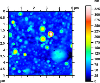
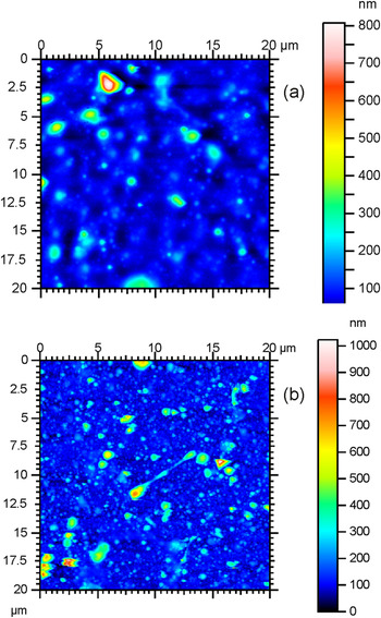
In Fig. 1 we present a top view AFM image of RNase A immobilized on SiO2 substrate at 0.4 J/cm2 laser fluence. The deposited structure consists of nanoparticles formed most probably by coalescence of RNase A molecules during their transit from the target toward the substrate surface constituting a continuous film. Apart from the nanoparticles, larger particles of about 500 nm mean diameter and about 200 nm mean height are present in the film’s structure. Both mean diameter and mean height of particles increases with the increase of the incident laser fluence (Fig. 2). At the highest laser fluence, 0.7 J/cm2 [Fig. 2(b)], the film is practically covered by micrometric particles.
Top view AFM image of the RNase A thin film obtained from a frozen composite target containing 1% (w/v) biomaterial in buffer Hepes solvent, by irradiation with 1.5 × 10⁴ subsequent laser pulses at 0.4 J/cm2 laser fluence.

Top view AFM images of the RNase A thin films obtained from a frozen composite target containing 1% (w/v) biomaterial in buffer Hepes solvent, by irradiation with 1.5 × 10⁴ subsequent laser pulses at (a) 0.5 and (b) 0.7 J/cm2 laser fluence.

The presence of large, micrometer sized particulates on the surface of MAPLE deposited organic thin films were related, according to theoretical predictions, to droplet ejection from the target surface as a result of explosive evaporation or spallation mechanisms,Reference Dingus and Scammon44, Reference Cramer, Haglund and Hillenkamp45 termed cold laser ablation.Reference Leveugle, Ivanov and Zhigilei46, Reference Mercado, Allmond, Hoekstra and Fitz-Gerald47 This ablation process could explain the increase of particle size and density resulting in a nonuniform surface morphology of the films with the increase of incident laser fluence. On the contrary, at low laser fluence, the surface morphology, constituted by tens of nanometer-sized particulates, can be assigned to surface evaporation and cluster formation.Reference György, Pérez del Pino, Sauthier and Figueras28
B. Characterization of chemical composition
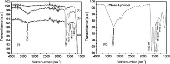
To obtain information about the molecular structure of the deposited layers, the RNase A thin films were investigated by FTIR spectroscopy (Fig. 3I). The films spectra were compared with the spectrum of the nonirradiated RNase A base material used for the preparation of the composite targets (Fig. 3II). As can be observed, the spectra reproduce all the characteristic bands assigned to structural elements of RNase A, namely the amide A band around 3300 cm–1 due to NH stretching, the band at 1632 cm–1 of the amide I region due to C=O stretching, the band at 1528 cm–1 attributed to tyrosine amino acid residues, the band at 1445 cm–1of the amide II region attributed to NH bending, and CN stretching, and the band around 1400 cm–1 corresponding to a random structure.Reference Georg, Wharton and Siebert48–Reference Neault, Diamantoglou, Bearegard, Nafisi and Tajmir-Riahi50 The band at wavenumbers lower than around 1200 cm–1 corresponds to the glass substrate.Reference György, Pérez del Pino, Sauthier and Figueras28
FTIR spectra of (I) RNase A thin films obtained from a frozen composite target containing 1% (w/v) biomaterial in buffer Hepes solvent, by irradiation with 1.5x10⁴ subsequent laser pulses at (a) 0.4, (b) 0.5, and (c) 0.7 J/cm2 laser fluence and (II) nonirradiated RNase A base material used for the preparation of the composite targets.

For all fluences regimes, the spectra of the obtained RNase A films are similar to the base material spectrum, reproducing the same absorption bands. We would like to notice that according to differential scanning calorimetry analyses,Reference Del Vecchio, Catanzano, de Paola and Barone51 thermal denaturation of RNase A takes place already at around 60 °C. Thus, decomposition could be expected during the thin films growth, under the action of the subsequent laser pulses incident on the target surface. Nevertheless, as confirmed by FTIR results, the deposited thin films preserved the structure of the base material used for the targets preparation. This feature is an evidence of the effectiveness of the special precautions characteristic for the MAPLE technique, frozen composite target preparation procedure and reduction of the incident laser fluence values as compared to those used in conventional PLD.
The intensity of the bands in the FTIR spectra increases with the increase of the laser fluence from 0.4 to 0.5 J/cm2 [Fig. 3I (a),(b)], most probably due to the increased amount of the enzyme molecules transferred to the collector surface. Nevertheless, the intensity of the bands decreases when the incident laser fluence was increased above 0.5 J/cm2 [Fig. 3I (c)]. This feature was attributed to the decomposition of the RNase A molecules under action of higher intensity incident laser pulses. The value of 0.5 J/cm2 was found as maximum threshold laser fluence value for which the spectra of the transferred and immobilized RNase A reproduce the bands of the nonirradiated base material, avoiding decomposition.
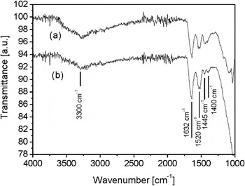
We analyzed the biomaterials solution of the composite targets before and after laser irradiation with different laser fluence values. To this purpose, drops from the nonirradiated and laser irradiated target solutions were deposited on glass substrates identical to those used for the laser immobilization of RNase A. The FTIR analyses were performed after the evaporation of the buffer Hepes solvent. The spectrum of the nonirradiated and laser irradiated target material with 0.5 J/cm2 laser fluence are shown in Figs. 4(a) and (b), respectively. The spectrum of the irradiated material still reproduces the bands of the initial RNase A base powder, suggesting that chemical composition and structure of the biomaterial were preserved even at the identified maximum laser fluence threshold value.
FTIR spectra of the target containing 1% (w/v) biomaterial in buffer Hepes solvent (a) before and (b) after laser irradiation at 0.5 J/cm2 fluence.

C. Enzymatic activity of the laser immobilized RNase A
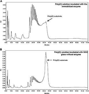
Fig. 5 shows the effect of the laser immobilized RNase A in the poly(C) substrate [Fig. 5(A)] and the control corresponding to the blank set of samples [Fig. 5(B)]. As is widely known, poly(C) can be considered to be a synthetic analog of single-stranded RNA and is commonly used in research laboratories for the determination of RNase enzymatic activity.Reference Moussaoui, Guasch, Boix, Cuchillo and Nogués43 The immobilization of the RNase A was performed using the established optimum laser irradiation conditions, 0.5 J/cm2 incident fluence on the composite target surface. The chromatographic profile corresponding to the products separation by reverse phase HPLC has previously been characterized.Reference Moussaoui, Guasch, Boix, Cuchillo and Nogués43 The last peak, with 38 min elution time, corresponds to the undigested poly(C) substrate, a high molecular mass polymer. The digestion products, smaller molecular mass oligonucleotides, elute in order of increasing molecular mass, from mononucleotides to polynucleotides, as a function of elution time from 1 to 34 min.
Reverse phase HPLC separation of the products obtained from the polymeric poly(C) substrate by RNase A activity: the elution profiles of (A) the poly(C) solution incubated with the RNase A enzyme immobilized at 0.5 J/cm2 incident laser fluence and (B) the poly(C) solution incubated with the SiO2 glass plate without enzyme. See Section II. C for details about the chromatographic procedure.

The profile corresponding to the poly(C) solution incubated with the immobilized enzyme [Fig. 5(A)] shows a large amount of low molecular mass digestion products and a reduced amount of the undigested poly(C) substrate. This result proves the enzymatic activity of the immobilized RNase A. In addition, the product formation pattern is in agreement with the products obtained by RNase A in solution under the same conditions,Reference Moussaoui, Guasch, Boix, Cuchillo and Nogués43 indicating the preference for endonuclease activity. The control sample consisting of poly(C) solution incubated with a SiO2 glass plates in the absence of RNase A [Fig. 5(B)] shows a large component corresponding to the high molecular mass undigested poly(C) substrate. The very low traces of smaller molecular mass components are due to a certain decomposition in time of the poly(C) substrate. In the same elution time interval, the peak corresponding to the undigested poly(C) substrate is nearly undistinguishable in the profile of the positive control sample.
IV. CONCLUSION
RNase A was immobilized on SiO2 glass collectors in the form of continuous thin films by the MAPLE technique. The films consisted of nanoparticles formed most probably by coalescence of RNase A molecules and large, micrometer sized particles ejected directly from the target’s surface. Both mean diameter and mean height of the particles increase with the increase of the incident laser fluence value. The FTIR spectra of the RNase A films were similar to the spectrum of the nonirradiated base material used for the preparation of the composite targets. The intensity of the bands increases with the increase of laser fluence until a maximum threshold value of 0.5 J/cm2. At higher fluence values, the band intensity diminishes due to the decomposition of part of the transferred material under the action of laser pulses. The immobilization process does not affect the RNase A behavior, since the enzyme maintains for the polymeric poly(C) substrate the cleavage pattern observed for the enzyme in solution.
ACKNOWLEDGMENTS
The authors acknowledge financial support from the Spanish National Research Council under Contract No. 200860I211 and the Romanian National University Research Council under Contract IDEAS No. 652.