




No CrossRef data available.
Article contents
PHACES: a neurocutaneous syndrome with anomalies of the aorta and supraaortic vessels
Published online by Cambridge University Press: 20 January 2005
Abstract
The acronym PHACES summarizes the most important manifestations of a rare neurocutaneous syndrome. Specifically, “P” accounts for malformation of the brain in the region of the posterior fossa, “H” stands for haemangiomas, “A” is for arterial anomalies, and “C” is for coarctation of the aorta along with cardiac defects, “E” is for abnormalities of the eye, and “S” for clefting of the sternum, and/or a supraumbilical abdominal raphe. Our objective is to introduce the syndrome to paediatric cardiologists. Our patient has stenosis of the aortic arch, multiple malformations of the great vessels arising from the aortic arch, intracranial vascular abnormalities, a sternal malformation with a supraumbilical raphe, and facial haemangiomas. We stress that it is important always to consider the existence of this syndrome in all patients with facial haemangiomas.
Information
- Type
- Brief Report
- Information
- Copyright
- © 2004 Cambridge University Press
Haemangiomas are the most common tumours encountered in infancy. The incidence in the general newborn population is between 1.0% and 2.6%, but can be as high as 10% in Caucasian populations.1 In contrast to other vascular malformations, haemangiomas are rarely associated with systemic anomalies.2 An association between capillary haemangiomas and craniocervical arterial anomalies, nonetheless, was reported by Pascual-Castroviejo3 as long ago as 1978. Other findings subsequently came to be added to this finding, indicating that the associations were far from random. It was in 1996 that Frieden et al.4 introduced the acronym “PHACE” to describe the association of malformations of the brain in the region of the posterior fossa, haemangiomas, arterial anomalies, coarctation of the aorta and cardiac defects, and abnormalities of the eye. It was also suggested that the acronym should be expanded to “PHACES” when there were also developmental defects involving the anterior abdominal and thoracic wall, such as sternal clefting or supraumbilical raphe.4
The syndrome differs from other neurocutaneous syndromes that simultaneously involve the skin and the central nervous system in that a great variety of extracutaneous manifestations can be found in the setting of PHACES. Cardiac defects, and/or malformation of the aorta and its branches, have been reported in more than one-third of cases. The pathogenesis of the syndrome, thus far, is unknown. Suggestions have been made for X-linked dominant heredity,5 or a field defect developing either between the third and sixth,6 or eighth and tenth,7 weeks of gestation. We describe here a patient with near complete manifestations of the syndrome, and discuss the impact of the syndrome for the paediatric cardiologist.
Case report
Our patient was born at term by vaginal delivery after an uncomplicated pregnancy. There was no known family history of cardiovascular disease. Antenatal ultrasonic scans were not performed. On initial examination, she was noted to have a complete defect of the sternum and a supraumbilical raphe. At the age of two weeks, a haemangioma involving the region of the lower lip was detected. At the age of three months, she presented with inspiratory stridor, and laryngoscopy showed a subglottic stenosis, while magnetic resonance imaging revealed a paratracheal haemangioma. The sternal defect was closed by surgery, without complications, when she was 6 months of age. Recurrent growth of the paratracheal haemangioma led to severe respiratory problems so that, in addition to treatment with systemic and local corticosteroids, endotracheal laser coagulation had to be performed several times.
At the age of six years (Fig. 1), she was admitted to our unit of paediatric cardiology because of a systolic murmur. Echocardiography revealed stenosis of the aortic arch. Subsequent magnetic resonance angiography not only confirmed this diagnosis, but also showed extensive abnormalities of the aorta and brachiocephalic vessels (Fig. 2). The aortic coarctation was repaired by resection and end-to-end anastomosis. The brachiocephalic artery was widened by insertion of a patch, and reimplanted into the aortic arch. Our patient is now seven years old, and shows normal neurological development. Ophthalmologic examination, and magnetic resonance imaging of the brain, has shown no pathologic findings.

Figure 1. In our patient, at the age of six years, and after surgical closure of the sternal defect at the age of six months, the facial haemangioma had involuted during treatment with systemic corticosteroids during the second year of life. The involuted rest of the haemangioma in the lower lip, shown in this picture, was the only external visible manifestation of the syndrome.

Figure 2. The frontal projection of the magnetic resonance angiogram (a) shows stenosis of the aorta next to the origin of the brachiocephalic artery, stenosis of the brachiocephalic artery itself, the right subclavian artery, the right external and internal carotid arteries, and hypoplasia of the right carotid artery. The right anterior cerebral artery is filled via the left internal carotid artery. All of the findings are confirmed by the antero-lateral view of the magnetic resonance angiogram (b).
Discussion
The syndrome known with the acronym “PHACES” accounts for a great variety of associated malformations. In many cases, only one extracutaneous manifestation is present (Table 1). Facial haemangiomas are the hallmark of the syndrome. Most such haemangiomas are large and impressive lesions that will immediately attract the attention of the investigator, as will sternal clefting and/or a supraumbilical raphe. There seems a high likelihood, nonetheless, that patients having the syndrome, but with small haemangiomas and no other or minimal external manifestations, will remain undiagnosed during routine clinical investigation. It has been suggested therefore, that all patients with facial haemangiomas, regardless of the characteristics of the lesions, should undergo a minimal clinical investigation, including cardiologic, neurologic and ophthalmologic examinations.8 The cardiologic investigation should include measurements of blood pressure in all four limbs along with echocardiography. In addition to these diagnostic manoeuvres, we suggest that, in all patients with extensive plaque-like facial haemangiomas, and/or any other manifestation of the syndrome, magnetic resonance angiography should be undertaken to profile the aortic arch and its brachiocephalic vessels, along with the structure of the brain.
Table 1. Manifestations of the PHACES Syndrome.4,6,8–10

We believe that the cardiologic examination should be undertaken during the first month of life when the haemangiomas first become clinically evident. In contrast to the cardiac malformations, other lesions, including vascular abnormalities, could develop later in life. It remains unclear, therefore, if all the components of the syndrome can be detected during the first month of life. Follow-up studies on affected young individuals will be needed to resolve this question.
When cardiovascular surgery is required, special attention should be paid to the possible presence of cerebral vascular abnormalities or a subglottic haemangioma. Cerebral vascular abnormalities may elevate the risk of an intraoperative cerebral ischemic event, and should therefore be excluded preoperatively by magnetic resonance angiography. Haemangiomas of the subglottic components of the airways, on the other hand, may cause respiratory problems and complicate the perioperative management. Careful investigation, therefore, is needed for symptoms suggestive of subglottic obstruction of the airways, such as cough, hoarseness, in- and expiratory stridor, and cyanosis.
References
Metry D, Herbert AA. Benign cutaneous vascular tumors of infancy. When to worry, what to do. Arch Dermatol 2000; 136: 905–914.Google Scholar
Burns AJ, Kaplan LC, Mulliken JB. Is there an association between hemangioma and syndromes with dysmorphic features? Pediatrics 1991; 88: 1257–1267.Google Scholar
Pascual-Castroviejo I. Vascular and nonvascular intracranial malformations associated with external capillary hemangiomas. Neuroradiology 1978; 16: 82–84.Google Scholar
Frieden IJ, Reese V, Cohen D. PHACE syndrome. The association of posterior fossa brain malformations, hemangiomas, arterial anomalies, coarctation of the aorta and cardiac defects, and eye abnormalities. Arch Dermatol 1996; 132: 307–311.Google Scholar
Gorlin RJ, Kantaputra P, Aughton DJ, Mulliken JB. Marked female predilection in some syndromes associated with facial hemangiomas. Am J Genet 1994; 52: 130–135.Google Scholar
Pascual-Castroviejo I, Viano J, Moreno F, Palencia R, Martinez-Fernandez V, Pascual-Pascual SI, Martinez-Bermejo A, Garcia-Penas JJ, Roche MC. Hemangiomas of the head, neck, and chest with associated vascular and brain anomalies: a complex neurocutaneous syndrome. Am J Neuroradiol 1996; 17: 461–471.Google Scholar
Drolet BA, Esterly NB, Frieden IJ. Hemangiomas in children. N Engl J Med 1999; 341: 173–181.Google Scholar
Metry DW, Dowd CF, Barkovich AJ, Frieden IJ. The many faces of PHACE syndrome. J Pediatr 2001; 139: 117–123.Google Scholar
Coats DK, Paysse EA, Levy ML. PHACE: A neurocutaneous syndrome with important ophthalmologic implications: case report and literature review. Ophthalmology 1999; 106: 1739–1741.Google Scholar
James PA, McGaughran J. Complete overlap of PHACE syndrome and sternal malformation–vascular dysplasia association. Am J Med Genet 2002; 110: 78–84.Google Scholar

In our patient, at the age of six years, and after surgical closure of the sternal defect at the age of six months, the facial haemangioma had involuted during treatment with systemic corticosteroids during the second year of life. The involuted rest of the haemangioma in the lower lip, shown in this picture, was the only external visible manifestation of the syndrome.

The frontal projection of the magnetic resonance angiogram (a) shows stenosis of the aorta next to the origin of the brachiocephalic artery, stenosis of the brachiocephalic artery itself, the right subclavian artery, the right external and internal carotid arteries, and hypoplasia of the right carotid artery. The right anterior cerebral artery is filled via the left internal carotid artery. All of the findings are confirmed by the antero-lateral view of the magnetic resonance angiogram (b).
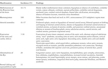
Table 1.
 You have
Access
- 20
- Cited by