Symptomatic neonates with tetralogy of Fallot remain a therapeutic challenge. The children may present with duct-dependent pulmonary circulation, unfavourable pulmonary artery anatomy, and critical pre-operative condition mainly related to refractory or repetitive hypercyanotic spells after closure of the patent arterial duct. Some centres have presented good results with primary repair in asymptomatic neonates; however, these neonates present with increased peri-operative morbidity and there is ongoing debate on the long-term effects on right ventricular performance. Traditionally, the insertion of aortopulmonary shunts is performed, and often this requires emergency operations.Reference Kanter, Kogon, Kirshbom and Carlock 1 There is evidence that palliative surgical enlargement of the right ventricular outflow tract especially in those patients with diminutive pulmonary arteries seems to be an adequate treatment strategy promoting adequate arterial oxygenation and pulmonary artery growth.Reference Kreutzer, Perry, Jonas, Mayer, Castañeda and Lock 2 – Reference Seipelt, Vazquez-Jimenez, Sachweh, Seghaye and Messmer 4 Balloon dilatation of the right ventricular outflow tract may be effective in those patients with predominant valvular obstruction,Reference Godart, Rey and Prat 5 , Reference Sluysmans, Neven and Rubay 6 but additional shunt insertion may be necessary especially in those with hypercyanotic spells indicating a predominantly infundibular component of the disease.Reference Wu, Wang, Lee, Chang and Wu 7 Stenting of the right ventricular outflow tract is reported to be successful in selected cases,Reference Shenoy, Padmakumar and Subashchandra 8 resulting in adequate oxygenation and increase in pulmonary artery size.Reference Dohlen, Chaturvedi and Benson 9 In the reported cases so far, stent implantation across the pulmonary valve resulted in free pulmonary regurgitation. It may be discussed whether the creation of a long stented infundibular segment with free pulmonary regurgitation may adversely affect right ventricular function. Finally, if stenting of the right ventricular outflow tract is performed in cases with only mild pulmonary annulus hypoplasia and a more dynamic obstruction at the infundibular level, this could potentially result in an acute ventricular left-to-right shunting with pulmonary overcirculation and secondary pulmonary hypertension.Reference Fraisse 10
We report our technique in two neonates presenting during the last year with severe hypercynaotic spells in tetralogy of Fallot. In both patients, stenting of the right ventricular outflow tract after balloon dilatation of the pulmonary valve was performed without overstenting the pulmonary valve.
Patients and methods
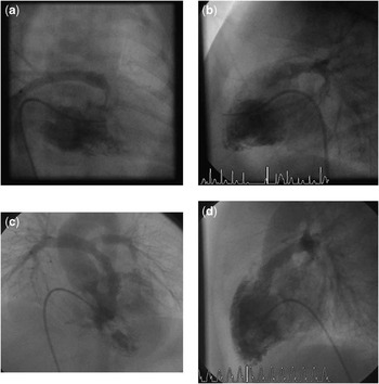
Patient 1 was a 10-day-old male (weight 3000 g, length 50 cm) who presented with tertralogy of Fallot, a small patent duct, and hypercyanotic spells. Echocardiographic examination revealed moderate overriding of the aorta (about 50%), a pulmonary valve annulus of 6 mm (Z-score: −0.5), adequate sized pulmonary arteries – left pulmonary artery 3.5 mm (Z-score: 0); right pulmonary artery 4.5 mm (Z-score: 1); Nakata-Index 125 mm2/m2 – but a severe infundibular hypertrophy and stenosis. A catheter investigation using deep conscious sedation was performed, and after balloon dilatation (7 × 20 mm) of the right ventricular outflow tract short segment infundibular stenosis (8 mm) remained the leading pathology resulting in cyanosis. Therefore, a premounted stent (7 × 12 mm, Palmaz® Blue™; Cordis, Bridgewater, New Jersey, United States of America) was implanted into the right ventricular outflow tract beneath the pulmonary valve, resulting in prompt and adequate pulmonary blood flow without significant pulmonary regurgitation (see Fig 1). The patient was discharged within 1 week and complete repair was performed at the age of 9 months. Catheterisation before surgery revealed no stenosis, no stent compression, and no fractures. Surgery was performed with explantation of the stent, transatrial and transpulmonary infundibulectomy without ventriculotomy, no transannular patch (pulmonary valve annulus 7.5 mm, Z-score: −1.5), closure of the ventricular septal defect, and patch augmentation of a stenosis of the left pulmonary artery.
( a ) Patient 1. Right ventricular angiography, frontal view, 20° cranial angulation. Note the complete long-segment obstruction of the lumen of the RVOT. The pulmonary valve is clearly visible and of adequate size; the left pulmonary artery showed some stenosis and is therefore not visible in this picture. ( b ) Patient 1. Right ventricular angiography, lateral view. Note the complete long-segment obstruction of the lumen of the RVOT. The pulmonary valve is clearly visible and of adequate size. ( c ) Patient 1. Right ventricular angiography, frontal view after stent implantation (Palmaz® Blue™). Note the adequate antegrade perfusion across the RVOT. The pulmonary valve is distal to the stent. ( d ) Patient 1. Right ventricular angiography, lateral view after stent implantation (Palmaz® Blue™). Note the adequate antegrade perfusion across the RVOT. The pulmonary valve is distal to the stent. RVOT = right ventricular outflow tract.

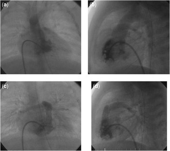
Patient 2 was a 4-week-old boy (2900 g, 48 cm) with severe tetralogy of Fallot and respiratory syncytial virus bronchiolitis requiring mechanical ventilation. He developed profound and sustained cyanosis (transcutaneous saturation between 45% and 60%) despite adequate supportive measures and ventilation strategy. Echocardiography showed a moderate overriding of the aorta (about 50%), a pulmonary valve annulus of 5 mm (Z-score: −1), moderate sized pulmonary arteries – left pulmonary artery 3 mm (Z-score: −1); right pulmonary artery 4 mm (Z-score: 0); Nakata-Index 97 mm2/m2 – but a severe and long (13 mm) infundibular hypertrophy and stenosis. An emergency catheter investigation was performed, and after balloon dilatation (6 × 20 mm) of the right ventricular outflow tract infundibular stenosis remained the leading pathology resulting in severe cyanosis. Therefore, a premounted stent (7 × 18 mm, Palmaz® Blue™) was implanted into the right ventricular outflow tract beneath the pulmonary valve, resulting in prompt and adequate pulmonary blood flow without significant pulmonary regurgitation (see Fig 2). The transcutaneous saturation increased to levels of 85% and the oxygen could be reduced from 100% to 30%. The patient was weaned from ventilation within 1 week and discharged home 1 week thereafter. He underwent surgical correction at the age of 6 months (pulmonary valve annulus 6.5 mm, Z-score: −1.5). Catheterisation before surgery revealed no stenosis, no stent compression, and no fractures. Surgery was performed with explantation of the stent, transatrial and transpulmonary infundibulectomy without ventriculotomy, no transanular patch, and closure of the ventricular septal defect.
( a ) Patient 2. Right ventricular angiography, frontal view. There is predominant blood flow to the aorta via the VSD and only diminutive flow to the pulmonary arteries. ( b ) Patient 2. Right ventricular angiography, lateral view. There is predominant blood flow to the aorta via the VSD; the RVOT is completely obstructed and there is only diminutive flow to the pulmonary arteries. ( c ) Patient 2. Right ventricular angiography, frontal view. After RVOT stening (Palmaz® Blue™), blood flow is now directed to the pulmonary arteries. ( d ) Patient 2. Right ventricular angiography, lateral view after stent implantation (Palmaz® Blue™). There is adequate antegrade perfusion across the right ventricular outflow tract. The pulmonary valve is distal to the stent. RVOT = right ventricular outflow tract; VSD = ventricular septal defect.

Stenting technique
After biplane RV-angiography, selective intubation of the right ventricular outflow tract is performed using a modified right Amplatzer coronary artery catheter, and the right ventricular outflow tract is crossed with a 0.014′′ floppy guide wire (Hi-Torque Floppy II Guide wire, 300 cm, Abbott Vascular Interventional BVBA, Diegem, Belgium). Then a 5-Fr-long sheath is positioned in the main pulmonary artery for stent delivery (Check-Flow Performer R Introducer, COOK, Bloomington, United States of America). Then a premounted bare metal stent (Palmaz® Blue™, 0.18 peripheral stent system, Cordis Europa, Roden, The Netherlands) is placed in the right ventricular outflow tract under repetitive control angiography via the side port of the long sheath. We thought that the great radial strength of this cobalt stent and its easy steerability made it an ideal stent in that situation. Finally, the stent is inflated to 7 mm with high pressure (12 atmospheres according to the manufacturer's manual). The stent size chosen was guarded by the measurements of the pulmonary valve annulus and the distal main pulmonary artery. Control angiography revealed a good position of the stent and adequate flow to the pulmonary arteries.
Discussion
Since the first surgical repair of Tetralogy of Fallot in 1956, there has been an ongoing debate and progress in the management of this often complex cardiac malformation. In some centres, neonatal primary repair is advocated in asymptomatic neonates, with the exception in the setting of small pulmonary arteries. In this specific group of patients, besides surgical palliation with shunt insertion, interventional palliation by balloon dilation of the pulmonary valve and arteries,Reference Sluysmans, Neven and Rubay 6 antegrade surgical palliation, or even repair with ventricular septal defect patch fenestration is performed.Reference Korbmacher, Heusch and Sunderdiek 3 , Reference Seipelt, Vazquez-Jimenez, Sachweh, Seghaye and Messmer 4
However, the need for early intervention in children with tetralogy of Fallot – at the neonatal age – compared with later intervention – at the age of 6 to 9 months – is usually related to factors that may also increase the risk of surgery in neonates, that is, respiratory or systemic infections, other systemic problems such as necrotising enterocolitis, and, most importantly, hypercyanotic episodes with potential neurological injury or even death. In those children with tetralogy of Fallot repaired in the neonatal age, long-term outcome was not as favourable as in those repaired at a later stage. In addition, poor pulmonary artery anatomy is related to adverse outcome and increased morbidity and mortality.Reference Fraisse 10
In many cases, the complete right ventricular outflow tract is narrowed and obstructed by displacement and hypertrophy of the infundibular septum and by hypertrophy of the free wall of the infundibulum. These patients tend to present with hypercyanotic spells. In addition, the pulmonary valve is usually abnormal with thickened leaflets and a variably hypoplastic annulus. Finally, but less frequently, the main and branch pulmonary arteries may be hypoplastic and have additional peripheral stenosis. Palliative therapy was previously limited to balloon valvulotomy for the rare patients in whom the obstruction is predominantly at the pulmonary valve level. In addition to that, stenting of the right ventricular outflow tract is a helpful additional tool to relieve the infundibular stenosis in those patients who present with hypercyanotic spells.Reference Godart, Rey and Prat 5 , Reference Shenoy, Padmakumar and Subashchandra 8 , Reference Dohlen, Chaturvedi and Benson 9 Adequate relief of the valvular component by balloon dilatation combined with the subvalvular stent implantation clearly optimises the antegrade perfusion in these patients. Placement of the stent clearly below the pulmonary valve annulus may be helpful in reducing the subsequent need for transannular patching when ultimately corrective surgery is performed. On the basis of the results in two patients only, it is, however, not possible to predict whether stenting of the outflow tract promotes pulmonary valve growth and pulmonary artery growth in all neonates with symptomatic tetralogy of Fallot.
The stents were not placed in the right ventricular outflow tract as a potential permanent treatment but as a transient solution. Basically, stent fractures may occur, but owing to the great radial strength of the stents used, potentially the relatively soft tissue of the neonatal cardiac musculature, and the relatively short duration of the stent placement no stent fractures were detected. In our experience, surgical extraction of the stents usually does not complicate total repair in these patients, because the stents placed for a shorter period of time did not produce severe adhesions or ingrowth.
As reported by others, palliation may minimise or obviate the need for surgical augmentation of pulmonary arteries at the time of repair. It has been postulated, that creating right ventricular to pulmonary artery continuity with an adequate size continuity and antegrade flow through central pulmonary arteries could promote a uniform increase in vessel size and even angiogenesis of distal microvessels, in particular when this occurs early in life. Furthermore, an additional practical advantage is that it allows easy access to peripheral pulmonary arteries for balloon dilatation of stenotic vessels if required. Additional advantages of early intervention may include early abolishment of cyanosis and minimisation of right ventricular hypertrophy and fibrosis. Finally, a longer intensive care unit stay is reported when repair is performed under 3 months of age and the neonatal brain may be more prone to surgery-related neurological injury.
In conclusion, stenting of the right ventricular outflow tract in symptomatic high-risk neonates is a feasible option that can be considered as an attractive alternative to early surgical repair or palliative surgical measures. The advantages of increased antegrade pulmonary blood flow may serve as an additional argument in those cases with diminutive or stenotic pulmonary arteries. In maintaining the function of the pulmonary valve, additional flow to the pulmonary arteries is supported by elevated diastolic pressure. Whether this procedure has an additional potential to support pulmonary valve growth and thereby avoid transannular patch repair later on is unclear.
Acknowledgements
None.
Financial Support
Hospital grant.
Conflicts of Interest
None.
Ethical Standards
The authors assert that all procedures contributing to this work comply with the ethical standards of the relevant German guidelines on good medical practice and human experimentation and with the Helsinki Declaration of 1975, as revised in 2008, and has been approved by the institutional committees of the Heart and Diabetes center.