Introduction
The genus Cryptosporidium comprises obligate protozoan parasites that predominantly inhabit the gastrointestinal epithelium of humans and other vertebrate animals (Fayer, Reference Fayer2010). Cryptosporidium has been under intensive investigation for more than 40 years and the enormous diversity in the genus has been revealed by genotyping studies conducted over the past 20 years. Studies on Cryptosporidium in humans and livestock have predominated due to the clinical and economic importance of cryptosporidiosis in these hosts (Robertson et al., Reference Robertson, Björkman, Axén, Fayer, Cacciò and Widmer2014; Kváč et al., Reference Kváč, McEvoy, Stenger, Clark, Cacciò and Widmer2014b). Research on Cryptosporidium spp. in wild animals has increased significantly in the last decade, expanding our knowledge of genetic diversity in the genus, but the biological properties of these parasites in wildlife remain poorly studied (Ren et al., Reference Ren, Zhao, Zhang, Ning, Jian, Wang, Lv, Wang, Arrowood and Xiao2012; Li et al., Reference Li, Pereira, Larsen, Xiao, Phillips, Striby, McCowan and Atwill2015; Kváč et al., Reference Kváč, Vlnatá, Ježková, Horčičková, Konečný, Hlásková, McEvoy and Sak2018; Tan et al., Reference Tan, Low, Ng, Ibrahim, Wang, Tan, Chellappan and Lim2019; Wei et al., Reference Wei, Liu, Zhao, Jiang, Zhang, Zhao, Jing, Lu and Qi2019). Recent studies indicate that rodents, which represent about 40% of the mammalian diversity, are predominantly parasitized by host-specific Cryptosporidium spp. with unknown biology (Lv et al., Reference Lv, Zhang, Wang, Jian, Zhang, Ning, Wang, Feng, Wang, Ren, Qi and Xiao2009; Feng et al., Reference Feng, Lal, Li and Xiao2011; Ng-Hublin et al., Reference Ng-Hublin, Singleton and Ryan2013; Stenger et al., Reference Stenger, Horčičková, Clark, Kváč, Čondlová, Khan, Widmer, Xiao, Giddings, Pennil, Stanko, Sak and McEvoy2017; Čondlová et al., Reference Čondlová, Horčičková, Havrdová, Sak, Hlásková, Perec-Matysiak, Kicia, McEvoy and Kváč2019). Today, 45 valid Cryptosporidium species and a similar number of genotypes have been reported (Holubová et al., Reference Holubová, Tůmová, Sak, Hejzlerová, Konečný, McEvoy and Kváč2020). Cryptosporidium muris and C. proliferans have a broad host range in the order Rodentia. In contrast, a narrow host specificity has been reported for C. alticolis and C. microti in voles, C. apodemi and C. ditrichi in apodemus mice, C. homai and C. wrairi in guinea pigs, C. tyzzeri in house mice, C. rubeyi in ground squirrels and C. occultus in rats (Tyzzer, Reference Tyzzer1910; Vetterling et al., Reference Vetterling, Jervis, Merrill and Sprinz1971; Ren et al., Reference Ren, Zhao, Zhang, Ning, Jian, Wang, Lv, Wang, Arrowood and Xiao2012; Li et al., Reference Li, Pereira, Larsen, Xiao, Phillips, Striby, McCowan and Atwill2015; Kváč et al., Reference Kváč, Havrdová, Hlásková, Daňková, Kanděra, Ježková, Vítovec, Sak, Ortega, Xiao, Modrý, Chelladurai, Prantlová and McEvoy2016, 2018; Zahedi et al., Reference Zahedi, Durmic, Gofton, Kueh, Austen, Lawson, Callahan, Jardine and Ryan2017; Čondlová et al., 2018; Horčičková et al., Reference Horčičková, Čondlová, Holubová, Sak, Květoňová, Hlásková, Konečný, Sedláček, Clark, Giddings, McEvoy and Kváč2018). Additionally, a large number of Cryptosporidium genotypes have been reported in rodents (Kváč et al., Reference Kváč, McEvoy, Stenger, Clark, Cacciò and Widmer2014b).
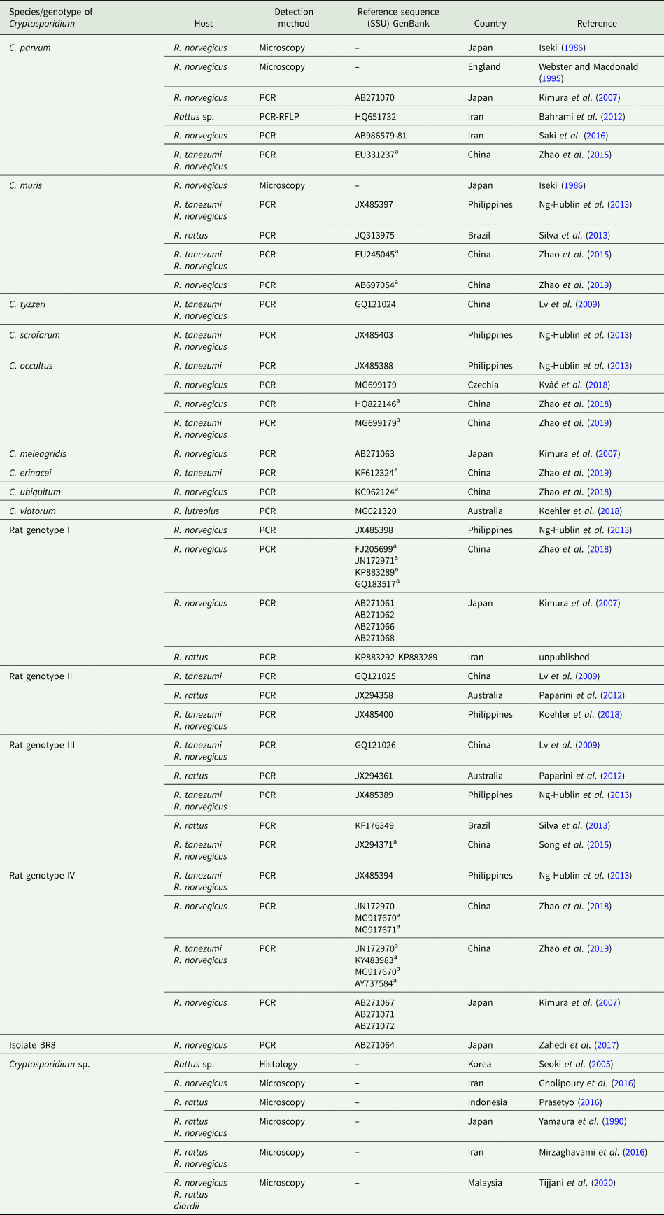
Representatives of the genus Rattus, which are globally distributed, with the exception of the polar region (Reid, Reference Reid2007; Thomson et al., Reference Thomson, Wiewel, Chinen, Maryanto, Sinaga, How, Aplin and Suzuki2018), have been reported as hosts of several Cryptosporidium spp. (Table 1). The recently described C. occultus is specific for rat hosts (Kváč et al., Reference Kváč, Vlnatá, Ježková, Horčičková, Konečný, Hlásková, McEvoy and Sak2018). Other species, C. muris, C. parvum, C. tyzzeri, C. scrofarum, C. meleagridis, C. erinacei, C. ubiquitum and C. viatorum, reported in rats are host-specific for other hosts. Rats probably represent minor host or the presence of these Cryptosporidium species is the result of the mechanical transmission of oocysts through the digestive tract (Kváč et al., Reference Kváč, Hanzlíková, Sak and Květoňová2009; Lv et al., Reference Lv, Zhang, Wang, Jian, Zhang, Ning, Wang, Feng, Wang, Ren, Qi and Xiao2009; Ng-Hublin et al., Reference Ng-Hublin, Singleton and Ryan2013; Tan et al., Reference Tan, Low, Ng, Ibrahim, Wang, Tan, Chellappan and Lim2019). Rats are frequently parasitized with Cryptosporidium rat genotypes I-IV, which have been reported in rats in Asia, Australia and South America (Table 1). As yet, there is no comprehensive genotyping study from Europe or North America and there is no knowledge of their biological properties including oocyst size, course and location of infection, or pathogenicity, etc. In the course of the study, we obtained an isolate of Cryptosporidium rat genotype I and examined its biological, morphological and genetic characteristics in detail. Our data showed that Cryptosporidium rat genotype I is genetically and biologically distinct from valid Cryptosporidium species and we propose to name it as a Cryptosporidium ratti n. sp.
Diversity of Cryptosporidium spp. in rat (Rattus sp.), brown rat (Rattus norvegicus), Asian house rat (Rattus tanezumi), Australian swamp rat (Rattus lutreolus), and Malayan black rat (Rattus rattus diardii) based on microscopic and molecular detection

a Indicates the sequence obtained in the paper has not been stored in the GenBank database and was identical to a sequence published previously.
Materials and methods
Area and specimens studied
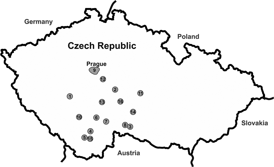
A total of 343 wild rats (Rattus norvegicus) were trapped using metal pedal or life traps at 16 localities in the Czech Republic over the period 2016–2019 (Fig. 1). Traps were checked every 3 hours and trapped animals were removed and transported to the Institute of Parasitology, Biology Centre CAS (PaU). Faecal samples from deceased rats were collected from the rectum during dissection. Live rats were individually housed with sterilised bedding, food and water. The feces of alive rats were collected individually for several days, each sample was individually examined for the presence of Cryptosporidium oocysts by the aniline–carbol–methyl violet staining (Miláček and Vítovec, Reference Miláček and Vítovec1985) followed by microscopic examination at 1000 × magnification (light microscope Olympus BX51, Tokyo, Japan), and specific DNA, by polymerase chain reaction (PCR)/sequencing targeting the small subunit ribosomal RNA gene (SSU) (below). If at least one sample was Cryptosporidium positive, the rat was considered positive. Alive rats that were negative for Cryptosporidium spp. were sacrificed humanly. Cryptosporidium positive rats were kept for several weeks and their feces were collected daily.
Sampling locations across the study area in the Czech Republic. Sample site numbers indicate the name of locations and coordinates are in brackets: (1) Březnice (49.556628, 13.954390), (2) Chyšná (50.545694, 13.437376), (3) Cizkrajov (49.0303555, 15.390124), (4) České Budějovice (48.974749, 14.453704), (5) Český Krumlov (48.813194, 14.321542), 6) Hodětín (49.251090, 14.547873), (7) Kardašova Řečice (49.18.2636, 14.848994), (8) Lidéřovice (49.064462, 15.373599), (9) Praha (50.074130, 14.522609), (10) Protivín (49.196654, 14.216850), (11) Přibyslav (49.580047, 15.739454), (12) Pyšely (49.875659, 14.680111), (13) Řevnov (49.475599, 14.632047), (14) Telč (49.184339, 15.472545), (15) Věžovatá Pláně (48.776780, 14.408550) and (16) Zmišovice (49.496220, 15.188810).

Molecular characterization
Total genomic DNA was extracted from 200 mg of feces or 100–200 mg of tissue specimens using a PSP spin stool DNA Kit (Invitek, Stratec, Berlin, Germany) followed by bead disruption for 60 s at 5.5 m s−1 using 0.5 mm glass beads in a FastPrep®-24 Instrument (MP Biomedicals, CA, USA). Purified DNA was stored at −20 °C prior to amplification by PCR. Fragments of the SSU, actin and the 70 kDa heat shock protein (HSP70) genes were amplified by nested PCR using published protocols and primers (Xiao et al., Reference Xiao, Morgan, Limor, Escalante, Arrowood, Shulaw, Thompson, Fayer and Lal1999; Sulaiman et al., Reference Sulaiman, Morgan, Thompson, Lal and Xiao2000, Reference Sulaiman, Lal and Xiao2002; Jiang et al., Reference Jiang, Alderisio and Xiao2005). Some PCR conditions were slightly modified from their original publications as previously described by Holubová et al. (Reference Holubová, Zikmundová, Limpouchová, Sak, Konečný, Hlásková, Rajský, Kopacz, McEvoy and Kváč2019). DNA of C. proliferans and molecular grade water were used as positive and negative controls, respectively. The secondary PCR products were separated by electrophoresis on a 1.5% agarose gel and visualized following staining with ethidium bromide. Amplicons were purified using the GenElute™ Gel Extraction Kit (Sigma-Aldrich, St. Louis, MO, USA) and sequenced in both directions using the secondary PCR primers at a commercial laboratory (SEQme, Dobříš, Czech Republic).
Phylogenetic analysis
The nucleotide sequences of each gene obtained from naturally- and experimentally-infected animals were verified by BLAST analysis (https://blast.ncbi.nlm.nih.gov/Blast.cgi), edited using Chromas Pro 2.1.4 (Technelysium, Pty, Ltd., South Brisbane, Australia) and aligned with reference sequences obtained from GenBank using BioEdit v.7.0.5 (Hall, Reference Hall1999). The alignments were end-trimmed and used in the phylogenetic analyses. Phylogenetic trees were inferred using the maximum likelihood (ML) method, with the substitution model that best fits the alignment selected using the Bayesian information criterion in MEGAX software. The robustness of the phylogeny was tested with 1000 bootstraps. Phylograms were edited for style using CorelDrawX7. Sequences have been deposited in GenBank under the Accession Numbers (Acc. nos.): MT504538-MT504544 for SSU, MT507482-MT507485 for HSP70, and MT507486-MT507491 for actin.
Origin of Cryptosporidium ratti n. sp. isolate
An isolate of C. ratti n. sp. was obtained from a wild-caught rat (isolate 29 356; Rat 0) trapped at locality no. 16 (Telč). The rat was individually housed with sterile bedding and provided with sterile food and water. The bedding was changed every second day. Oocysts were purified using cesium chloride gradient centrifugation (Arrowood and Donaldson, Reference Arrowood and Donaldson1996) and used for morphometry and phylogenetic analysis (SSU, actin and HSP70 genes). Oocysts obtained from Rat 0 were used to infect a single 1-week-old rat (Rat 1). Oocysts of C. ratti n. sp. obtained from Rat 1 were purified using cesium chloride gradient centrifugation and their viability was examined using propidium iodide (PI) staining by a modified assay of Sauch et al. (Reference Sauch, Flanigan, Galvin, Berman and Jakubowski1991). They were used for morphometry and phylogenetic analysis and for experimental infection of other animals (see the transmission studies section). The oocysts were stored in PBS at 4–8 °C for a maximum of 3 weeks.
Transmission studies
Five 1-week-old and 8-week-old severe combined immunodeficiency (SCID) mice (strain C.B-17), BALB/c mice (Mus musculus), Mongolian gerbils (Meriones unguiculatus) and laboratory rats (Rattus norvegicus, strain Wistar Han) were used for transmission studies. Three animals from each host species/strain were used as negative controls. All experimental 1-week- and 8-week-old animals were inoculated by oesophageal tube with 10 000 purified oocysts of C. ratti n. sp. (Rat 1 origin) suspended in 50 and 200 μL of sterile PBS, respectively. Animals used as negative controls were inoculated with the same volume of sterile PBS. For a week prior to infection, faecal samples from all experimental animals were screened daily for the presence of Cryptosporidium oocysts and DNA using aniline–carbol–methyl violet staining and nested PCR targeting the SSU gene, respectively. To prevent environmental contamination with oocysts, laboratory rodents were housed in plastic cages and supplied with a sterilized diet (TOP-VELAZ, Prague, Czech Republic) and sterilized water ad libitum. Starting on the second-day post infection, faecal samples from each animal were screened daily for the presence of Cryptosporidium oocysts and DNA using aniline–carbol–methyl violet staining and nested PCR targeting the SSU gene, respectively. One animal from each experimental group was euthanatized at 10 and 20 days post infection (DPI). Tissue specimens from the oesophagus, stomach, small intestine and large intestine (the entire tract was divided into 1 cm-long sections), trachea, lungs, liver and kidney were sampled and processed for PCR targeting the SSU gene, histology and scanning electron microscopy. Specimens for histology and electron microscopy were processed according to Holubová et al. (Reference Holubová, Zikmundová, Limpouchová, Sak, Konečný, Hlásková, Rajský, Kopacz, McEvoy and Kváč2019). All experiments were terminated at 30 DPI. Faecal consistency, faecal colour and animal behaviour were examined daily. Animals received standard care at the Institute of Parasitology (IP) (Holubová et al., Reference Holubová, Zikmundová, Limpouchová, Sak, Konečný, Hlásková, Rajský, Kopacz, McEvoy and Kváč2019). All housing, feeding and experimental procedures were conducted under protocols approved by the IP and the Central Commission for Animal Welfare, Czech Republic (protocol nos. 55/2014, 35/2018 and MZP/2019/630/1411).
Morphometric analysis
Oocyst size was determined using digital analysis of images (Olympus cellSens Entry 2.1 software, Olympus Corporation, Shinjuku, Tokyo, Japan) collected using an Olympus Digital Colour Camera DP73 (Olympus). The length and width of C. ratti n. sp. oocysts from naturally- (Rat 0) and experimentally-infected animals (20 oocysts from each isolate) were examined using differential interference contrast (DIC) microscopy at 1000 × magnification (Olympus IX70, Tokyo, Japan). These measurements were used to calculate the length-to-width ratio. Samples containing purified C. parvum oocysts (calf origin) were used as a size control. Oocyst size was measured using the same microscope and by the same person. Each slide was screened using a meandering path to prevent repeated measurement of an oocyst. Additionally, faecal smears with oocysts of C. ratti n. sp. and C. parvum (data not shown) were stained by modified Ziehl-Neelsen (ZN; Henriksen and Pohlenz, Reference Henriksen and Pohlenz1981) and labelled with a Cy3-labeled mouse monoclonal antibody targeting the Cryptosporidium oocyst outer wall antigenic sites (A400Cy2R-20X, Crypt-a-Glo, Waterborne, Inc, New Orleans, LA, USA).
Statistical analysis
Differences in Cryptosporidium spp. oocysts size were tested using Hotelling's multivariate version of the 2 sample t-test, package ICSNP: Tools for Multivariate Nonparametrics (Nordhausen et al., Reference Nordhausen, Sirkia, Oja and Tyler2018) in R 4.0.0. (R Core Team, 2019). The hypothesis tested was that two-dimensional mean vectors of measurement are the same in the two populations being compared.
Results
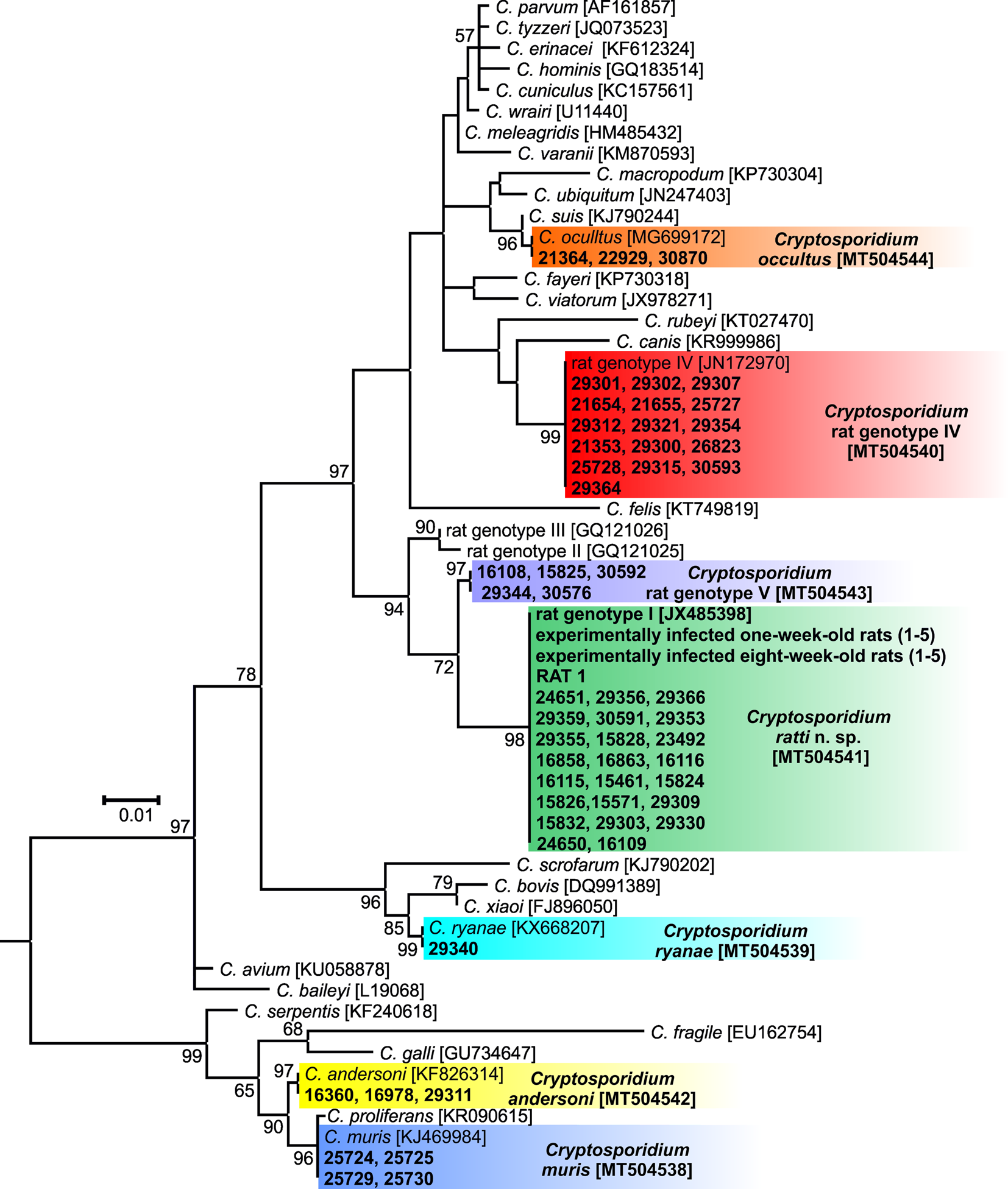
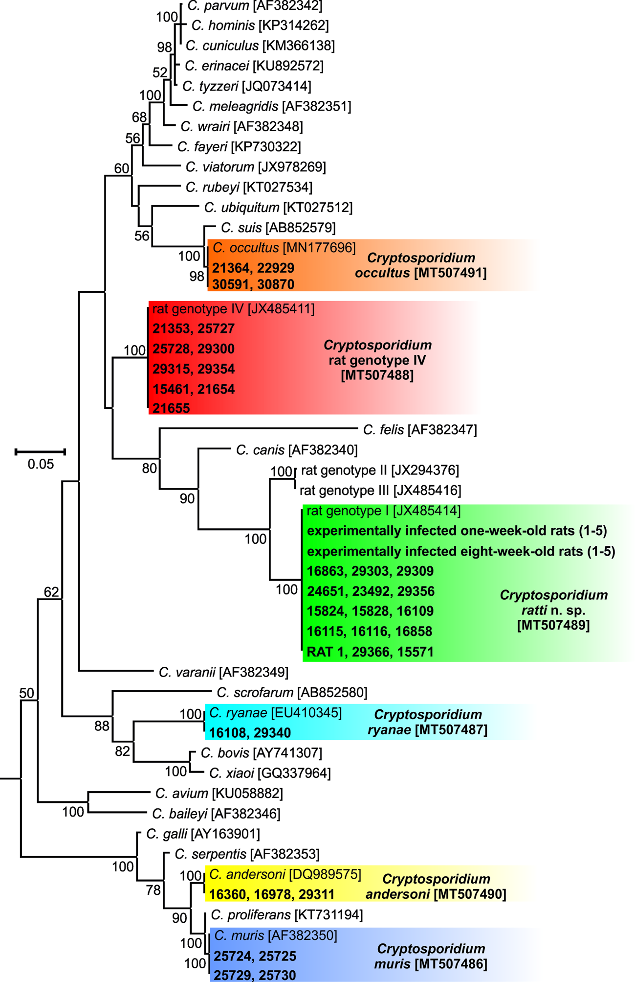
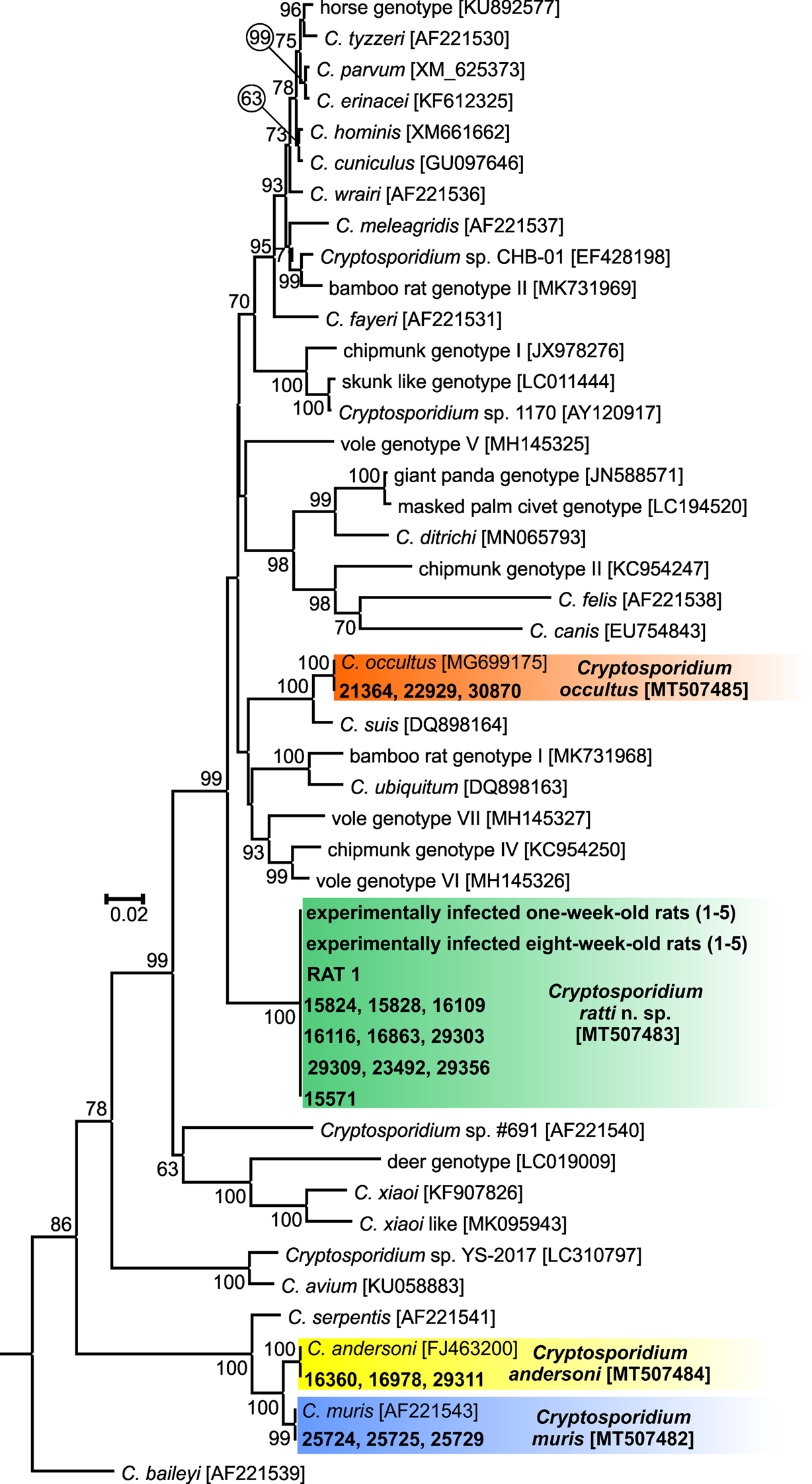
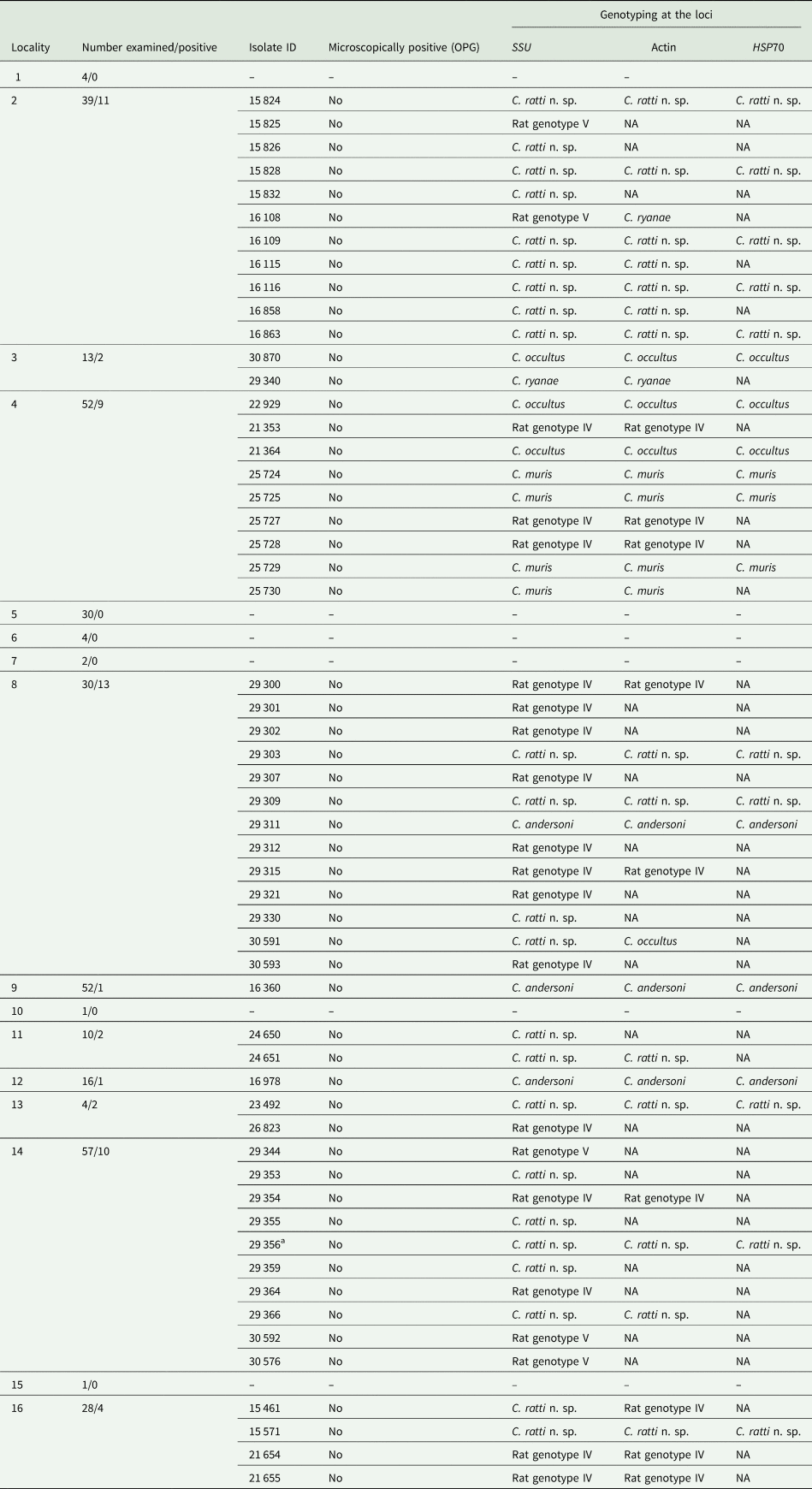
A total of 343 faecal samples were obtained from trapped brown rats at 16 localities were tested for the presence of Cryptosporidium spp. (Table 2). Cryptosporidium-specific DNA was detected in 55 samples by nested PCR targeting the SSU gene. None of the samples was positive for Cryptosporidium oocysts by microscopy. Out of the 55 Cryptosporidium-positive rats, 55, 36 and 19 were genotyped by sequence analysis of the SSU, actin and HSP70 genes, respectively. The remaining positive samples failed to amplify at the actin (n = 19) and HSP70 (n = 36) loci (Table 1). ML trees constructed from SSU sequences showed the presence of C. muris (n = 4), C. andersoni (n = 3), C. ryanae (n = 1), C. occultus (n = 3), C. ratti n. sp. (n = 23) and Cryptosporidium rat genotype IV (n = 16). Five isolates clustered in a novel group, which we have named Cryptosporidium rat genotype V. This group was closely related to C. ratti n. sp. and Cryptosporidium rat genotypes II and III (Fig. 2, Table 2). For the actin gene, isolates of C. occultus, C. ryanae, C. muris, C. andersoni, C. ratti n. sp. and Cryptosporidium rat genotype IV shared 100% sequence identity with sequences of Cryptosporidium spp. previously reported (Fig. 3). Actin sequences were not detected in any of the samples that were positive for Cryptosporidium rat genotype V at the SSU locus. A mixed infection was detected in three samples – isolate 16 108 was positive for C. ryanae at actin and for Cryptosporidium rat genotype IV at SSU; isolate 30 591 was positive for C. occultus at actin and for C. ratti n. sp. at SSU; and isolate 15 461 was positive for Cryptosporidium rat genotype IV at actin and for C. ratti n. sp. at SSU (Table 1, Fig. 3). None of the samples with mixed infection were successfully sequenced at the HSP70 locus. At the HSP70 gene, none of the isolates positive for Cryptosporidium rat genotype IV or V was amplified. Likewise, 13 of the 23 positive for C. ratti n. sp. and one of the four positive for C. muris failed to be amplified at the HSP70 gene (Table 2, Fig. 4). The sequences of individual Cryptosporidium species and genotypes detected in this study were identical to each other (Figs 2–4).
Maximum likelihood tree based on partial sequences of the gene encoding the small subunit rRNA (SSU), including sequences obtained from naturally- and experimentally-infected hosts in this study. Tamura's 3-parameter model was applied, using a discrete Gamma distribution and invariant sites. The robustness of the phylogeny was tested with 1000 bootstraps and the numbers at the nodes represent the bootstrap P values with more than 50% bootstrap support. The branch length scale bar, indicating the number of substitutions per site, is included. Sequences obtained in this study are identified by isolate number (e.g. 29 356). The GenBank Accession number is in the bracket. Cryptosporidium species and genotypes detected in this study are colour-coded. The tree was rooted with the SSU sequence of Plasmodium falciparum (JQ627151) and the root was removed from the figure.

Maximum likelihood tree based on partial sequences of the actin gene. The General Time Reversible model was applied, using a discrete Gamma distribution and invariant sites. The robustness of the phylogeny was tested with 1000 bootstraps and the numbers at the nodes represent the bootstrap P values with more than 50% bootstrap support. The branch length scale bar, indicating the number of substitutions per site, is included. Sequences obtained in this study are identified by isolate number (e.g. 29 356). The GenBank Accession number is in the bracket. Cryptosporidium species and genotypes detected in this study are colour-coded. The tree was rooted with the actin sequence of Eimeria maxima (XM013478337) and the root was removed from the figure.

Maximum likelihood tree based on partial sequences of the Heat Shock Protein 70 (HSP70) gene. The General Time Reversible model was applied, using a discrete Gamma distribution. The robustness of the phylogeny was tested with 1000 bootstraps and the numbers at the nodes represent the bootstrap P values with more than 50% bootstrap support. The branch length scale bar, indicating the number of substitutions per site, is included. Sequences obtained in this study are identified by isolate number (e.g. 29 356). The GenBank Accession number is in the bracket. Cryptosporidium species and genotypes detected in this study are colour-coded. The tree was rooted with the HSP70 sequence of Eimeria maxima (Z46964) and the root was removed from the figure.

Cryptosporidium spp. in wild brown rats (Rattus norvegicus) at localities in the Czech Republic

Oocysts were quantified by microscopy and reported per gram of feces (OPG). Fragments of the small subunit rDNA (SSU), actin and heat shock protein 70 (HSP70) genes were amplified by PCR. NA indicates PCR amplification failure.
a Isolate of Cryptosporidium rat genotype I used for experimental studies.
Purified oocysts of C. ratti n. sp. from Rat 0 (isolate 29 356) trapped at locality no. 14 did not infect 8-day-old BALB/c mice (n = 3); whereas, an 8-day-old rat (Rat 1) was successfully infected. The oocysts purified from experimentally-infected 1-week- and 8-week-old rats (below) were morphometrically identical to oocysts recovered from RAT 0 and RAT 1. The sequences of the SSU, actin and HSP70 genes obtained from Rat 1 were identical to those of Rat 0 (isolate 29 356). Oocysts recovered from Rat 1 were used for the description of oocyst morphometry, as well as transmission and molecular studies.
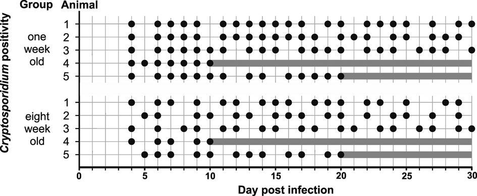
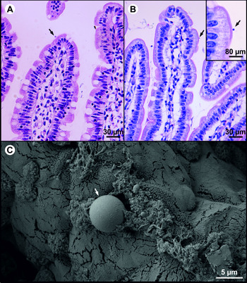
Cryptosporidium ratti n. sp. oocysts (Rat 1 origin) were only infectious for 1-week- and 8-week-old rats (Fig. 5). All rats started to shed Cryptosporidium oocysts detectable by PCR at 4–5 DPI. Microscopically detectable infection was not observed in any rat. The presence of specific C. ratti n. sp. DNA in faecal specimens was more often detected in rats infected at 1-week-old (21 times during the experiment) compared to rats infected at 8-weeks-old (16–18 times, Fig. 5). All rats remained infectious until the end of the experiment (Fig. 5). Examination of the gastrointestinal tract tissue of 1-week- and 8-week-old rats at 10, 20 and 30 DPI by PCR, histology and electron microscopy revealed the presence of specific DNA and developmental stages of C. ratti n. sp. in the jejunum and ileum. Developmental stages were scattered on an isolated villus (Fig. 6). The lamina propria in the jejunum was sporadically slightly edematous, but these changes were probably not related to the Cryptosporidium infection. A slight multiplication of goblet cells on infected villi was observed in the posterior part of the ileum. One-week- and 8-week-old BALB/c and SCID mice, as well as gerbils experimentally inoculated with oocysts of C. ratti n. sp. (Rat 1 origin), did not develop infections detectable in feces by microscopy or PCR. These animals also had no endogenous stages detectable by histology or electron microscopy. All groups of rats, mice and gerbils used as negative controls remained uninfected.
Course of infection of Cryptosporidium ratti n. sp. in 1-week- and 8-week-old rats (Rattus norvegicus) based on microscopic and molecular (SSU) examination of feces. Grey circle indicates the detection of oocysts by microscopy, black circle indicates the detection of specific DNA by PCR. Grey line represents absence of rat due to sacrificing at 10 or 20 days post infection.

Presence of developmental stages of Cryptosporidium ratti n. sp. (arrow) on jejunal mucosal epithelium in rat (Rattus norvegicus) infected at 1-week-old and sacrificed 10 days post infection. (A) and (B) histological sections stained by hematoxylin eosin, (C) scanning electron microphotograph. Scale bar is included in each figure.

Taxonomic summary
Family Cryptosporidiidae Léger, 1911
Genus Cryptosporidium Tyzzer, 1907
Cryptosporidium ratti n. sp.
Syn: Cryptosporidium rat genotype I ex Rattus norvegicus of Zhao et al. (Reference Zhao, Wang, Ren, Yang, Yang, Zhang, Xu, Liu and Ling2018), Japan Kimura et al. (Reference Kimura, Edagawa, Okada, Takimoto, Yonesho and Karanis2007) and Philippines Ng-Hublin et al. (Reference Ng-Hublin, Singleton and Ryan2013); Cryptosporidium sp. rat genotype rat193 ex Rattus norvegicus (Gen Bank no. JN172971, unpublished); Cryptosporidium environmental sequence clone ECUST628 from wastewaters of Feng et al. (Reference Feng, Li, Duan and Xiao2009); Cryptosporidium sp. 2162 ex Boa constrictor subsp. ortoni of Xiao et al. (Reference Xiao, Ryan, Graczyk, Limor, Li, Kombert, Junge, Sulaiman, Zhou, Arrowood, Koudela, Modrý and Lal2004); Cryptosporidium sp. rat genotype from raw water of Chalmers et al. (Reference Chalmers, Robinson, Elwin, Hadfield, Thomas, Watkins, Casemore and Kay2010), Cryptosporidium sp. 18 and 23 ex Rattus rattus (Gen Bank no. KP883292 and KP883289, respectively, unpublished).
Type-host: Rattus norvegicus (Berkenhout, 1769) (Rodentia: Muridae), brown rat.
Other natural hosts: Rattus rattus (Linnaeus, 1758), black rat.
Type-locality: Telč (49.184339N, 15.472545E), Czech Republic.
Other localities: Chyšná (50.545694N, 13.437376E), Czech Republic; Lidéřovice (49.064462N, 15.373599E), Czech Republic; Přibyslav (49.580047N, 15.739454E), Czech Republic; Řevnov (49.475599N, 14.632047E), Czech Republic; Zmišovice (49.496220N, 15.188810E), Czech Republic.
Type-material: Histological sections of infected jejunum (nos. 181–183/2016) and ileum (nos. 184–189/2016); scanning electron microscopy specimens of infected jejunum (nos. 181–183/2016) and ileum (nos. 184–189/2016); genomic DNA isolated from faecal samples of naturally- (isolate 29 356) and experimentally- (isolate 16 848) infected rats; genomic DNA isolated from jejunum and ileum of experimentally-infected rat (isolate 44 331); faecal smear slides with oocysts stained by ACMV and ZN staining (nos. 6/16848 and 15/16853). Specimens deposited at the Institute of Parasitology, Biology Centre of the Czech Academy of Sciences, Czech Republic.
Site of infection: Jejunum and ileum (present study, Fig. 6).
Distribution: As Cryptosporidium rat genotype I ex Rattus norvegicus: China (Zhao et al., Reference Zhao, Wang, Ren, Yang, Yang, Zhang, Xu, Liu and Ling2018), Japan (Kimura et al., Reference Kimura, Edagawa, Okada, Takimoto, Yonesho and Karanis2007) and Philippines (Ng-Hublin et al., Reference Ng-Hublin, Singleton and Ryan2013); Cryptosporidium sp. 2162 ex Boa constrictor subsp. ortoni in USA (Xiao et al., Reference Xiao, Ryan, Graczyk, Limor, Li, Kombert, Junge, Sulaiman, Zhou, Arrowood, Koudela, Modrý and Lal2004); Cryptosporidium sp. 18 and 23 ex Rattus rattus in Iran; Cryptosporidium sp. rat genotype from raw water in the UK (Chalmers et al., Reference Chalmers, Robinson, Elwin, Hadfield, Thomas, Watkins, Casemore and Kay2010).
Prepatent period: Rattus norvegicus: 4–5 DPI.
Patent period: At least 30 DPI in all experimentally infected rats (Rattus norvegicus)
Representative DNA sequences: Representative nucleotide sequences of SSU (MT504541), actin (MT507489) and HSP70 (MT507483) genes were saved in the GenBank database.
ZooBank registration: To comply with the regulations set out in Article 8.5 of the amended 2012 version of the International Code of Zoological Nomenclature (ICZN, Reference ICZN2012), details of the new species have been submitted to ZooBank. The Life Science Identifier (LSID) of the article is urn:lsid:zoobank.org:pub:59E724AA-5CBB-4E81-96C3-397D858E782D. The LSID for the new name Cryptosporidium ratti is urn:lsid:zoobank.org:act:C42A2AFD-7DB1-4B2E-AA37-3FAC0B069A26.
Etymology: The species name ratti is derived from the Latin noun “rattus” (meaning rat).
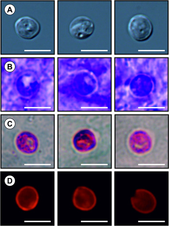
Description: Oocysts obtained from fresh feces specimens ex Rattus norvegicus (isolate 29 356) were spherical measuring 4.4–5.4 × 4.3–5.1 μm (4.9 ± 0.2 × 4.6 ± 0.2 μm) with a length to width ratio of 1.0–1.1 (1.1 ± 0.1) (Fig. 7). The oocyst wall was smooth and colourless, composed of a single layer. Micropyle and polar granule were absent, oocyst residuum was present, composed of numerous small granules and one spherical globule. Four sporozoites were present within each oocyst. Morphology and morphometry of other developmental stages are unknown.
Oocysts of Cryptosporidium ratti n. sp. (A) differential interference contrast microscopy, (B) aniline-carbol-methyl violet staining, (C) Ziehl-Nielsen staining, (D) labelling with antibody reagent consisting of a Cy3-labeled mouse monoclonal antibody made against Cryptosporidium oocyst outer wall antigenic sites. Bar = 5 μm.

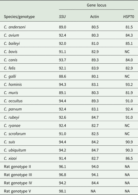
Remarks: Oocysts of Cryptosporidium ratti n. sp. showed typical Cryptosporidium ACMV and ZN staining characteristics and cross-react with immunofluorescence reagents developed primarily for C. parvum. There were no statistically significant size differences between oocysts from naturally infected rat and oocysts obtained from experimentally infected rat which measured 4.5–5.4 × 4.5–5.0 μm (4.9 ± 0.3 × 4.7 ± 0.2 μm) with a length/width ratio of 1.0–1.1 (1.1 ± 0.1) (T 2 = 4.26, df 1 = 2, df 2 = 35.62, P = 0.1408). Oocysts of C. ratti n. sp. are smaller than those of C. parvum (T 2 = 18.88, df 1 = 2, df 2 = 27.88, P = 0.009) and C. occultus (T 2 = 30.38, df 1 = 2, df 2 = 28.24, P < 0.0001). Cryptosporidium ratti n. sp. can be differentiated genetically from other Cryptosporidium species based on the sequences of SSU, actin and HSP70 genes. Percentage of nucleotide similarities at the SSU locus between C. ratti n. sp. and the rat derived C. occultus and Cryptosporidium rat genotype II, III, IV and V was 94.4, 96.1, 96.8, 94.2 and 98.1%, respectively (Table 3). At the actin locus, C. ratti n. sp. shared 89.3, 94.0, 94.1 and 84.4% sequence identity, respectively, with C. occultus and rat derived Cryptosporidium genotype II, III and IV (Table 3). At the HSP70 locus, C. ratti n. sp. exhibited 91.0% sequence identity with C. occultus (Table 3).
Percentage of nucleotide similarities between Cryptosporidium ratti n. sp. and selected closest and furthest Cryptosporidium species and Cryptosporidium rat genotypes II-V at small subunit ribosomal RNA (SSU), actin and 70 kDa heat-shock protein (HSP70) genes

NA, sequences are not available; NC, partial sequence does not cover sequence of C. ratti n. sp.
Discussion
At least 17 Cryptosporidium spp. has been detected in rats worldwide (Kimura et al., Reference Kimura, Edagawa, Okada, Takimoto, Yonesho and Karanis2007; Lv et al., Reference Lv, Zhang, Wang, Jian, Zhang, Ning, Wang, Feng, Wang, Ren, Qi and Xiao2009; Ng-Hublin et al., Reference Ng-Hublin, Singleton and Ryan2013; Zhao et al., Reference Zhao, Wang, Zhao, Qi, Zhao, Zhang, Li and Liu2015; Koehler et al., Reference Koehler, Wang, Haydon and Gasser2018; Kváč et al., Reference Kváč, Vlnatá, Ježková, Horčičková, Konečný, Hlásková, McEvoy and Sak2018). The high number of detected species and genotypes in rats compared to other vertebrates may be explained by the frequent presence of non-rat-host-specific Cryptosporidium spp. It is possible that in cases of the presence of non-rat-host-specific Cryptosporidium spp., we detected only DNA from the mechanical transmission, as has been previously reported in other studies (Crawshaw and Mehren, Reference Crawshaw, Mehren, Ippen and Schroder1987; Graczyk et al., Reference Graczyk, Cranfield, Fayer and Anderson1996; Kváč et al., Reference Kváč, Kestřánová, Květoňová, Kotková, Ortega, McEvoy and Sak2012). This presumption is supported by the fact that most of the non-rat-host-specific species come from either farm animals or from animals that are the prey of rats. In this study, we found C. ryanae and C. andersoni in rats trapped on dairy farms (data not shown). Similarly, Ng-Hublin et al. (Reference Ng-Hublin, Singleton and Ryan2013) consider the occurrence of C. scrofarum in rats in the Philippines to be mechanical transmission, as pig entrails are present at the markets and pigs are raised in the villages close to the rice fields where the rats were trapped. Also, detection of C. ratti n. sp., rat-specific Cryptosporidium, in Boa constrictor subsp. ortoni by Xiao et al. (Reference Xiao, Ryan, Graczyk, Limor, Li, Kombert, Junge, Sulaiman, Zhou, Arrowood, Koudela, Modrý and Lal2004) probably represent mechanical passage after the snake caught the infected rat. It is worth noting that all non-rat-host-specific Cryptosporidium species – C. meleagridis, C. erinacei, C. ubiquitum, C. tyzzeri and C. viatorum – have only been found in some studies and rarely to a high degree within them. For the most part, one to three positive rats were detected, as in this study (Kimura et al., Reference Kimura, Edagawa, Okada, Takimoto, Yonesho and Karanis2007; Koehler et al., Reference Koehler, Wang, Haydon and Gasser2018; Zhao et al., Reference Zhao, Wang, Ren, Yang, Yang, Zhang, Xu, Liu and Ling2018, 2019). These results show a random distribution of these Cryptosporidium spp. rather than adaptation to the host (Tan et al., Reference Tan, Low, Ng, Ibrahim, Wang, Tan, Chellappan and Lim2019). The presence of C. parvum in most of the studies is not surprising, as it lacks host specificity. This shows that rats are susceptible, although not the typical hosts, which are livestock (Nydam et al., Reference Nydam, Wade, Schaaf and Mohammed2001). Comparable to previous studies, we found a low occurrence of C. muris, a species with broad host specificity within rodents, which suggests, as in the case of C. parvum, that rats are natural but not typical hosts. The frequent occurrence and high prevalence of Cryptosporidium ratti n. sp. (previously known as Cryptosporidium rat genotype I) and Cryptosporidium rat genotypes II-IV in previous as well as this study and the fact that these Cryptosporidium spp. have very rarely or never been detected in other hosts could imply that this species is host-specific for rats (Kimura et al., Reference Kimura, Edagawa, Okada, Takimoto, Yonesho and Karanis2007; Lv et al., Reference Lv, Zhang, Wang, Jian, Zhang, Ning, Wang, Feng, Wang, Ren, Qi and Xiao2009; Paparini et al., Reference Paparini, Jackson, Ward, Young and Ryan2012; Ng-Hublin et al., Reference Ng-Hublin, Singleton and Ryan2013; Silva et al., Reference Silva, Richtzenhain, Barros, Gomes, Silva, Kozerski, de Araujo Ceranto, Keid and Soares2013; Kváč et al., Reference Kváč, Vlnatá, Ježková, Horčičková, Konečný, Hlásková, McEvoy and Sak2018; Zhao et al., Reference Zhao, Wang, Ren, Yang, Yang, Zhang, Xu, Liu and Ling2018). Additionally, the finding that rats are susceptible to C. ratti n. sp. infection under experimental conditions, while mice and gerbils are not, supports the narrow host specificity of this species.
In contrast to other studies from Asia, Australia and South America, we did not detect any Cryptosporidium rat genotypes II and III (Lv et al., Reference Lv, Zhang, Wang, Jian, Zhang, Ning, Wang, Feng, Wang, Ren, Qi and Xiao2009; Paparini et al., Reference Paparini, Jackson, Ward, Young and Ryan2012; Ng-Hublin et al., Reference Ng-Hublin, Singleton and Ryan2013; Silva et al., Reference Silva, Richtzenhain, Barros, Gomes, Silva, Kozerski, de Araujo Ceranto, Keid and Soares2013; Zhao et al., Reference Zhao, Zhou, Huang, Xu, Rao, Wang, Wang, Yi, Zhou, Wu, Ma, Wang, Hu, Peng, Yin and Lu2019). Given that this work is the first comprehensive study from Europe, it would not be appropriate to draw conclusions regarding the absence of these genotypes in the Czech Republic. Further studies are needed. Similarly, Čondlová et al. (Reference Čondlová, Horčičková, Havrdová, Sak, Hlásková, Perec-Matysiak, Kicia, McEvoy and Kváč2019) detected Cryptosporidium apodemus genotypes I and II across Europe, including the Czech Republic, in a 2019 study, although both genotypes were missing in their study performed in the Czech Republic in 2018 (Čondlová et al., 2018). On the other hand, the absence of Cryptosporidium rat genotypes II and III may be suggestive of patterns of geographical distribution of these genotypes.
The novel Cryptosporidium rat genotype V, which we found in five animals from two locations, has never been detected in other hosts or wastewater. Repeated detection in independent samples more than t2 years apart (data not shown) may indicate that Cryptosporidium rat genotype V is infectious to rats. More studies are needed to confirm that this genotype is specific for rats and to explain why it was not detected in previous studies.
Although the diagnostic methods using microscopy are still frequently used for differentiation among species due to their simplicity and low cost, it is difficult to distinguish among the various Cryptosporidium species and genotypes because the size variability of the oocysts is small and the oocyst size of most Cryptosporidium genotypes is unknown. Although, oocyst size of C. ratti n. sp. difference from other C. occultus (5.2 × 4.9 μm), C. parvum (5.3 × 4.7 μm), it would be difficult to differentiate it microscopically from these and other Cryptosporidium species reported in rats. For example, C. tyzzeri (4.6 × 4.2 μm), C. meleagridis (5.2 × 4.6 μm), C. erinacei (4.9 × 4.4 μm), C. ubiquitum (5.0 × 4.7 μm) and C. viatorum (5.4 × 4.7 μm) have morphometrically similar oocysts (Lindsay et al., Reference Lindsay, Blagburn, Sundermann and Hoerr1989; Fayer et al., Reference Fayer, Santín and Macarisin2010; Elwin et al., Reference Elwin, Hadfield, Robinson, Crouch and Chalmers2012; Ren et al., Reference Ren, Zhao, Zhang, Ning, Jian, Wang, Lv, Wang, Arrowood and Xiao2012; Kváč et al., Reference Kváč, Hofmannová, Hlásková, Květoňová, Vítovec, McEvoy and Sak2014a; Kváč et al., Reference Kváč, Vlnatá, Ježková, Horčičková, Konečný, Hlásková, McEvoy and Sak2018).
Cryptosporidium ratti n. sp. is genetically distinct from valid Cryptosporidium species at SSU, actin and HSP70 and did not exhibit sequence heterogeneity. At the SSU locus, C. ratti n. sp. formed a separate cluster with Cryptosporidium rat genotype II, III and V and was closely related to C. felis with nucleotide similarities of 96.2, 96.0, 98.1, and 92.1%, respectively. These genetic variations were greater than that observed between close related species, i.e. C. occultus and C. suis (99.5%) or C. muris and C. andersoni (99.0%), and similar to that observed between distinct related species, i.e. C. parvum and C. erinacei (93.2%) or C. alticolis and C. ditrichi (96.1%). At actin locus, C. ratti n. sp. clustered together with C. canis, C. felis, and Cryptosporidium rat genotypes II and III with nucleotide similarity of 89.3, 83.9, 94.0 and 94.1%, respectively. These genetic variations are greater than those between i.e. C. parvum and C. eriancei (99.5%) and similar to those between C. ryanae and C. bovis (88.9%). There are missing nucleotide sequences of several Cryptosporidium species at the HSP70 locus. Analyses of the HSP70 locus indicate that C. ratti n. sp. and C. occultus, the rat-specific Cryptosporidium species, shared a nucleotide similarity of 91.0%. In comparison, i.e. C. parvum and C. erinacei share 99.2% similarity and i.e. C. parvum and C. andersoni 88.0%.
The prepatent period of C. ratti n. sp. was 4–5 DPI, which is consistent with C. occultus in rats (4–5 DPI) and other intestinal Cryptosporidium spp.: for example, C. alticolis in voles (3–4 DPI), C. parvum in calves (2–7 DPI), C. tyzzeri in mice (4–7 DPI), C. xiaoi in sheep (7–8 DPI), and C. scrofarum in pigs (4–6 DPI) (Tzipori et al., Reference Tzipori, Smith, Halpin, Angus, Sherwood and Campbell1983; Fayer and Santín, Reference Fayer and Santín2009; Ren et al., Reference Ren, Zhao, Zhang, Ning, Jian, Wang, Lv, Wang, Arrowood and Xiao2012; Kváč et al., Reference Kváč, Kestřánová, Pinková, Květoňová, Kalinová, Wagnerová, Kotková, Vítovec, Ditrich, McEvoy, Stenger and Sak2013, 2018; Horčičková et al., Reference Horčičková, Čondlová, Holubová, Sak, Květoňová, Hlásková, Konečný, Sedláček, Clark, Giddings, McEvoy and Kváč2018). Unlike C. occcultus, which causes a massive infection of the colonic epithelium but low shedding of oocysts, C. ratti n. sp. causes a weak infection of the small intestine and the intensity of oocyst shedding matches the intensity of the developmental stages observed in the epithelium (Kváč et al., Reference Kváč, Vlnatá, Ježková, Horčičková, Konečný, Hlásková, McEvoy and Sak2018). A similar relationship between oocyst secretion and gastrointestinal involvement has been observed in other Cryptosporidium species infecting the small intestine (Ren et al., Reference Ren, Zhao, Zhang, Ning, Jian, Wang, Lv, Wang, Arrowood and Xiao2012; Kváč et al., Reference Kváč, Kestřánová, Pinková, Květoňová, Kalinová, Wagnerová, Kotková, Vítovec, Ditrich, McEvoy, Stenger and Sak2013; Li et al., Reference Li, Pereira, Larsen, Xiao, Phillips, Striby, McCowan and Atwill2015; Čondlová et al., 2018; Holubová et al., Reference Holubová, Zikmundová, Limpouchová, Sak, Konečný, Hlásková, Rajský, Kopacz, McEvoy and Kváč2019).
Cryptosporidium spp. are often considered to be a cause of diarrheal diseases of humans and animals (Naciri et al., Reference Naciri, Lefay, Mancassola, Poirier and Chermette1999; Morgan-Ryan et al., Reference Morgan-Ryan, Fall, Ward, Hijjawi, Sulaiman, Fayer, Thompson, Olson, Lal and Xiao2002; Rašková et al., Reference Rašková, Květoňová, Sak, McEvoy, Edwinson, Stenger and Kváč2013; Chappell et al., Reference Chappell, Okhuysen, Langer-Curry, Lupo, Widmer and Tzipori2015). The faecal samples from trapped wild rats and from those experimentally infected with C. ratti n. sp. had solid consistency and none of the animals exhibited gastrointestinal symptoms related to Cryptosporidium infection. This is consistent with the results of previous studies that have found that rats and other wild animals rarely develop clinical cryptosporidiosis (Kimura et al., Reference Kimura, Edagawa, Okada, Takimoto, Yonesho and Karanis2007; Ren et al., Reference Ren, Zhao, Zhang, Ning, Jian, Wang, Lv, Wang, Arrowood and Xiao2012; Ng-Hublin et al., Reference Ng-Hublin, Singleton and Ryan2013; Silva et al., Reference Silva, Richtzenhain, Barros, Gomes, Silva, Kozerski, de Araujo Ceranto, Keid and Soares2013; Li et al., Reference Li, Pereira, Larsen, Xiao, Phillips, Striby, McCowan and Atwill2015; Song et al., Reference Song, Kim, Chang, Abdelkader, Han, Kim, Oh, Lee, Kim, Kim, Oh, Hur, Suh and Park2015; Ježková et al., Reference Ježková, Horčičková, Hlásková, Sak, Květoňová, Novák, Hofmannová, McEvoy and Kváč2016; Stenger et al., Reference Stenger, Horčičková, Clark, Kváč, Čondlová, Khan, Widmer, Xiao, Giddings, Pennil, Stanko, Sak and McEvoy2017).
Based on the results of this and previous studies, it has been shown that Cryptosporidium rat genotype I is biologically and molecularly different from other Cryptosporidium species and represents a separate species within the genus Cryptosporidium. Therefore, we propose the name Cryptosporidium ratti n. sp.
Acknowledgements
We acknowledge the Laboratory of Electron Microscopy, Institute of Parasitology, Biology Centre, CAS supported by the MEYS CR (LM2015062 Czech-Bioimaging).
Financial support
This work was supported by the Ministry of Education, Youth and Sports of the Czech Republic (LTAUSA17165), National Natural Science Foundation of China (31820103014) and the Grant Agency of the University of South Bohemia (028/2019/Z).
Conflict of interest
None of the authors has any competing interests in the manuscript.
Ethical standards
Not applicable.