Congenital cardiac masses are rare in the general population of children, albeit recognized as unusual causes of cardiomegaly and extrasystoles. We present the case of an isolated cystic lesion of the atrioventricular component of the membranous septum. The lesion is of unclear aetiology, but responsible for cardiomegaly and benign disturbances of cardiac rhythm.
Case report
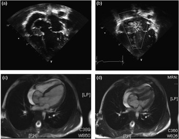
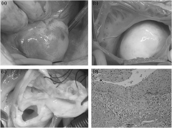
S.H. was a healthy five year old male, referred to our institution for evaluation of a cardiac murmur noted on a recent physical examination associated with mild cardiomegaly as demonstrated in a chest radiograph. His initial electrocardiogram documented frequent premature atrial contractions, but was otherwise unremarkable. On physical examination, he was found to have an active praecordium with slight left ventricular heave. Auscultation revealed a few extrasystoles, normal first and second heart sounds, an ejection click, and a short systolic ejection murmur graded at 2 to 3 out of 6. The murmur was vibratory in nature, and radiated along his left sternal border toward the base whenever in the supine position, but disappeared when he was standing. Examination of the abdomen revealed normal findings, with no hepatosplenomegaly. Peripheral pulses were normal and symmetrical. The echocardiogram showed marked left ventricular dilation, with an aneurysmal mass arising from the postero-basal aspect of the left ventricular outflow tract. This outpouching protruded posteriorly and inferiorly towards the crux of the heart, extending into the right atrium at the base of the atrial septum, and exhibiting a to-and-fro pattern of flow within its cavity (Fig. 1). Cardiac magnetic resonance imaging (Fig. 1) confirmed the presence of a multiloculated mass measuring 23.6 by 36.9 by 31.5 millimetres that originated from the left ventricle along the antero-inferior margin of the aortic leaflet of the mitral valve, and extended into the region of the antero-inferior portion of the atrial septum. The neck of this outpouching measured 11.7 by 21.2 millimetres in size. A thrombus was also noted within the anterior portion of the mass. The non-thrombosed portion demonstrated enlargement during ventricular systole, with protrusion into the left atrium, the right atrium, and the inferior portion of the pericardial sac, adjacent to the junction of the inferior caval vein and the right atrium. The aneurysmal sac had delayed enhancement, consistent with a make-up of fibrous tissue. The inferior papillary muscle of the mitral valve gave off tendinous cords that extended to the antero-inferior margin of the neck of the aneurysm. The sac also protruded into the right atrium adjacent to the septal leaflet of the tricuspid valve, albeit without extension through the valve during any portion of the cardiac cycle. Surgery was undertaken to resect the aneurysm, and revealed a reddish pulsatile mass prolapsing externally in the inferior atrioventricular groove at the junction of the inferior caval vein and the right atrium (Fig. 2a). The decision was made to excise the mass through the right atrium. Upon opening of the right atrium, a giant-sized egg-shaped lesion was visible, which was whitish in appearance and very fibrotic, jutting up into the inferior portion of the right atrium, nearly obstructing the coronary sinus, and obstructing the junction of the inferior caval vein with the right atrium (Fig. 2b). The mass was opened, yielding yellowish, almost serum-like fluid and no clot. Its neck was identified underneath the annulus of the mitral valve, and was closed using a Prolene® suture and a Gore-Tex® patch. After removal, the mass was sent to pathology for further delineation of its aetiology. The entire specimen was submitted for microscopic evaluation and was described as consisting of a cystic structure lined by benign epithelium. The epithelium was predominantly of squamous nature, but had focal areas of simple cuboidal to pseudostratified epithelium. The wall displayed oedema and mixed inflammation, but contained no teratomatous elements. The histologic features of this lesion were not consistent with an aneurysm, a diverticulum, or a vascular malformation. Albeit that the patient initially experienced postoperative heart block, requiring implantation of a pacemaker, he ultimately returned to sinus rhythm. Since discharge, he has remained in sinus rhythm, and is otherwise well.
Imaging studies. (a) Four chambered view of the heart tipped posterior showing the large mass arising from the left ventricle. (b) Colour Doppler showing flow within the mass. (c) Cardiac magnetic resonance imaging revealing the size of the mass and (d) its connection to the left ventricle.

Intracardiac mass. (a) Pulsatile mass visualized externally in the inferior atrioventricular groove at the junction of the inferior caval vein with the right atrium. (b) View through the right atrium revealing whitish, fibrotic mass jutting up into the inferior portion of the right atrium. (c) The opened mass with the neck connecting to the left ventricle. (d) A haematoxylin and eosin stained tissue section demonstrating cyst wall lined by squamous epithelium with underlying granulation tissue.

Discussion
Our patient is very unusual in terms of the anatomy and aetiological classification of his cardiac mass, which to the best of our knowledge has not previously been described. Initially, the mass was deemed to be a congenital aneurysm, or a diverticulum protruding through the crux of the heart. There is frequently debate over distinguishing these two types of outpouching. A review of the literature indicates that a sac with a short communicating neck, composed of well differentiated muscular layers, and showing synchronous contraction with the ventricular mass as documented by the presence of a systolic pattern of flow from the mass to the ventricular cavity,Reference Teske, McGovern and Allen1, Reference Tissot, Pache and da Cruz2 as well as an association with midline defects,Reference Gueron, Hirsch, Opschitzer and Mogel3 would favor the diagnosis of a ventricular diverticulum. In contrast, aneurysms frequently have a wider neck, lack muscular layers, and show a paradoxal or absent pulsatilty, resulting in systolic flow from the ventricular cavity into the mass. In lesions of this type described thus far, there is a stronger association with congenital or acquired abnormalities, such as a perimembranous ventricular septal defect or coronary arterial disease.Reference Cay, Tufekcioglu and Ozturk4 The pathological findings from our specimen, rather than supporting either the diverticular or aneurysmal aetiology, suggest the differential diagnosis to be between a defect in embryogenesis or a monophasic teratoma. Cardiac teratomas are uncommon at any age, but nearly two-thirds of those reported have been diagnosed in infants.Reference Isaacs5, Reference Ou, Dorrière, Sidi, Bonnet and Vouhé6 Most have been described as located in the pericardium or near the base of the great vessels. Intracardiac teratomas are consistent with similar tumours in other locations, frequently consisting of a cystic area lined by a variety of epithelium and solid areas that are composed of neuralgic tissue, pancreas, thyroid, bone, or muscle.Reference Isaacs5 Although this mass was consistent with a monophasic teratoma, it is difficult to establish a distinction from a defect in embryogenesis. Its unusual composition, as well as its location, makes it atypical for an intracardiac teratoma. Consequently, a defect in embryogenesis may be the most likely cause of this mass.
A pre-operative concern for our patient was the possibility of rupture of the mass. Due to its unusual location, had the mass not remained intact, rupture could have occurred either into the pericardial cavity or to the right atrium. Had it ruptured to the right atrium, it would have produced the variant of atrioventricular septal defect also known as the Gerbode defect, namely a left ventricular-right atrial communication through the fibrous atrioventricular septum which may be either congenital or acquired. Whether acquired or congenital, this defect differs from the atrioventricular shunting frequently found in atrioventricular septal defects with common atrioventricular junction in that the essence of the Gerbode defect is the presence of discrete right and left atrioventricular junctions.Reference McKay, Battistessa and Wilkinson7 The congenital form of the defect occurs in less than 0.1% of all patients with congenitally malformed hearts.Reference Wasserman, Fann and Atwood8 The acquired Gerbode defect, also exceedingly rare, has been described as the result of endocarditis, aneurysm, trauma, myocardial infarction, and replacement of either the mitral or aortic valves.Reference Wasserman, Fann and Atwood8 We have been unable, however, to find a report of an intact or ruptured defect comparable to the arrangement observed in our patient. Fortunately, in our patient, the mass remained intact until surgery. Resection has now resolved both the cardiomegaly and extrasystolic contractions, even if not answering all of the questions regarding the aetiology of the mass.
Acknowledgement
We acknowledge Eduardo da Cruz, MD for his assistance in the preparation of the manuscript.