INTRODUCTION
Schistosomiasis, a fresh water-borne disease considered the second most prevalent after malaria, affects 200 million people worldwide, with 600 million at risk of the infection. The great majority of human infections are caused by Schistosoma mansoni, endemic in Sub-Saharan Africa, the Nile Valley and South America, and Schistosoma haematobium, endemic in the Middle East and most African Countries (Gryzeels et al. Reference Gryzeels, Polman, Clerinx and Kestens2006). Schistosomes are blood-dwelling flukes, which may survive for years in the mammalian host, unscathed by any element of the immune effectors they vigorously elicit (Pearce, Reference Pearce2003; Gryzeels et al. Reference Gryzeels, Polman, Clerinx and Kestens2006). The outer surface of the developing and adult schistosomes, the tegument, is the critical site of host-parasite interaction. The outermost tegument is a 2–4 μm thick syncytium. The innermost membrane of the tegument is a conventional membrane (the basal membrane), whereas the outer membrane on the syncytium surface consists of 2 tightly apposed bilayers, the inner and outer bilayers, each of which is composed of inner and outer leaflets (Hockley, Reference Hockley1973; Hockley and McLaren, Reference Hockley and McLaren1973; McLaren and Hockley, Reference McLaren and Hockley1977). The schistosome ability to evade the host immune mechanisms was attributed to the impermeability of the outer double lipid bilayer protecting the parasite tegument, and entirely excluding specific antibody binding to apical membrane antigens (Hockley, Reference Hockley1973; Hockley and McLaren, Reference Hockley and McLaren1973; McLaren and Hockley, Reference McLaren and Hockley1977; Kusel and Gordon, Reference Kusel and Gordon1989; Abath and Werkhauser, Reference Abath and Werkhauser1996). Lipids extracted from worm tegumental outer membranes of guinea pig-derived 5-day, 2-, 3- and 6-week-old schistosomes have been analysed by thin layer chromatography, and were shown to consist, as for typical biological membranes, of neutral phosphoglycerides, sphingomyelin, and cholesterol (Rogers and McLaren, Reference Rogers and McLaren1987). Membranes, isolated from the tegument of mature schistosomes by spontaneous release into phosphate-buffered saline, or by removal from the parasite's surface using poly-lysine beads, showed a typical plasma membrane-like profile with high sphingomyelin content (approximately 20%), and cholesterol to phospholipid molar ratio of 0·7–1·1 (Allan et al. Reference Allan, Payares and Evans1987). Few other reports have documented the lipid composition of the membranes at the host parasite interface (Furlong, Reference Furlong1991), probably because of the extreme difficulty of isolating intact outer membranes, free from underlying tegument structures (McDiarmid et al. Reference McDiarmid, Dean and Podesta1983; Roberts et al. Reference Roberts, MacGregor, Vojvodic, Wells, Crabtree and Wilson1983). We have recently been able to extract cholesterol from the apical lipid bilayer of lung-stage schistosomula using the membrane-impermeable, cholesterol-extracting, methyl-β-cyclodextrin (MBCD) drug (El Ridi et al. Reference El Ridi, Tallima, Mohamed and Montash2004; Tallima and El Ridi, Reference Tallima, Salah and El Ridi2005; Tallima et al. Reference Tallima and El Ridi2005). Analyses involving cholesterol extraction with MBCD and staining by filipin indicated that the content of apical lipid bilayer cholesterol is probably higher in S. mansoni than in S. haematobium worms (Tallima and El Ridi, Reference Tallima, Salah and El Ridi2005; Tallima et al. Reference Tallima and El Ridi2005). Alterations in the absorption spectra of filipin following interaction with cholesterol allowed wide use of filipin staining as a probe for the detection, localization, and quantitation of cholesterol in biological membranes (Blanchette-Mackie et al. Reference Blanchette-Mackie, Dwyer, Amende, Kruth, Butler, Sokol, Comly, Vanier, August, Brady and Pentchev1988; Mukherjee et al. Reference Mukherjee, Zha, Tabas and Maxfield1998). We believed that more quantitative methods should be established to assess the cholesterol content of the parasite's apical membrane, in order to devise more effective strategies for drug and vaccine development. We herein report on the quantification of the cholesterol content in the apical membrane of adult schistosomes, and the impact of cholesterol depletion on exposure of surface membrane molecules to specific antibody binding.
MATERIALS AND METHODS
Parasites
Adult worms were recovered from Syrian hamsters, at the time-intervals specified in the text, after percutaneous infection with 400±25 cercariae of Schistosoma mansoni or Schistosoma haematobium. Infection and hepatic perfusion were performed under anaesthesia, following the recommendations of the current edition of the Guide for the Care and Use of Laboratory Animals, Institute of Laboratory Animal Resources, National Research Council, USA, at the Schistosome Biological Materials Supply Program, Theodore Bilharz Research Institute (SBSP/TBRI). Adult worms were transported from SBSP/TBRI to the laboratory in ice-cold Rosewell Park Memorial Institute (RPMI)-1640 medium (BioWhittaker Europe, Verviers, Belgium), supplemented with 200 U penicillin, and 200 μg streptomycin/ml, 25 mmN-2-hydroxyethylpiperazine-N′−2-ethanesulfonic acid, 2 mm L-glutamine, and 1 mm sodium pyruvate (RPMI medium), washed 5 times taking great care of worm integrity, and 1, 3, or 5 male or female worms placed in wells of sterile, flat bottom, 96-well microculture plates. Worms were assessed for viability, contractility, and tegument integrity by 40× light microscopy (Olympus Inverted Microscope Model IX70, Olympus, Tokyo, Japan).
Treatment with methyl-β-cyclodextrin and filipin staining
Live worms were incubated for 2 h at 37°C in a total volume of 100 μl of RPMI medium supplemented with 0, 5, 10, 20 and 40 mm MBCD (Sigma Chemical Co., St Louis, Missouri). At the end of the incubation period, the medium was removed from each well, using sterile tips, into labelled sterile microcentrifuge tubes (Corning, Acton, Massachusetts), and used for assessment of cholesterol and triglycerides immediately or after overnight storage at −70°C. The possibility that regurgitated red blood cells contributed to the released lipids was discarded, since no haemoglobin was detected in the supernatants examined by the quantitative, colorimetric method based on the catalytic action of haemoglobin on the oxidation of 3,3′,5,5′-tetramethybenzidine by hydrogen peroxide (Welch and Young, Reference Welch and Young1983).
Worms in each well were washed 3 times in RPMI medium, and stained with filipin as described (Blanchette-Mackie et al. Reference Blanchette-Mackie, Dwyer, Amende, Kruth, Butler, Sokol, Comly, Vanier, August, Brady and Pentchev1988; Neufeld et al. Reference Neufeld, Cooney, Pitha, Dawidowicz, Dwyer, Pentchev and Blanchette-Mackie1996; Mukherjee et al. Reference Mukherjee, Zha, Tabas and Maxfield1998; Leventhal et al. Reference Leventhal, Chen, Tall and Tabas2001; McCabe and Berthiaume, Reference McCabe and Berthiaume2001; Tallima and El Ridi, Reference Tallima, Salah and El Ridi2005; Tallima et al. Reference Tallima and El Ridi2005). Briefly, worms were fixed in 100 μl of 3% paraformaldehyde (Sigma) in 50 mm phosphate buffer, pH 7·2, overnight at 4°C, washed 3 times in RPMI medium-2% FCS, and incubated with 50 μm filipin (Sigma) in RPMI medium-2% FCS for 2 h at room temperature. Worms were inspected by ultraviolet (u.v.) microscopy (Olympus) using u.v. filter Model U-MWU2, for visualization of filipin-cholesterol fluorescence intensity (5+ to 0+).
Quantitative determination of cholesterol and triglycerides
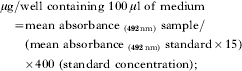
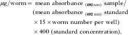
Enzymatic colorimetric (Multiskan EX, Labsystems, Helsinki, Finland) quantitative determination of cholesterol (CHOD-POD. Enzymatic colorimetric, Spinreact, S.A., Santa Colona, Spain), and triglycerides (GPO-POD. Enzymatic colorimetric, Spinreact), was performed following the manufacturer's instructions, with some modifications. Briefly, 100 μl of working reagent were mixed with 15·0 μl of test samples, in duplicate wells of 96-well microplates (Costar, Cambridge, Massachusetts). Quadruplicate standard wells contained 100 μl of working reagent, 2·0 μl of lipid standard, and 13·0 μl of RPMI medium. Cholesterol and triglyceride content was evaluated using the following formulae:
Estimation of worm surface area
Worm length and width were measured with a digital curvimeter (Comcurve-5, Koizumi Sokki, Mfg. Co., Nagaoka, Niigata, Japan), following drawing under stereomicroscope/camera lucida (Wild M8, Heerbrugg, Switzerland) and a microprojector (Micrprojector X1000-1, Ken-A-Vision, Mfg., Inc., Raytown, Missouri). Evaluation of the schistosome surface area was calculated as for Onchocerca volvulus microfilariae (Kron and MacKenzie, Reference Kron and MacKenzie1991) with modifications, assuming the worm body is rather cylindrical, and the schistosome pitted surface area has an area of about 10× that of a flat surface of the same basic size (Hockley, Reference Hockley1973), using the formula: Approximate surface area=Π×width×length×10.
Indirect membrane immunofluorescence (IF)
Adult worms were incubated in 500 μl of RPMI medium supplemented with 0, 10, 20 or 40 mm MBCD in wells of 24-well plates for 2 h at 37°C, washed 3 times in RPMI medium supplemented with 5% fetal calf serum (RPMI medium-5% FCS), and distributed in 3 parallel wells of 96-well microculture plates. Worms were incubated, overnight at room temperature in a humidified atmosphere, with 100 μl of RPMI medium-5% FCS containing 1·0 μl of serum from C57BL/6 naïve mice (control serum), or from mice 6 weeks after infection with 150 unattenuated cercariae by the tail exposure method (infection serum), or 4 weeks following two immunizations at 4-week intervals, with u.v. radiation-attenuated (RA) cercariae of S. mansoni (RA vaccine serum). It is of note that cercariae used for immunization were attenuated by exposure to 330 μW/cm2 u.v. light from an S-68 Mineralight Lamp (Ultra-Violet Products, San Gabriel, California) for 3 min, as described (El Ridi et al. Reference El Ridi, Tallima, Mohamed and Montash2004; Tallima and El Ridi, Reference Tallima, Salah and El Ridi2005; Tallima et al. Reference Tallima and El Ridi2005). Adult worms were then washed 3 times in RPMI medium-5% FCS, incubated with 1:100-diluted anti-mouse IgG (Fab-specific) fluorescein isothiocyanate-labelled goat immunoglobulins F(ab′)2 fragment of affinity-isolated antibody, adsorbed with bovine, horse, and human serum proteins (Sigma), washed 4 times in RPMI medium, and inspected by alternate light and u.v. microscopy.
Statistical analysis
Student's t-, Mann Whitney, and the Chi-square tests were used to analyse the statistical significance of differences between mean values from experimental and control values.
RESULTS
Qualitative evaluation of MBCD-mediated cholesterol extraction
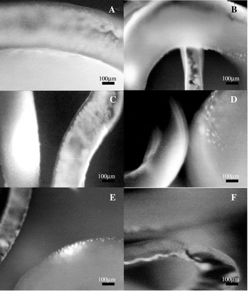
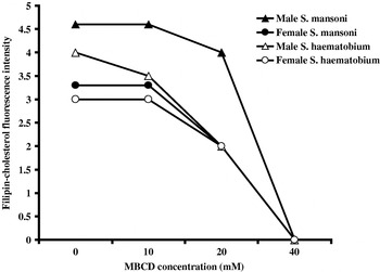
Incubation of live, adult S. mansoni or S. haematobium worms for 2 h in the presence of 0, 5, 10 or 20 mm MBCD did not impair viability or contractility. All worms were mobile and contractile for more than 100 min after exposure to 40 mm MBCD; after that, movement gradually diminished and then stopped. At the end of the 2 h incubation period, the worms excluded trypan blue and appeared intact, yet totally immobile. Staining with filipin indicated a gradual depletion of apical membrane cholesterol from male and female worms with an increase in MBCD concentration (Fig. 1). For both male and female worms, complete loss of surface membrane cholesterol occurred at MBCD concentrations of 40 mm (Fig. 2).
Filipin-cholesterol fluorescence intensity. Adult male and female schistosomes treated with different MBCD concentrations and stained with filipin showed filipin-cholesterol fluorescence intensity of 5+ (A), 4+ (B), 3+ (C), 2+ (D), 1+ (E) and 0+ (F).

Effect of MBCD treatment on filipin-cholesterol fluorescence intensity. Each point on the curve represents mean values for 3 independent experiments. In each experiment, 3 male or female Schistosoma mansoni or S. haematobium worms were incubated in the presence of 100 μl of MBCD-free or MBCD-supplemented medium for 2 h at 37°C before filipin staining. Filipin-cholesterol fluorescence intensity levels are illustrated in Fig. 1.

Quantitative evaluation of MBCD-mediated cholesterol extraction
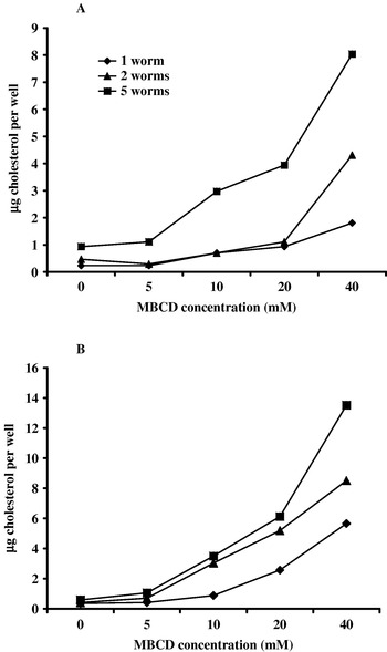
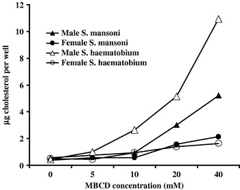
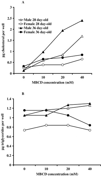
Cholesterol was detected in culture supernatants of worms maintained for 2 h in 100 μl of MBCD-free RPMI medium. Cholesterol content per well increased with increment in worm number and MBCD concentration (Fig. 3), and values were significantly (P<0·0001) higher for male than female worms (Figs 4 and 5). Cholesterol content per well also significantly (P<0·0001) increased with increase in adult worm age (Fig. 5A). It is of note that the triglyceride content in worm culture supernatants was rather similar whether worms were incubated in the presence of 0, 10, 20 or 40 mm MBCD (Fig. 5B).
MBCD-mediated release of cholesterol from the apical lipid bilayer of 47-day-old Schistosoma mansoni (A) or 120-day-old S. haematobium (B) male worms per well. Representative of 2 independent experiments.

MBCD-mediated release of cholesterol from the apical lipid bilayer of 3 male or female 47-day-old Schistosoma mansoni or 91-day-old S. haematobium worms per well. Each point represents mean values for 2 independent experiments with s.d. around the mean less than 5%.

MBCD-mediated release of cholesterol (A) and triglycerides (B) from the apical lipid bilayer of 3 Schistosoma mansoni worms per well. Each point represents mean values for 2 independent experiments with s.d. around the mean less than 5%.

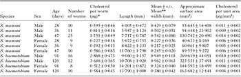
Calculation of the cholesterol content per worm and approximate estimation of worm surface area revealed that apical membrane cholesterol content is significantly (P<0·0001) higher in S. mansoni than S. haematobium worms, and is about twice higher in males than females for both species (Table 1).
Cholesterol content in apical lipid membranes of adult schistosomes

* Calculations based on worms incubated for 2 h at 37°C in 100 μl RPMI medium supplemented with 40 mm MBCD.
** Mean of 3 measurements (anterior, middle, and posterior parts) for each worm.
Effect of cholesterol extraction on surface membrane antigen exposure
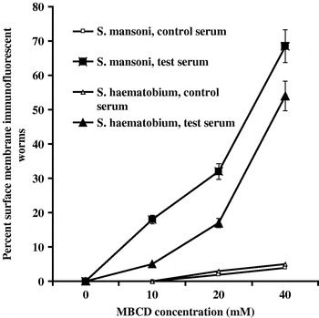
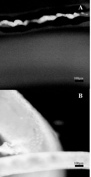
Adult S. mansoni and S. haematobium worms treated with 0, 10, 20 or 40 mm MBCD failed to bind any antibody from control serum (Fig. 6). Worms incubated in the presence of MBCD-free medium were also entirely negative whether exposed to infection or RA vaccine serum (Fig. 7A). Treatment with MBCD was associated with binding of infection and RA vaccine serum antibodies to the surface membrane of a proportion of male and female worms (Fig. 7B). An increase in the percentage of IF reactive worms paralleled an increment in cholesterol depletion; however, total loss of apical membrane cholesterol was associated with surface membrane exposure to specific antibody binding in only 50% and 70% of S. haematobium and S. mansoni worms, respectively (Fig. 6).
Effect of MBCD treatment on worm apical membrane antigen exposure to specific antibody binding in indirect membrane immunofluorescence (IF). Each point on the curve represents mean values and vertical bars denote s.d. about the mean for 3 independent experiments. In each experiment, 15 Schistosoma mansoni or S. haematobium worms were incubated in the presence of 500 μl of MBCD-free or MBCD-supplemented medium for 2 h at 37°C, washed, distributed in 3 parallel wells for IF reactivity with control, infection, or RA vaccine serum. Values for infection and RA vaccine sera were similar and, therefore, pooled (test serum).

Reactivity of adult schistosomes in indirect membrane immunofluorescence (IF). Adult male and female Schistosoma mansoni worms were treated with different concentrations of MBCD and incubated in the presence of 1:100-diluted infection serum. Worms exposed to 0 mm MBCD were consistently IF negative (A), in contrast to a substantial majority of worms treated with 40 mm MBCD (B).

DISCUSSION
Cholesterol is an essential structural component of eukaryotic cellular membranes and an important factor in determining membrane functions (Balut et al. Reference Balut, Steels, Radu, Ameloot, Van Driessche and Jans2006). Cholesterol helps to pack phospholipids, thus lessening phospholipid lateral diffusion and giving more rigidity and impermeability to the membranes (Yeagle, Reference Yeagle1985; Brown and Goldstein, Reference Brown and Goldstein1999; Simons and Ikonen, Reference Simons and Ikonen2000). In schistosomes, the outer double lipid cholesterol to phospholipid molar ratio reaches 1·1 (Allan et al. Reference Allan, Payares and Evans1987). Cholesterol was assumed to be critical for the rigidity and impermeability of the double lipid bilayer of developing and adult worms (Kusel and Gordon, Reference Kusel and Gordon1989; Abath and Werkhauser, Reference Abath and Werkhauser1996). We have previously reported that MBCD more readily extracts cholesterol from the apical membrane of Schistosoma haematobium than S. mansoni lung-stage schistosomula, yet allows exposure of surface membrane antigens to antibody binding in S. mansoni but not S. haematobium larvae. It was, hence, surmised that cholesterol content is higher and plays a more predominant role in antigen sequestration in S. mansoni than S. haematobium (El Ridi et al. Reference El Ridi, Tallima, Mohamed and Montash2004; Tallima and El Ridi, Reference Tallima, Salah and El Ridi2005; Tallima et al. Reference Tallima and El Ridi2005). No reports, however, were available regarding the cholesterol content of the outer membrane of schistosomes, likely because of the extreme difficulty of isolating the outer lipid bilayer free from underlying structures, either by cationic beads (Allan et al. Reference Allan, Payares and Evans1987) or subcellular fractionation (McDiarmid et al. Reference McDiarmid, Dean and Podesta1983; Roberts et al. Reference Roberts, MacGregor, Vojvodic, Wells, Crabtree and Wilson1983; Rogers and McLaren, Reference Rogers and McLaren1987; Braschi and Wilson, Reference Braschi and Wilson2006). Cholesterol content was determined in the plasma membrane of mammalian cells by cholesterol oxidase treatment of whole cells, preferably after exposure of the cells to glutaraldehyde fixation or to low ionic strength buffers (Lange, Reference Lange1992). Alternatively, cells were incubated in the presence of radioactive cholesterol, and the amount of [3H] cholesterol in the incubation medium and remaining in the cells following MBCD treatment was estimated by liquid scintillation counting (Keller and Simons, Reference Keller and Simons1998; Burgos et al. Reference Burgos, Klattenhoff, de la Fuente, Rigotti and Gonzalez2004).
We have herein incubated the worms in FCS-free medium in the presence of different concentrations of MBCD, assessed cholesterol depletion in the schistosome outer surface membrane by filipin staining, and measured the amount of cholesterol released in the medium by commercially available, enzymatic colorimetric assay. Worms incubated in MBCD-free medium were found to release negligible, yet detectable, amounts of cholesterol and triglycerides in agreement with reports documenting the low turnover of adult worm outer membranes (Saunders et al. Reference Saunders, Wislon and Coulson1987; Brouwers et al. Reference Brouwers, Skelly, Van Golde and Tielens1999). The amounts of released triglycerides did not vary with increase in MBCD concentration, thus indicating that the worms remained essentially intact even following exposure to 40 mm MBCD. In contrast to triglycerides, the amount of cholesterol released in the medium increased with an increase in MBCD concentration, in support of the well-documented ability of MBCD to specifically extract cholesterol from plasma membranes (Neufeld et al. Reference Neufeld, Cooney, Pitha, Dawidowicz, Dwyer, Pentchev and Blanchette-Mackie1996). The positive correlations between the amount of released cholesterol, MBCD concentration, and worm number and age point to the reliability of the newly developed method for assessing cholesterol content in the apical membrane of parasites. This simple method could be used for the estimation of the amount of cholesterol in the plasma membranes of mammalian cells such as sperms, and normal and cancerous blood cells.
The amount of cholesterol per worm was calculated based on the values obtained with 40 mm MBCD, since filipin staining revealed total loss of cholesterol from the outer membrane. It is of note that following exposure to this drug concentration, the worms were immobile, yet the outer tegument and membranes were intact, as judged by lack of increment in released triglyceride levels. Transmission electron microscopical analysis also indicated that incubation of S. mansoni lung-stage larvae in the presence of MBCD for 6 h did not elicit disorganization in the tegument structure, pits and fold (El Ridi et al. Reference El Ridi, Tallima, Mohamed and Montash2004). The amount of cholesterol per worm appeared much higher in S. haematobium than S. mansoni adult worms and in males than in females. Adult S. haematobium worms were, however, older than S. mansoni. Therefore, we have estimated the length and width of individual worms and attempted to have an approximate estimate of the surface area, taking into consideration the parasite tubular shape, and the factor calculated by Hockley (Reference Hockley1973) for the pits, folds and invagination-related increase in adult worm surface area. Based on these calculations, the amount of cholesterol per approximate surface area unit was found to be higher in S. mansoni than S. haematobium for both male and female worms, in agreement with our prediction concerning the lung-stage schistosomula (Tallima and El Ridi, Reference Tallima, Salah and El Ridi2005). The approximate calculations took into consideration the adult schistosome outer lipid bilayer pits, folds, and invaginations, which show few differences between male and female worms (Hockley, Reference Hockley1973; Hockley and McLaren, Reference Hockley and McLaren1973; McLaren and Hockley, Reference McLaren and Hockley1977). Accordingly, the conclusion that males have a 2-fold higher cholesterol content per unit area than females may well hold, even if the exact surface areas of schistosomes were calculated.
It is of note that the amount of cholesterol per mm2 in the outer membrane of male and female schistosomes appears to be about 10 times higher than the cholesterol content per mm2 of the plasma membrane of human erythrocytes, fibroblasts and liver cells, which is reported to be 0·0009, 0·0010 and 0·0006 μg/mm2, respectively (Lange et al. Reference Lange, Swaisgood, Ramos and Steck1989).
Depletion of apical membrane cholesterol was associated with an increase in exposure of surface membrane antigens to antibody binding, providing strong support for the role of cholesterol, when present in large amounts, in acting as a permeability barrier for the eukaryotic membrane (Bloom et al. Reference Bloom, Evans and Mouritsen1991; Corvera et al. Reference Corvera, Mouritsen, Singer and Zuckerman1992). The present findings strongly corroborate data recently reported for the lung-stage schistosomula (El Ridi et al. Reference El Ridi, Tallima, Mohamed and Montash2004; Tallima and El Ridi, Reference Tallima, Salah and El Ridi2005; Tallima et al. Reference Tallima and El Ridi2005). These findings together indicate that antigen shedding (Pearce et al. Reference Pearce, Basch and Sher1986) or masking by host proteins (Chiang and Caulfield, Reference Chiang and Caulfield1989) are not the basis for lack of antibody binding to the outer membrane of intact schistosomes, and argue against any role for antibody-dependent, cell-mediated cytotoxicity in the demise of developing or adult worms (Keating et al. Reference Keating, Wilson and Skelly2006). Cholesterol, however, does not appear to be the major player in schistosome surface membrane antigen sequestration. This is because total loss of cholesterol was associated with apical membrane antigen exposure in a substantial majority, but not all, test worms, in agreement with our hypothesis implicating sphingomyelin as the principal responsible for protecting the outer lipid bilayer from the action of host effector molecules (El Ridi and Tallima, Reference El Ridi and Tallima2006).
This research work was supported by the Arab Science and Technology Foundation, Project No. BT 05 2 05.