Introduction
Protein kinase R (PKR), also called eukaryotic translation initiation factor 2-alpha kinase 2 (EIF2AK2), is an interferon (IFN)-inducible double-stranded RNA (dsRNA) protein kinase with multiple effects in cells (reviewed in Refs Reference Garcia1, Reference Garcia, Meurs and Esteba2). PKR actively contributes to the cellular response to numerous types of stress with a critical role in the antiviral defence mechanism of the host induced by IFNs, which are cytokines with a wide range of biological functions, including antiviral, antiproliferative and immunomodulatory properties (Refs Reference Samuel3–Reference Metz and Esteban5). PKR is expressed constitutively in mammalian cells and has an IFN-stimulated response element (ISRE) in its determined promoter, required for transcriptional induction by type I IFN, but also a kinase conserved sequence motif with an important role in basal transcription in absence of cytokine treatment (Ref. Reference Kuhen and Samuel6). PKR is a first-line defence mechanism against infection and a regulator of cell growth and cell proliferation, and recently, it has also been involved in metabolism, inflammatory processes and age-related diseases (reviewed in Refs Reference Williams7, Reference Marchal8).
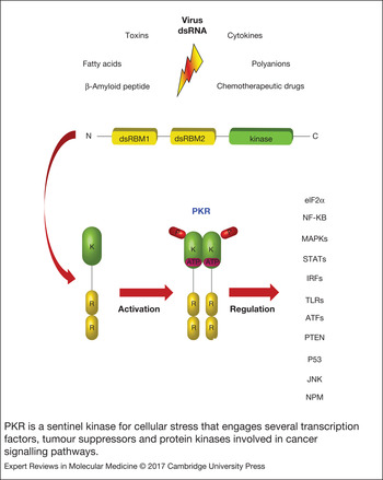
PKR is a serine–threonine kinase composed of a kinase domain, shared by the members of the eukaryotic initiation factor 2α (eIF2α) kinase family and two dsRNA-binding domains that constitute the regulatory domain (Fig. 1) (Refs Reference Patel9, Reference Donnelly10). Although the primary PKR activator is dsRNA (produced during infection by several viruses and detected at low doses in mammalian cells), PKR is also activated by a variety of cellular stresses including cytokines, calcium stress, oxidative stress, endoplasmic reticulum stress, lipo-stress, amyloid-β (Aβ) peptide accumulation, polyanions such as heparin, and several drugs (Refs Reference Barreto-de-Souza11–Reference Anderson15), or through the PKR associated activator (PACT) (Refs Reference Marques16, Reference Singh and Patel17). PKR, in response to specific stress signals, is activated by autophosphorylation and leads to the phosphorylation of eIF-2α impairing its activity, which results in the inhibition of protein synthesis and the induction of apoptosis (Refs Reference Lee and Esteban18, Reference Der19). In addition to its translational regulatory function, PKR has a role in signal transduction and transcriptional control through the regulation of the κB inhibitor (IκB)/nuclear factor κB (NF-κB) pathway (Refs Reference Kumar20, Reference Gil, Alcami and Esteban21). PKR also regulates some tumour suppressors and protein kinases involved in cancer pathways such as the signal transducers and activators of transcription factors (STATs), activating transcription factors (ATFs), tumour suppressor p53 (Tp53), the phosphatase and tensin homologue tumour suppressor (PTEN), the mitogen-activated protein kinases (MAPKs) and the toll-like receptors (TLRs) (Fig. 1) (reviewed in Refs Reference Garcia1, Reference Garcia, Meurs and Esteba2, Reference Marchal8). As predicted for a translation regulator, PKR is associated with ribosomes, mainly 40S subunits. Ribosomal association of PKR appears to be mediated by its dsRNA-binding domains (dsRBDs), implying a key role of these domains in the regulation of PKR activity (Refs Reference Wu, Kumar and Kaufman22, Reference Zhou23). In addition, a recent study identified a PKR nuclear interactome formed by a complex of proteins involved in ribosome biogenesis, RNA splicing, mRNA stability, gene expression, cell cycle, or chromatin organisation, suggesting PKR roles in ribosome biogenesis, mRNA processing and cell division (Ref. Reference Blalock24). These PKR-associated processes support the relevance of PKR's regulation; in fact, numerous studies have found PKR to be dysregulated in most type of cancers, neurodegenerative diseases, metabolic disorders and other pathologies (Refs Reference Marchal8, Reference Sud25). Whereas PKR is recognised for its negative effects on neurodegenerative diseases, the role of PKR activation in cancer is complex and remains controversial (reviewed in Ref. Reference Marchal8). Alternatively, recent studies have shown that PKR is activated by chronically elevated levels of specific nutrients and deletion of PKR has a number of effects on whole-body metabolism including protection against high-fat diet-induced obesity, glucose intolerance, insulin resistance and inflammation (review in Refs Reference Marchal8, Reference Koromilas26).
PKR is a sentinel kinase for cellular stress that engages several transcription factors, tumour suppressors and protein kinases involved in cancer signalling pathways. PKR is a serine-threonine kinase from the eIF2α kinase family composed of a kinase domain and two dsRNA-binding domains that constitute the regulatory domain. Although the main direct PKR activator is the dsRNA produced during infection by several viruses and detected at low doses in mammalian cells, PKR is also activated by a variety of forms of cellular stress including cytokines, calcium stress, oxidative stress, endoplasmic reticulum stress, lipo-stress, Aβ peptide accumulation, polyanions such as heparin, several drugs such as 5-FU and DOX, and fatty acids such as ceramide and palmitate. PKR is activated by autophosphorylation and leads to the phosphorylation of eIF-2α resulting in the inhibition of protein synthesis. PKR has been also implicated in NF-κB, IRFs, STATs, ATFs, p53, PTEN, MAPKs and TLRs signalling pathways. Aβ, amyloid-β; ATF, activating transcription factors; DOX, doxorubicin; 5-FU, 5-Fluorouracil; eIF2α, eukaryotic initiation factor 2α; IRF, interferon regulatory factor; MAPKs, mitogen-activated protein kinases; NF-κB, nuclear factor κB; PKR, protein kinase R; PTEN, phosphatase and tensin homolog; STATs, signal transducers and activators of transcription factors; TLRs, toll-like receptors.

This review discusses the dual role of PKR in cancer and the involvement of this kinase in metabolic disorders. Moreover, PKR is proposed as a relevant therapeutic target and biomarker.
PKR role in cancer
In addition to its well-established role in the IFN response, PKR is involved in many cellular pathways exerting various functions on cell growth and tumourigenesis (reviewed in Refs Reference Garcia1, Reference Garcia, Meurs and Esteba2, Reference Marchal8). The role of PKR activation in cancer is complex and remains controversial. In general, PKR is considered to have a tumour suppressor function, and some clinical data show a correlation between suppressed or inactivated PKR and a poor prognosis for several cancers. The outcome of eIF2α phosphorylation by PKR in cancer is complex where a single phosphorylation event can act as an integrator of diverse stress stimuli and orchestrate different biological outcomes in stressed cells (Ref. Reference Koromilas26). In fact, increased eIF2α phosphorylation has been associated with several pathophysiological conditions including cancer, neurodegeneration, virus infection, inflammation, diabetes and obesity (Ref. Reference Koromilas26). Furthermore, the apoptosis induced by PKR activation in cancer cells, in response to several antitumour drugs, indicates a role in potentiating the response to chemotherapy. However, other studies have shown high PKR expression and elevated PKR activation in various cancers, suggesting that PKR might be contributing to neoplastic progression (Ref. Reference Marchal8). It is well-known that PKR is a potent activator of NF-κB, via interaction with TRAF family modulators; inducing IFN transcription in concerted action with IFN regulatory factors 3 and 7 (IRF-3/7) (Ref. Reference Gil27, Reference Kumar28). Aberrant activation of NF-κB has been linked to inflammatory and autoimmune diseases, infection and cancer (Refs Reference Marchal8, Reference Gil27). Therefore, dissecting the regulatory contribution of PKR and identifying components involved in its deregulation have clinical and therapeutic importance.
PKR involvement in different signalling pathways implicated in cancer
PKR is involved in various pathways that activate and engage a number of transcription factors controlling the expression of multiple genes. PKR has been implicated in the activation of IFN regulatory factor 1 (IRF 1), which acts mainly as a transcriptional activator of IFN-α/β gene expression, and it has been proposed as an inhibitor of oncogenesis in melanoma patients (Refs Reference Kumar28, Reference Mirjačić29). Moreover, PKR controls several pathways by modulation of STAT1 and STAT3 transcription factors. In fact, PKR-knockout cells are defective in STAT1 phosphorylation on Ser727, resulting in a significant decrease in the activation function of STAT1 gene (Ref. Reference Ramana30). STAT1 is also a target for PKR-mediated activation in response to lipopolysaccharide in glial cells (Ref. Reference Lee31). Recently, PKR has been described to be involved in the differentiation of chondrocytes through the modulation of STAT1 and the transcription factor Sox-9 expression (Ref. Reference Morimoto32). Furthermore, PKR also associates with STAT3, and is required for full STAT3 activation in response to platelet-derived growth factor (PDGF) involved in regulation of ERK activation and ultimately involved in STAT3 phosphorylation (Ref. Reference Deb33). The transcription factor STAT3 plays an important role in cancer cells through the abnormal activation of cell cycle progression, and the deregulation of survival and senescence pathways. A novel tumour suppressor function for PKR was suggested by Koromillas's group via the inhibition of the hypoxia-inducible factor-1α (HIF-1α) expression through STAT3 in an eIF2α phosphorylation independent manner (Ref. Reference Papadakis34). Interactions between PKR and STAT3 have been shown to regulate autophagic pathways in the cellular metabolism control of fatty acids (Refs Reference Jonchère35–Reference Shen37).
Further, PKR is an activator for signalling cascades involved in stress-activated protein kinases, and mediates Jun kinase (JNK) and mitogen-activated protein kinase p38 (MAPK) activation in response to specific stimuli (Refs Reference Williams7, Reference Gourmaud38). Thus, the activation of both p38 and JNK after lipopolysaccharide or cytokine treatments such as IFN-γ, interleukin (IL)-1, or tumour necrosis factor (TNF)-α, depend, in part, on PKR presence (Ref. Reference Goh, de Veer and Williams39). Moreover, PKR interacts with and activates mitogen-activated protein kinase kinase 6 (MKK6) in response to dsRNA stimulation (Ref. Reference Silva40).
We used human cDNA microarrays to identify genes differentially expressed after PKR expression (Ref. Reference Guerra41). Most regulated genes were classified according to biological function, including apoptosis, stress, defence and immune response. Interestingly, we found upregulation of the ATF3 transcription factor by PKR activation. ATF3 induced apoptosis in cancer cells in response to different stimuli including antitumour drugs (Ref. Reference Udayakumar42), and was suggested to be a suppressor of invasion and metastasis (Ref. Reference Liu43). Although the activation of ATF3 induced by PKR has been analysed in the context of viral infection, this link suggests an interesting mechanism to be further explored in cancer. A connection between ATF4 and PKR has been recently discovered to mediate the anti-tumour activity of a small molecule, currently tested in early phase clinical trials, called ONC201 (also known as TIC10) (Ref. Reference Kline44).
PKR is also involved in the tumour suppressor function of p53. A bidirectional and complex regulatory relationship between PKR and p53 has been demonstrated in cancer cells (reviewed in Ref. Reference Marchal8). PKR interacts directly with the C-terminal part of p53 and phosphorylates p53 on Ser392 (Ref. Reference Cuddihy45). On the other hand, PKR is able to promote the proteosomal degradation of p53 in association with glycogen synthase kinase 3 and mouse double minute 2 homologue, independently of translational control (Ref. Reference Raven46). Moreover, it has been demonstrated that the ability of p53 to cause cell cycle arrest and regulate transcription of target genes was impaired in PKR-knockout cells (Ref. Reference Cuddihy45). In fact, p53-mediated tumour suppression can be attributed, at least in part, to the biological functions of PKR induced by p53 in some genotoxic conditions, and PKR could be transcriptionally regulated by p53 activity in response to DNA damage (Ref. Reference Yoon47). Recently, it has been proposed that activated PKR inhibits pancreatic β-cell proliferation through sumoylation-dependent stabilisation of p53, implicating both proteins also in the metabolic regulation of diabetes (Ref. Reference Song48), as we discuss in the next section. Moreover, a sumoylation-dependent mechanism involving p53 phosphorylation through the PACT-PKR stress response induces translational activation leading to G1 cell cycle arrest (Ref. Reference Bennett49).
The alternative reading frame (ARF) encoded by the INK4a-ARF locus is one of the most frequently mutated genes in cancer (Ref. Reference Garcia50). The control of cellular proliferation by this tumour suppressor is crucial to restrict tumour development. Several reports describe the activation of ARF after the expression of viral proteins, type I IFN treatment, or after virus infection, suggesting a physiological role for ARF during virus infection (Ref. Reference Garcia50). We have revealed that ARF can be induced by viral infection and that the expression of ARF reduces viral infectivity. We also have shown that this antiviral effect depends, in part, on PKR activation mediated by its release from inhibitory complexes with nucleophosmin (NPM) (Ref. Reference Garcia51), a protein commonly overexpressed, mutated, rearranged and sporadically deleted in cancer (Ref. Reference Box52). Although the role of the PKR-NPM complex has been only studied in the viral context, at present we are analyzing its involvement in cancer. Tumour suppressor retinoblastoma protein, involved in cell cycle control, differentiation and inhibition of oncogenic properties (Ref. Reference Classon53), was required for the activation of the NF-κB pathway in response to virus infection, also in a PKR-dependent manner (Ref. Reference Garcia54). These results provided a new link between tumour suppression, PKR and the antiviral host defence, indicating an important tumourogenic role of viruses and a crucial potential issue in the forthcoming use of viruses as therapeutic agents. In fact, selective tumour cell replication of oncolytic viruses is thought to depend on infection of neoplastic cells, which harbour low levels of PKR and dysfunctional tumour suppressors and type I IFN signalling elements (Ref. Reference Muñoz-Fontela55).
PKR is a potent pro-apoptotic protein
Cell death by apoptosis is a genetic program in multicellular organisms that conditions the ordered removal of damaged or unwanted cells during development and in adult life. Deregulation of the apoptotic process can lead to pathological conditions such as cancer, autoimmunity and neurodegeneration (Ref. Reference Driver56). Moreover, apoptosis is an important event during some chemotherapy treatments in cancer.
PKR mediates apoptosis induced by several viruses such us poxviruses, influenza, etc., probably through dsRNA production (Refs Reference Kibler57, Reference Takizawa, Ohashi and Nakanishi58). However, PKR also regulates apoptosis in the absence of viral infection. Some examples are the apoptosis observed during Alzheimer's disease (Ref. Reference Onuki59), the apoptosis induced by oncogenes such as IRF1 or E2F-1, or triggered in response to dsRNA, TNFα, lipopolysaccharide, tunicamycin, serum starvation, or IL-3 withdrawal (reviewed in Refs Reference Garcia1, Reference Garcia, Meurs and Esteba2). Many of the stimuli that trigger PKR-dependent apoptosis in the absence of viral infection rely on PACT/RAX activation. PACT/RAX mediates PKR activation and subsequent apoptosis in response not only to cytokines and serum withdrawal, but also to chemotherapy, ethanol and viral infection (Refs Reference Marques16, Reference Singh and Patel17). However, PACT-knockout cells and mice did not exhibit significant differences in response to stressful stimuli compared with a wild-type phenotype (Ref. Reference Marques16). Analysis of the role of PKR effectors in mediating cell death suggests an intricate pathway, in which at least eIF-2α, NF-κB, ATF-3 and p53 have been implicated. PKR-induced apoptosis mainly involves the FADD/caspase 8 pathway and caspase 9 activation, which correlate with Bax protein translocation to the mitochondria and cytochrome c release to the cytoplasm, resulting in mitochondrion depolarisation (Refs Reference Gil60, Reference Gil61). Numerous works including ours have highlighted the importance of apoptosis induction in cancer mediated by PKR in response to different stimuli such as IL and antitumour drugs (Refs Reference Garcia62–Reference Chen65).
Does PKR induce cell death or cell survival in tumours?
The balance between cell survival and cell death is essential for cancer initiation and for the control of cancer outcome. PKR is able to induce cell survival through NF-κB activation or cell death via eIF2α/apoptosis induction in a complex temporally regulated manner balancing cell survival and cell death (Ref. Reference Donzé66). EIF2α phosphorylation has been associated with cell survival, whereas a long-term induction of this factor has been associated with cell death (Ref. Reference Koromilas26). Although NF-κB is often classified as a pro-survival factor that prevents apoptosis, there is evidence for NF-κB as either, a pro- or an antiapoptotic factor, depending on the stimulus that triggers the event (Ref. Reference Gil, Alcami and Esteban67). Probably, because of this dual ability to produce death or survival, the precise role of PKR in cancer biology remains controversial. Initially, PKR was thought to be a tumour suppressor. The overexpression of PKR in mammalian, insect and yeast cells indicated that PKR controls cell growth and, consequently, may function as an inhibitor of cell proliferation. In fact, the expression of several PKR dominant-negative mutants leads to malignant transformation of NIH 3T3 cells, and are able to cause tumourigenesis in nude mice (Refs Reference Koromilas68, Reference Barber69). The strong ability of PKR to induce the apoptotic machinery combining intrinsic and extrinsic routes of caspase activation agrees with the notion that this protein could be a tumour suppressor. Moreover, PKR has been suggested to play an essential role in the antitumour activity of tumour suppressors, such as p53 and PTEN (Refs Reference Yoon47, Reference Mounir70). Alternatively, data supporting the antitumour role of PKR comes from studies demonstrating the importance of PKR/eIF2α status in cancer response to chemotherapy (discussed above).
The evidence that PKR is suppressed or inactivated in tumours, and the correlation between increased PKR expression and improved prognosis have been shown in head, neck, melanoma, lung and colon cancer (Refs Reference Guo71–Reference Shimada76). However, other studies also suggest an antagonistic role of PKR in cancer, challenging the proposed function of PKR as a tumour suppressor. In fact, it has been reported that PKR is overexpressed and linked with malignancy in thyroid carcinoma, broncheoalveolar carcinoma, colon, melanoma, lung, breast and liver cancers (Refs Reference Marchal8, Reference Cheng77–Reference Roh79). Furthermore, it has been suggested that PKR is involved in leukaemia and could play a negative role in some haematological disorders (Ref. Reference Blalock80). Increased expression of PKR has been related to the promotion of genomic instability in a mouse model of myelodysplastic syndrome (Ref. Reference Cheng81) and high PKR expression in CD34(+) cells of acute myeloid leukaemia patients has been correlated with decreased survival and shortened remission duration (Ref. Reference Oshima82). More recently, PKR-p38 signalling pathway has been negatively implicated in the suppression of gastric cancer metastasis induced by Gelsolin, an actin-binding protein that regulates cytoskeletal turnover, which is crucial for the migration and invasion of tumour cells (Ref. Reference Yuan83). Finally, PKR translocation to the nucleus has been involved in lung cancer as a mediator of radiation resistance (Ref. Reference Hao84).
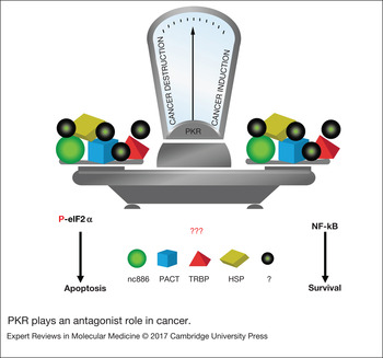
It is now well established that activation of PKR leads to the induction of pro-survival as well as pro-death pathways depending on the intensity and nature of the activating stimulus, as well as, the activation or level of expression of PKR modulators (Fig. 2). Several direct and indirect modulators of PKR protein have been described (reviewed in Refs Reference Garcia1, Reference Garcia, Meurs and Esteba2, Reference Marchal8). For instance, PACT, the trans-activation response RNA-binding protein (TRBP), chaperones heat shock protein (HSP)90 and HSP70 and the noncoding RNA nc886, among others, have been well characterised as PKR modulators (reviewed in Refs Reference Garcia1, Reference Garcia, Meurs and Esteba2, Reference Marchal8). The specific role that those modulators play in diseases where PKR is deregulated still needs to be elucidated in a clinical context (Fig. 2). For example, the finding of different expression patterns of PKR/eIF2α/NF-κB activity in cancer cell lines derived from different cholangiocarcinoma patients indicates the complexity of the role of PKR and its regulation by the pre-mir-nc886 in cancer (Ref. Reference Kunkeaw85). Consistent with these data, an elegant model proposes the escape of virus and cancer cells from the PKR/eIF2α cell death pathway (Ref. Reference Lee86).
PKR plays an antagonist role in cancer. PKR activation can induce both pro-survival and pro-death pathways. The nature and the intensity of activation stimuli as well as the level of expression of PKR modulators (PACT, TRBP, HSPs, nc886 among others) could influence the balance between cancer cell survival or destruction by apoptosis of tumour cells. Understanding the cellular factors and signals that regulate PKR in the different diseases would be extremely valuable from a clinical point of view. HSP, heat shock protein; PKR, protein kinase R; PACT, PKR associated activator; TRBP, trans-activation response RNA binding protein.

In summary, clinical studies related to PKR regulation are still insufficient. Furthermore, the few studies that have been conducted did not follow a standardised protocol, differing in the number of tumour samples analysed and in the inclusion criteria of patients, which makes difficult to compare results between studies. Therefore, it is necessary dissect in vivo the involvement of PKR in different cellular pathways, and the importance of its compartment localisation in the presence or absence of certain regulator factors that may affect its activity in several diseases.
PKR as a molecular target of chemotherapeutics and novel antitumour drugs
The chemotherapeutic drug 5-Fluorouracil (5-FU) is widely used in the treatment of a range of cancers, being the first and second line of combination treatments in colorectal cancer patients, and the third line in the palliative care of numerous cancer types. However, adverse effects and resistance to the drug remain a major clinical problem. Since defects in mediators of apoptosis may account for chemoresistance, the identification of new targets involved in 5-FU-induced apoptosis is of great clinical interest. The p53 tumour suppressor has been reported to be an important protein involved in 5-FU-induced apoptosis (Ref. Reference Longley87). However, several studies have shown that apoptosis can also occur in mutant p53 cell lines by a mechanism still unknown (Refs Reference Garcia62, Reference Longley87).
Given the heterogeneous response to 5-FU induced apoptosis and 5-FU sensitivity or resistance, the finding of biomarkers that could predict 5-FU behaviour in patients is of vital relevance to improve personalised cancer therapies. In this respect, we have identified PKR as a key molecular target of 5-FU involved in apoptosis induction in human colon and breast cancer cell lines (Ref. Reference Garcia62). We analysed PKR distribution and activation, apoptosis induction and cytotoxic effects during 5-FU and 5-FU/IFNα treatment in several colon and breast cancer cell lines with different p53 status. PKR protein was activated by 5-FU treatment in a p53-independent manner, inducing phosphorylation of eIF-2α and cell death by apoptosis. Furthermore, PKR interference promoted a decreased response to 5-FU treatment and those cells were not affected by the synergistic anti-tumour activity of a 5-FU/IFNα combination. In addition, we showed that PKR is activated in absence of p53 expression and, whereas PKR knockdown decreased 5-FU-mediated apoptosis, cell death was completely abolished in absence of both PKR and p53 proteins. These results suggest the importance of both proteins in 5-FU-induced apoptosis and the role of PKR in tumour cells where p53 is mutated, considering that more than 50% of colon tumours are deficient in p53 activity (Ref. Reference Garcia62).
PKR has been also implicated in the anti-tumour activity of anthracycline doxorubicin (DOX), a commonly used treatment for a wide range of cancers. Following DOX application, PKR induces apoptosis in cancer cell lines by mechanisms dependent on eIF2α phosphorylation, p53 phosphorylation and JNK activation (Ref. Reference Peidis88). Importantly, in a mouse xenograft model, colon cancer cells with reduced PKR expression induced early tumourigenesis and were resistant to DOX or etoposide treatment when compared with control cells (Ref. Reference Yoon47). Moreover, PKR negatively regulated leukaemia progression in association with PP2A activation, Bcl-2 inhibition and increased apoptosis in response to DOX treatment in a xenograft model (Ref. Reference Cheng77). Taken together these results indicate that increased PKR expression in cancer tissues may serve as a biomarker for response to 5-FU or DOX-containing chemotherapy regimens. Future therapeutic approaches to promote PKR expression/activation and eIF2α phosphorylation may be beneficial for the treatment of some cancers.
In this regard, we have analysed PKR/ eIF2α activation in response to new antitumour drugs. For instance, Bozepinib [(RS)-2,6-dichloro-9-[1-(p-nitrobenzenesulfonyl)-1,2,3,5-tetrahydro-4,1- benzoxazepin-3-yl]-9H-purine] a potent antitumour compound has been shown to induce apoptosis in breast and colon cancer cells (Refs Reference Lopez-Cara89, Reference Marchal90). We identified PKR as a target of Bozepinib, being upregulated and activated by the drug. However, p53 was not affected and was not necessary for Bozepinib induction of apoptosis in either breast or colon cancer cells. In addition, the efficacy of Bozepinib was improved when it was combined with IFNα cytokine, supporting a role of PKR in Bozepinib-induced apoptosis (Refs Reference Marchal90, Reference Ramírez91).
PKR role in metabolism
The balance between glucose production and utilisation is finely controlled by pancreatic cells secreting insulin, which increases glucose uptake by skeletal muscles and inhibits hepatic glucose production. When the diet provides excess of lipids, the capacity of adipose tissue to store fatty acids is overwhelmed, and fatty acids are accumulated in collateral tissues inducing the insulin resistance observed during obesity or type 2 diabetes. Recently, it was observed that PKR is involved in the induction of insulin resistance in peripheral tissues (Refs Reference Nakamura92–Reference Nakamura95), and antiproliferative activities in pancreatic β cells (Ref. Reference Chen96), indicating a novel role of PKR in metabolism regulation and type 2 diabetes mellitus. Sustained phosphorylation of eIF2α has been observed as a feature of obesity and insulin resistance in experimental systems and humans (Refs Reference Gregor97, Reference Oyadomari98). As in the case of pathogen exposure, PKR coordinates eIF2α phosphorylation-mediated suppression of protein translation with the induction of inflammatory responses upon exposure to excess of nutrients and energy. These functions of PKR are attributed to its kinase catalytic activity. Interestingly, an effective therapeutic strategy targeting PKR was confirmed using small-molecule inhibitors of the kinase activity that improved insulin sensitivity and glucose clearance in a mouse model of obesity and insulin resistance (Ref. Reference Lundh99).
PKR is also a critical regulator of metabolic homeostasis, placing this enzyme at a critical juncture in immunometabolic pathways (Refs Reference Nakamura92, Reference Carvalho-Filho93). In fact, recently it has been suggested that the association between PKR and TRBP protein integrates metabolism with translational control and inflammatory signalling, playing important roles in metabolic homeostasis and disease. The formation of a complex between PKR and TRBP during metabolic and obesity-induced stress has been critical for the regulation of eIF2α phosphorylation and JNK activation (Ref. Reference Nakamura100). Therefore, exposure of cells and tissues to lipo-toxicity also leads to PKR activation and PKR-dependent JNK activation, raising the possibility that PKR serves as a sensor for metabolic stress signals (Ref. Reference Hage Hassan101). Indeed, PKR activity and JNK phosphorylation are elevated in multiple tissues in obese humans (Refs Reference Carvalho94, Reference Gregor97, Reference Boden102), strongly suggesting that this role of PKR is conserved and relevant to human disease.
The translational consequences of the involvement of PKR in metabolism and inflammation are currently present in neurodegenerative diseases such as Alzheimer. For example, it has been suggested that β-amyloid oligomers and Alzheimer-associated toxins activate PKR in a TNF-α-dependent manner, resulting in eIF2α phosphorylation, neuronal insulin receptor substrate (IRS-1) inhibition, synapse loss, and memory impairment (Ref. Reference Lourenco103). Moreover, cognitive scores in Alzheimer's participants showed a significant positive relationship with IKKβ and a negative correlation with IRS1, JNK and PKR status (Ref. Reference Taga104).
The complexity of the different activity of PKR found in cancer has also been shown in the context of metabolism and inflammation. The involvement of PKR in metabolism homeostasis is attributed to its kinase catalytic activity; however, PKR can function as an adaptor protein via its protein-binding domain, but not via its regulatory dsRNA-binding domain. In fact, under the conditions where the kinase catalytic activity was defective, the protein-binding function of PKR was shown to promote β-cell proliferation, suggesting a role of the PKR protein-binding domain distinct from its kinase activity, and a pivotal role for PKR protein-binding function on the proliferation of pancreatic β cells through TRAF2/RIP1/NF-κB/c-Myc pathways (Ref. Reference Gao105). Nevertheless, the importance of a functional dsRNA-binding domain in PKR has also been revealed by a study showing that endogenous snoRNAs can activate PKR under metabolic stress (Ref. Reference Youssef106). Thus, the therapeutic inhibition of PKR at all levels is complicated, and most inhibitor compounds act only on the catalytic domain. Some strategies have implied the use of two structurally distinct small-molecule inhibitors of PKR in the treatment of insulin resistance and type 2 diabetes in both cells and mouse models. These strategies were able to reduce in vitro and in vivo stress-induced JNK activation and IRS-1 serine phosphorylation (Ref. Reference Nakamura95). In addition, treatment with both PKR inhibitors reduced adipose tissue inflammation, and improved insulin sensitivity, and glucose intolerance in mice after the establishment of obesity and insulin resistance (Ref. Reference Nakamura95). These findings suggest that pharmacologically targeting PKR may be an effective therapeutic strategy for the treatment of insulin resistance and type 2 diabetes. However, the questioned specificity and toxicity of PKR inhibitors (Refs Reference Naz107, Reference Weintraub108) together with the ubiquitous and multifunctional properties of PKR, in a catalytic-dependent and independent manner, are still major challenges for future clinical application of these inhibitor compounds.
Are metabolism and cancer connected by PKR?
The process of transformation in cancer cells is accompanied by profound alterations of cellular metabolism that guarantee the energy required for cancer cell growth and proliferation (Ref. Reference Hanahan and Weinberg109). A recent systematic analysis of metabolic gene expression across several cancer types showed that besides glycolysis, other metabolic pathways, including nucleotides, enzymatic activities and protein synthesis modulation, are activated in cancer (Ref. Reference Hu110). Obesity is a risk factor for many cancers, but the mechanisms by which it contributes to cancer development and patient outcome have yet to be fully elucidated. We have shown the effects of co-culturing human-derived adipocytes with established and primary breast cancer cells on tumourigenic potential. We found that the interaction between adipocytes and cancer cells increased the secretion of pro-inflammatory cytokines, increasing the proportion of cells expressing stem-like markers and increasing the metastasis potential in vivo (Ref. Reference Picon-Ruiz111). Since PKR is able to induce cytokines under multiple stress conditions, we are currently exploring its potential role in this process. Previous works have demonstrated that the deregulated inflammation process is a common pathological feature that may negatively contribute to cancer, neurodegeneration and metabolism pathogenesis (reviewed in Ref. Reference Marchal8). Based on studies demonstrating that PKR is induced by IFNs, and the pro-inflammatory NF-κB transcription factor is a target of the kinase and that it contributes to the inflammasome complex formation, the PKR implication in inflammatory processes has been suggested (Ref. Reference Lu112).
Activation of oncogenes and loss of tumour suppressors promote metabolic reprogramming in cancer, resulting in enhanced nutrient uptake for energetic and biosynthetic pathways. Within the hierarchy of pathways altered in cancer, glucose and glutamine metabolism are consistently reprogrammed by mutations in the tumour suppressor p53, the Myc and Ras-related oncogenes, and the AMP-acivated kinase (AMPK) and PI3 kinase (PI3K) signalling pathways, among others (Ref. Reference Boroughs and De Berardinis113). Although these metabolic features of cancer are now exploited for diagnostic and therapeutic purposes, their broader clinical implications are still under intense investigation. Epidemiologic studies suggest that people with diabetes, predominantly type 2 diabetes mellitus, had significantly higher risk to develop cancer and greater cancer mortality (Ref. Reference Hua, Yu and Hu114). Although, the mechanisms that underlie the associations between insulin resistance, type 2 diabetes mellitus and cancer risk remain far from understood, the insulin-like growth factor axis, inflammation, autophagy, endoplasmic reticulum stress, fatty acid excess stress and other mechanisms have been proposed to be important in this process. The deregulation of PKR is clearly involved in cancer and metabolism; however, its clinical involvement in the context of the two biological mechanisms is only now beginning to be explored. An interesting study showing the complexity of PKR in cancer and metabolism in lung cancer cells and tumours, suggests crosstalk among PKR, the AMPK and nutrient depletion (Ref. Reference Guo71). AMPK is activated by cellular stresses that deplete ATP and is a key factor in cancer metabolism (Ref. Reference Hardie, Ross and Hawley115). The authors showed that PKR causes nutrient depletion, which increases AMP levels and decreases ATP levels, causing AMPK phosphorylation. The inhibition of AMPK expression enhanced PKR-mediated cell death. However, the combination of PKR and p-AMPK expression in non-small cell lung cancer patients predicted a poor outcome for adenocarcinoma patients with high PKR expression and a better prognosis for those with low PKR expression (Ref. Reference Guo71).
On the other hand, besides the known role that PKR plays in the tumour suppressor function of p53, it has recently been demonstrated that PKR also can regulate metabolism via modulation of p53 stability by sumoylation. Activated PKR was found to inhibit pancreatic β-cell proliferation, partially through accumulation of p53 (Ref. Reference Chen96). Recently, it has been suggested that in the development of type 2 diabetes, glucolipotoxicity and pro-inflammatory cytokines promote the sumoylation-dependent stability of p53 via the ceramide/PKR/Ubc9 signalling pathway inducing the pancreatic-cell proliferation inhibition (Ref. Reference Song48). Therefore, the relationship between PKR and p53 needs to be further explored with in vitro as well in vivo studies to assess the role that both proteins play as potential biomarkers and therapeutic targets in cancer and metabolic related diseases.
In addition, PKR activation has been linked to cachexia, involving loss of muscle, insulin resistance and inflammation in cancer patients, indicating that the kinase may be implicated in metabolic pathways associated with cancer formation (Ref. Reference Tisdale116). Finally, in addition to the connection between the transcription factor STAT3 and PKR in cancer pathways, a link it has been established between both proteins in a metabolic context. STAT3 is able to regulate autophagic pathways interacting with PKR kinase in the cellular metabolism control of fatty acids (Ref. Reference Shen37). Several fatty acids, including palmitate, trigger autophagy via a pathway that involves the disruption of the STAT3-PKR complex as well as the phosphorylation of mitogen-activated protein kinase 8/c-Jun N-terminal kinase 1 and eIF2α phosphorylation (Ref. Reference Shen37). This study suggests an interesting crosslink between cellular metabolism (fatty acids), pro-inflammatory signalling (STAT3), innate immunity (PKR) and translational control (eIF2-α) that regulates autophagy and cancer processes suggesting translational and clinical potential.
Although these studies suggest that PKR is involved in signalling pathways common in both processes, the connection between cancer and metabolic disorders needs to be determined in order to support PKR as a relevant therapeutic candidate.
The PKR role in metabolic diseases
Although numerous studies have concluded that PKR deletion prevents the deleterious consequences of a high-fat diet, with considerable phenotypic variations (Refs Reference Nakamura92–Reference Nakamura95), recently it has been shown that PKR deletion has no effect on high-fat diet induced obesity or the development of impaired glucose metabolism, having only a modest effect on adipose tissue inflammation (Ref. Reference Lancaster117). Disappointing results have also emerged from the study of PKR role in cancer. Despite the demonstration that PKR inhibits cell growth and that mutated forms of PKR results in cellular transformation (Refs Reference Garcia1, Reference Koromilas68, Reference Barber69), other studies using two different mouse models bearing a homozygous disruption of the pkr gene, showed that PKR inactivation did not cause spontaneous tumour development (Refs Reference Yang118, Reference Abraham119). Curiously, cells derived from knockout mouse models express truncated forms of the protein PKR maintaining either, the catalytic activity (C-PKR KO) or the dsRNA-binding properties (N-PKR KO) depending on the mouse model used (Ref. Reference Baltzis, Li and Koromilas120). In fact, metabolic studies from in vivo analysis using the same two knockout models that were used before in cancer studies, suggested differences between groups that may account for the phenotypic differences observed between the two mouse models (Ref. Reference Lancaster117). Indeed, differences have been reported in the signalling responses of cells isolated from C-PKR KO and N-PKR KO mice exposed to specific stimuli (Refs Reference Yang118–Reference Baltzis, Li and Koromilas120). Since PKR may have an important role in both cancer and metabolism in either a catalytic- dependent and independent manner, the total deficiency of the protein PKR must be absolutely confirmed to minimise contradictory results obtained between groups in both pathologies.
Conclusions and outlooks
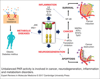
Numerous studies have confirmed the multifaceted and diverse roles of PKR. The involvement of PKR in IFN-mediated activities, the induction of apoptosis, the activation of inflammatory transcription factors, along with its role in signalling pathways linking numerous biological events, including cell growth and metabolism, are clear indicators of the great potential that this protein may have in the regulation of a variety of pathologies (Fig. 3). The identification of PKR as a target for both conventional chemotherapeutics and novel drugs highlights the need to carry out studies with patients to validate its potential as a biomarker in cancer diseases. Moreover, PKR deregulation in age-related diseases like cancer and Alzheimer's, in inflammatory processes and in metabolism disorders, like obesity and insulin resistance in patients, underlines the need to find applicable inhibitors of this protein. Since PKR is expressed in almost all cells and it is able to act in a catalytic- dependent or independent manner, its therapeutic potential depends on understanding and controlling its regulation factors. In fact, understanding the cellular factors and signals that regulate PKR in different diseases would be extremely valuable from a clinical point of view. The controversial results obtained from the two knockout mouse models available to study PKR in cancer and in metabolic diseases indicate the need to agree on another mouse model that guarantees the total deficiency of PKR expression. The use of CRISPR/Cas9 technologies to generate knockout models will allow future studies with human cells from various origins and animal models with complete deletion of PKR in combination with other related host genes to demonstrate beyond doubt its role in biological effects (i.e. cancer and metabolism) in a whole organism.
Unbalanced PKR activity is involved in cancer, neurodegeneration, inflammation and metabolism disorders. PKR coordinates the eIF2α phosphorylation-mediated suppression of protein translation with the induction of inflammatory responses upon exposure to excess of nutrients, energy and different cellular stresses. Sustained phosphorylation of eIF2α has been observed as a feature of obesity and insulin resistance. In addition, PKR is involved in inflammasome activation and cytokine liberation such as IL-6, TNF α and HMGB among others. Moreover, PKR activation can induce both pro-survival via NF-κB activation or cell death by apoptosis via eIF2α phosphorylation playing an antagonist role in cancer. Excess of PKR/eIF2α-P inducing apoptosis has been found in neurodegeneration. Furthermore, an unbalanced effect induced by dysregulated PKR activity could contribute to inflammatory diseases, neurodegeneration, cancer and metabolism disorders. Translational research will help to determine the clinical and therapeutic potential of this pleiotropic kinase. HMGB, high-mobility group box; IL, interleukin; NF-κB, nuclear factor κB; PKR, protein kinase R; TNF, tumour necrosis factor.

Activation of oncogenes and loss of tumour suppressors promote metabolic reprogramming in cancer, and the crosstalk among cancer and metabolic pathways are now exploited for diagnostic and therapeutic purposes. The present review has shown the importance to conduct continued basic molecular, preclinical and translational research to understand cancer, metabolism, inflammation and even neurodegeneration interplay. With personalised medicine looming in the present/future and the genetic and proteomic background of each patient determining specific approaches, PKR will play in the not too distant future a relevant clinical role.
Acknowledgements
The authors are thankful to the members of the Dr Esteban group who worked on the PKR project for their contribution. Special thanks to Dr Esteban for giving the opportunity to work in the exciting world of PKR kinase. This work was supported in part by grants from the Fundación Pública Andaluza Progreso y Salud, Consejería de Salud Junta de Andalucía in collaboration with JANNSSEN CILAG,S.A. (projects numbers PI-0533-2014, PI-0441-2014), from the Ministry of Economy and Competitiveness (MINECO, FEDER funds, grant number MAT2015-62644-C2-2-R), Instituto de Salud Carlos III (FEDER funds, project DTS15/00174) and from the Chair ‘Doctors Galera-Requena’ in cancer stem cell research.
Conflict of interest statement
The authors report no conflict of interest.