Introduction
Located in the eastern Betic Cordillera, southern Spain, the type level of Actinoporella campanensis new combination is dated by Granier (Reference Granier1987) from the Berriasian or basal Valanginian. Other specimens in thin section useful to detect the structure of the alga are illustrated by Sokač and Velić (Reference Sokač and Velić1978a) from the Valanginian of Croatia, while other specimens are reported in other areas, including Spain, France, Romania, Croatia, Serbia, Slovakia, Turkey, and Switzerland. In the following chronological account, we will pay special attention to the consecutive structural interpretations and taxonomy of this species, which, although seemingly seldom abundant, is nevertheless easily identifiable.
In 1972, Azéma and Jaffrezo established Likanella campanensis. The diagnosis and the reconstruction refer to a cylindrical axis bearing twin whorls of all equal, pyriform, and diverging laterals. The species is assigned to the Permian genus Likanella Milanović, Reference Milanovic1966, whose original diagnosis, potentially subject to various interpretations, is as follows:
The thallus is composed of loosely connected cylindrical segments, which at their lower end possess 3 whorls of branches. The long branches which at the top are open and non-ramified have separate calcareous walls. In the walls of the main stem and of branches there are to be found fine pores. (Milanović, Reference Milanovic1966, p. 9)
Later, Sokač and Velić (Reference Sokač and Velić1978b) implicitly transferred L. campanensis to the genus Selliporella Sartoni and Crescenti, Reference Sartoni and Crescenti1963 emend. Barattolo, De Castro, and Radoičić, Reference Barattolo, De Castro and Radoičić1988. Although not formally emended, Selliporella is newly described and assigned to the tribe Diploporeae.
An alternative structural view of L. campanensis was presented by Granier (Reference Granier1987). The whorls are simple. They consist of clusters (tufts) of three, all similar first-order fertile laterals, lined up along the axis of the thallus and arising from a proximal vestibule. Separately, the author noticed the special pattern of calcification, similar to Clypeina jurassica Favre in Favre and Richard, Reference Favre and Richard1927. Based on L. campanensis, the genus Falsolikanella was introduced with the following diagnosis (translated from French):
Alga with a continuous cylindrical main axis bearing simple verticils, more or less spaced out, protruding, composed of ramifications only of first order, grouped in tufts, and inserted in small number on a simple vestibule (metaspondyl type, with a vestibule). (Granier, Reference Granier1987, p. 208)
Falsolikanella was assigned to the tribe Diploporeae by implicit analogy with the arrangement of the laterals in the Triassic type species of the tribe, Diplopora annulata (Schafhäutl, Reference Schafhäutl1853) Schafhäutl, Reference Schafhäutl1863.
According to Berger and Kaever (Reference Berger and Kaever1992, p. 47), the genus Falsolikanella may not belong to the Diploporeae.
The structure of the alga is here reexamined. The analysis of specimens in thin section of the type material allows us to prospect a new interpretation of F. campanensis (Azéma and Jaffrezo, Reference Azéma and Jaffrezo1972), and consequently the validity of the genus Falsolikanella is discussed.
Materials and methods
This study is based on the type material of Likanella campanensis. It consists of three thin sections labeled as follows. FSL 420001: Likanella companensis, lame 3486, holotype (Azéma and Jaffrezo, Reference Azéma and Jaffrezo1972, pl. 1, fig. 1), Puig Campana (Alicante, Spain), Berriasien. FSL 420002: Likanella companensis, lame 3486-3 (Azéma and Jaffrezo, Reference Azéma and Jaffrezo1972, pl. 1, fig. 5), Puig Campana (Alicante, Spain), Berriasien. FSL 420003: Likanella campanensis, lame 3486-4, Puig Campana (Alicante, Spain), Berriasien.
Material with Praturlonella danilovae Radoičić, Reference Radoičić1975 and Actinoporella kukoci Radoičić, Reference Radoičić1974 belongs to the Rajka Radoičić collection (thin sections labeled RR).
Repositories and institutional abbreviations
Types examined in this study are deposited in the following institutions: (1) Likanella campanensis is in the collections of the University of Lyon, UCB Lyon 1 (inventory FSL 420001 = slide 3486; FSL 420002 = slide 3486.3; FSL 420003 = slide 3486.4). (2) Praturlonella danilovae Radoičić, Reference Radoičić1975 and Actinoporella kukoci Radoičić, Reference Radoičić1974 (labeled RR) are housed in R. Radoičić private collection at Kralja Petra 38, 11158 Stari Grad, Beograd, Serbia.
Structural constraints
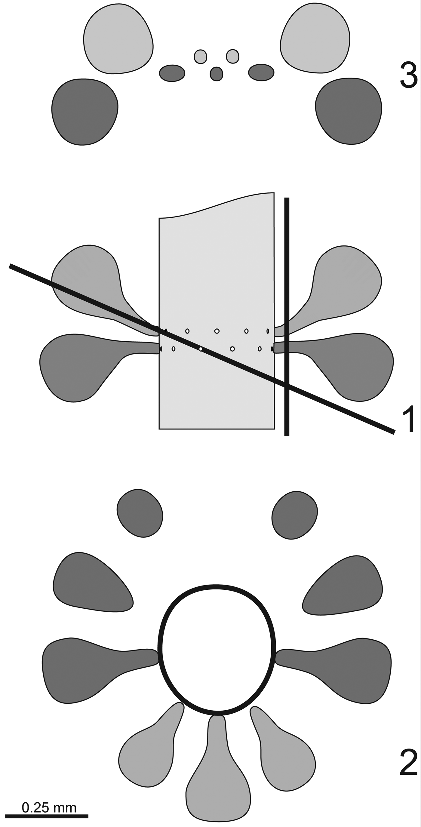
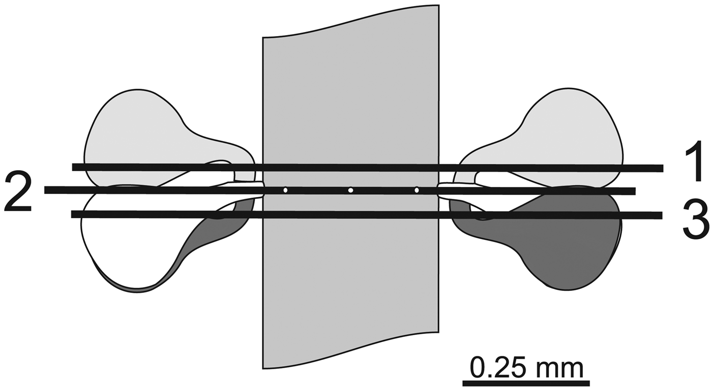
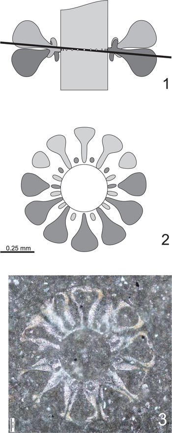
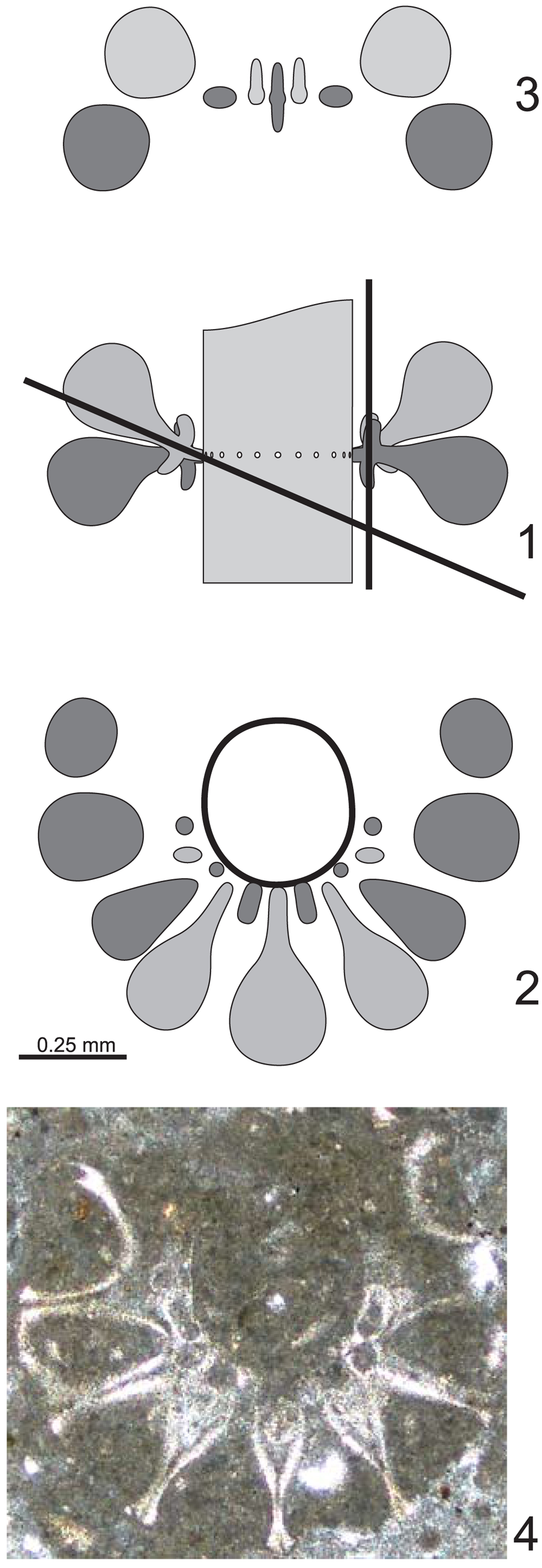
According to Azéma and Jaffrezo (Reference Azéma and Jaffrezo1972), the alga bears spaced whorls, each composed of two rows of 12 laterals set in alternation (here indicated as two-rows model, Fig. 1). The swollen outer part of the laterals is responsible for the downward and upward bending of the laterals in the lower and upper rows, respectively. The two authors are elusive on the significance of the two rows. Possibilities are as follows: (1) the rows are simply due to the heterocline (upward and downward) arrangement of the laterals; however, the whorl is single, making the number of laterals per whorl correspond to the sum of the two rows (i.e., w = 24); and (2) contrariwise, the two rows result from the presence of two separate whorls; in this case the value of w is 12. Figure 1 illustrates the second possibility.
The two-rows model: organization of Likanella campanensis sensu Azéma and Jaffrezo (Reference Azéma and Jaffrezo1972). (1) Axial view with traces of the oblique and tangential sections; (2) oblique section; (3) tangential section.

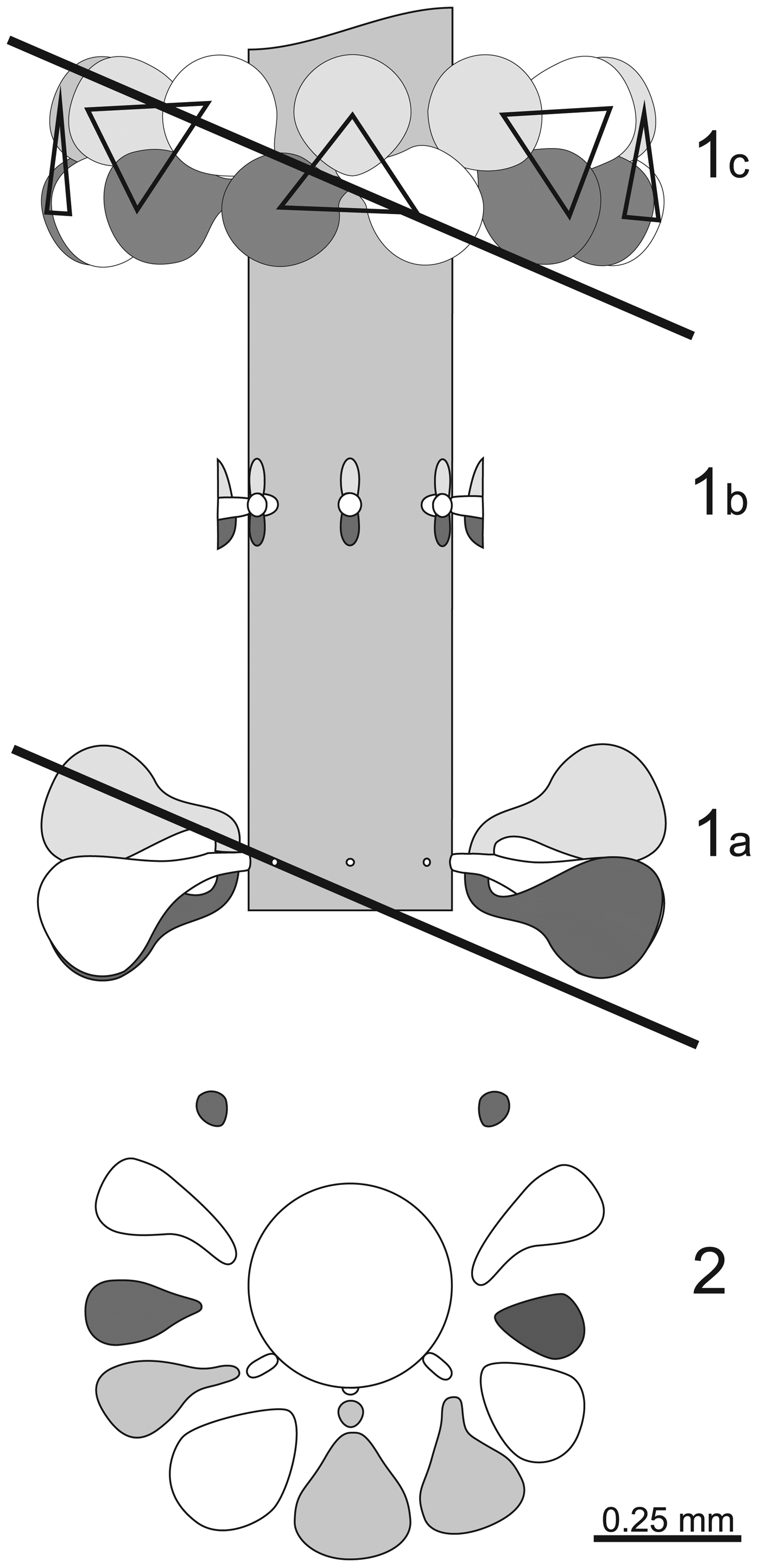
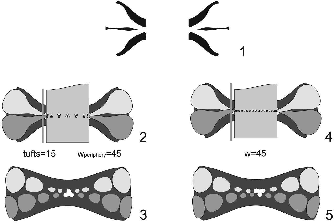
Drawing on new material originating from the type locality, Granier (Reference Granier1987) anticipated the presence of a tuft of three vertically aligned pores (Granier, Reference Granier1987, p. 207, pl. 6, fig. d). For this author, the structure corresponds to a metaspondyl tuft. In the table of biometrical values, the author did not indicate the number of laterals per whorl (Granier, Reference Granier1987, p. 209). In the reconstruction (Granier, Reference Granier1987, fig. 1b), the displayed number of vestibules is eight, corresponding to the value of w, each vestibule bearing three vesiculiferous, fertile primary laterals distally arranged chiefly in two rows as usual, such as already interpreted by Azéma and Jaffrezo (Reference Azéma and Jaffrezo1972). This model is here designated as the metaspondyl model (Fig. 2). Considering such a threefold structure, this metaspondyl model implies that the pores are proximally eight in number, distally corresponding to 24 in number. In case of arrangement in two rows, each row should contain 12 vesiculiferous laterals.
The metaspondyl model: organization of Falsolikanella campanensis sensu Granier (Reference Granier1987). (1a) Axial view with trace of an oblique section; (1b) perspective view of the tufts cut at branching level; (1c) perspective view of the vesiculiferous laterals arranged in two rows; triangles indicate the tufts; (2) oblique section.

Number and arrangement of pores per tuft
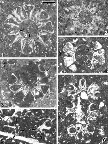
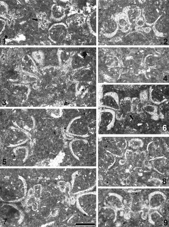
The occurrence of the threefold structure put forward by Granier (Reference Granier1987) (Figs. 3, 4) in the type material is herein confirmed. As illustrated by this author, three pores are present, axially aligned close to the central stem (Figs. 3.5, 4.1). As shown by Granier (Reference Granier1987, pl. 6, fig. d), the tufts are laterally very close each other (Fig. 4.2, 4.6–4.8); moreover, they are not completely aligned in a single row, but somewhat scalloped, shifted upward and downward by lateral compression.
Actinoporella campanensis (Azéma and Jaffrezo, Reference Azéma and Jaffrezo1972) n. comb. Type material. (1) Subtransversal section; upper coronae appear as elliptic pores between gametophores (white arrow); other equivalent pores show the tendency to split in two circular smaller pores aligned radially (black arrows), possibly corresponding to corona protuberances like in extant polyphysaceans; thin section n. 3486.3 (specimen already figured by Azema and Jaffrezo, Reference Granier1972, pl. 1, fig. 5); (2) transversal section, thin section n. 3486.3; (3) oblique section of an incomplete specimen, thin section n. 3486.4; (4) oblique section; notice the narrow circular pore (arrow) close to the thin calcification of the central stem corresponding to upper corona, thin section n. 3486 (specimen already figured by Azéma and Jaffrezo, Reference Azéma and Jaffrezo1972, pl. 1, fig. 4); (5) longitudinal section showing a fertile lateral bending downward (left) and another one directed upward (right); notice the arrows showing the basal part, the upper and lower coronae (left), and the basal part and lower corona (right); thin section n. 3486; (6) oblique section, thin section n. 3486 (specimen already figured by Azéma and Jaffrezo, Reference Azéma and Jaffrezo1972, pl. 1, fig. 1, the holotype). (1, 2) Scale bar shown in (1) = 0.25 mm; (3–6) scale bar shown in (3) = 0.25 mm.

Actinoporella campanensis (Azéma and Jaffrezo, Reference Azéma and Jaffrezo1972) n. comb. Type material. (1) Tangential section, thin section n. 3486; notice the threefold fertile laterals (arrow); (2) oblique section, thin section n. 3486.3; (3) oblique section, thin section n. 3486; (4) oblique-tangential section, thin section n. 3486.3; (5) longitudinal-oblique section, thin section n. 3486 (specimen already figured by Azéma and Jaffrezo, Reference Azéma and Jaffrezo1972, pl. 1, fig. 9); (6) tangential section; thin section n. 3486 (specimen already figured by Azéma and Jaffrezo, Reference Azéma and Jaffrezo1972, pl. 1, fig. 10); notice two adjacent pores not separated by calcareous wall (arrow); (7) tangential section, thin section n. 3486 (specimen already figured by Azéma and Jaffrezo, Reference Azéma and Jaffrezo1972, pl. 1, fig. 8 left); (8) tangential section, thin section n. 3486; (9) tangential section; thin section n. 3486.3. Scale bar = 0.25 mm.

Number of proximal pores per whorl (w)
Even taking into consideration the metaspondyl model, if a verticil is cut transversally (Fig. 5), the number of innermost pores corresponds to the value of w. In the type material, the values of w range from 16 to 22. As to the estimation of w from oblique sections, several methods are known (Pia, Reference Pia1920; De Castro, Reference De Castro1997). Based on the ‘couple of pores’ method (see Supplemental Data), the values range from 18 to 25, comparable to 16–22. Likewise, this calculation is confirmed in proximal-tangential sections (e.g., Fig. 4.6, 4.8), resulting in the distance between tufts (Δtuft) to average 76 µ (55–100 µ) and the estimated value of w to come to about 20 (w = d threefold* π/Δtuft). The diameter at the threefold level (d threefold) it is about 1.35 times the inner diameter (d = 0.37 mm in average, then d threefold = 0.50 mm). To sum up, the number of pores in a whorl (w) ranges within 16–25.
Transversal sections across the metaspondyl model (organization of Falsolikanella campanensis sensu Granier, Reference Granier1987). (1) Cut just above the ‘vestibule’; (2) cut in correspondence with the ‘vestibule’; (3) cut just below the ‘vestibule.’ In all cuts, the number of expected proximal pores is the same (i.e., eight).

Number of distal pores per whorl (wdist)
From the type material, the arrangement of the large distal pores prevalently set in two rows can be confirmed. The number of distal pores per row is 10–12, according to Azéma and Jaffrezo (Reference Azéma and Jaffrezo1972, p. 126–127). Although limited, the type material, allows to estimate the total number of distal pores (wdist) to 19–24. A method based on trigonometry aimed at calculating the inferred value of w and wdist from oblique sections is presented in the Supplemental Data. When applied to the holotype (Fig. 3.6), the estimated value of wdist is 22 (see Table 1 in Supplemental Data).
Metaspondility versus euspondility—geometrical constraints
It is usually easy to distinguish metaspondyl algae from euspondyl algae with heterocline laterals. However, under certain conditions this distinction may prove problematic, and some sections can lead to erroneous conclusions if taken alone, out of the context of accompanying sections. Figure 6.1 shows a typical axial section referable to a metaspondyl or euspondyl alga. The calcareous skeleton is poorly calcified, slightly intusannulated at the junction of the laterals and the central stem, which typically occurs when the laterals are closely packed. As displayed by the figure, the same axial section can be interpreted as metaspondyl (Fig. 6.2) or euspondyl (Fig. 6.4). The longitudinal section is not the only one to be problematic, also a tangential s.
Metaspondyl versus euspondyl arrangement. (1) Axial section of an ideal whorl, calcification (black); (2) metaspondyl model of (1) in axial view; black = calcification; (3) tangential cut marked by vertical line in (2); (4) euspondyl laterals model of (1); black = calcification; (5) tangential section marked by vertical line in (4). (3) and (5) are very similar to each other, but in the central part of (3), tufts can be still perceived.

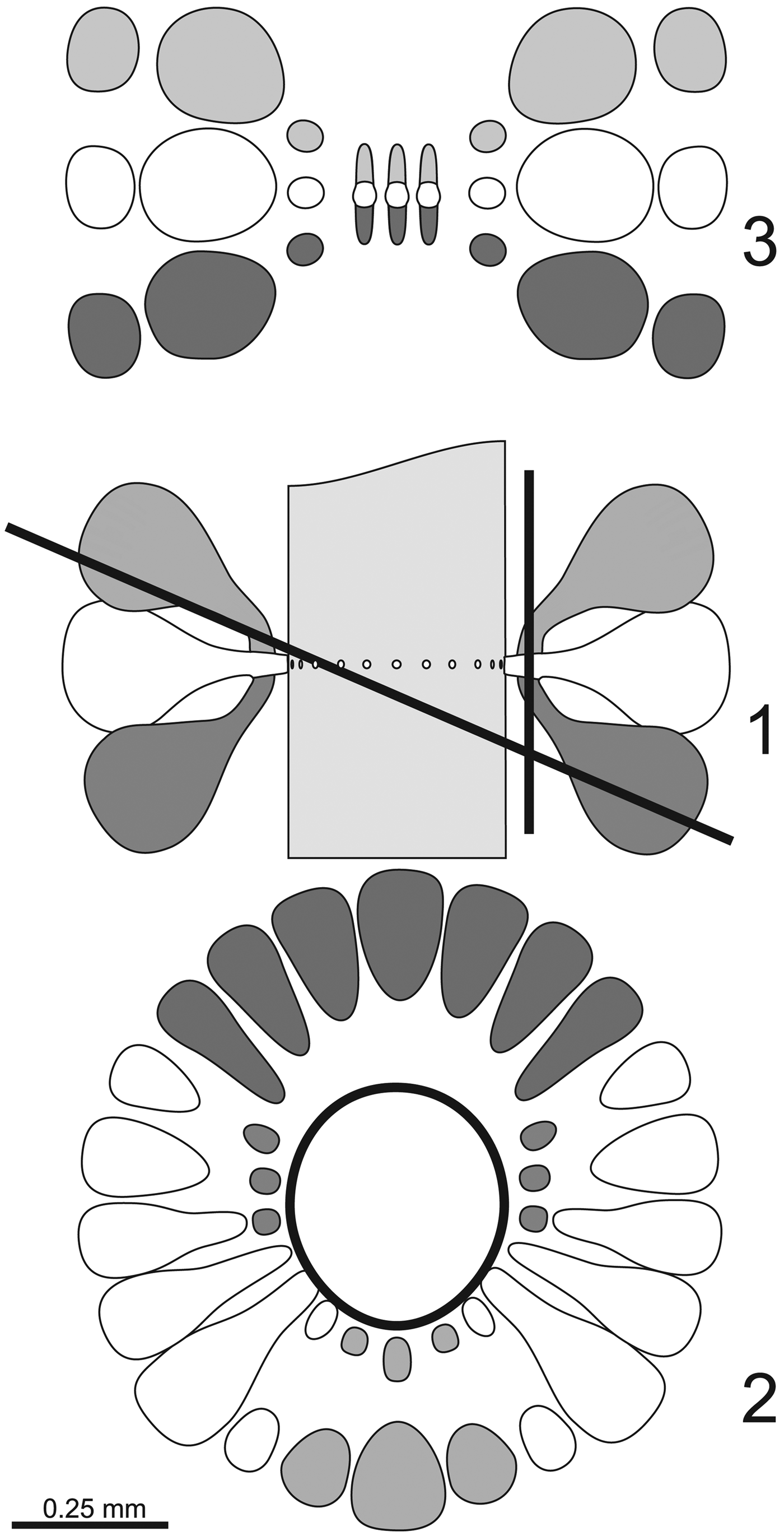
Subtransversal-oblique sections depicted in Figures 1.2 and 2.2 are particularly indicative to discern between the metaspondyl and euspondyl models. Such sections are present in Figure 3.3 and Granier (Reference Granier1987, pl. 7, figs. b, c). In the two-rows model (Fig. 1.2) sensu Azéma and Jaffrezo (Reference Azéma and Jaffrezo1972), there is no trace of proximal circular pores (no tuft of three pores vertically aligned), while in the metaspondyl model sensu Granier (Reference Granier1987) and Figure 2.2, the number of proximal pores is far fewer than observed (8 versus 24) while the value of the number of distal pores per row is the same (i.e., 12). If a w = 24 metaspondyl model sensu Granier (Reference Granier1987) is applied (Fig. 7), to correct the proximal pore framework, the number of distal pores (wdist) inevitably increases too much (72 instead of 24) as does the number of rows (at least three).
The metaspondyl model with w = 22. (1) Axial view with three rows of 22 laterals; (2) oblique section corresponding to oblique line in (1); (3) tangential section marked by vertical line in (1).

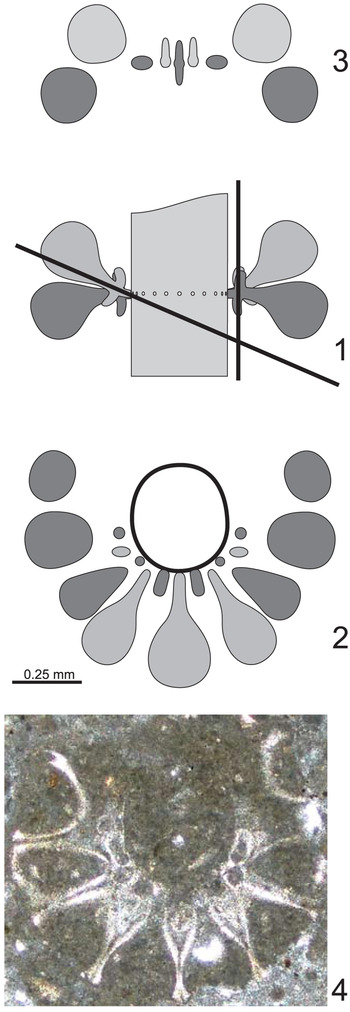
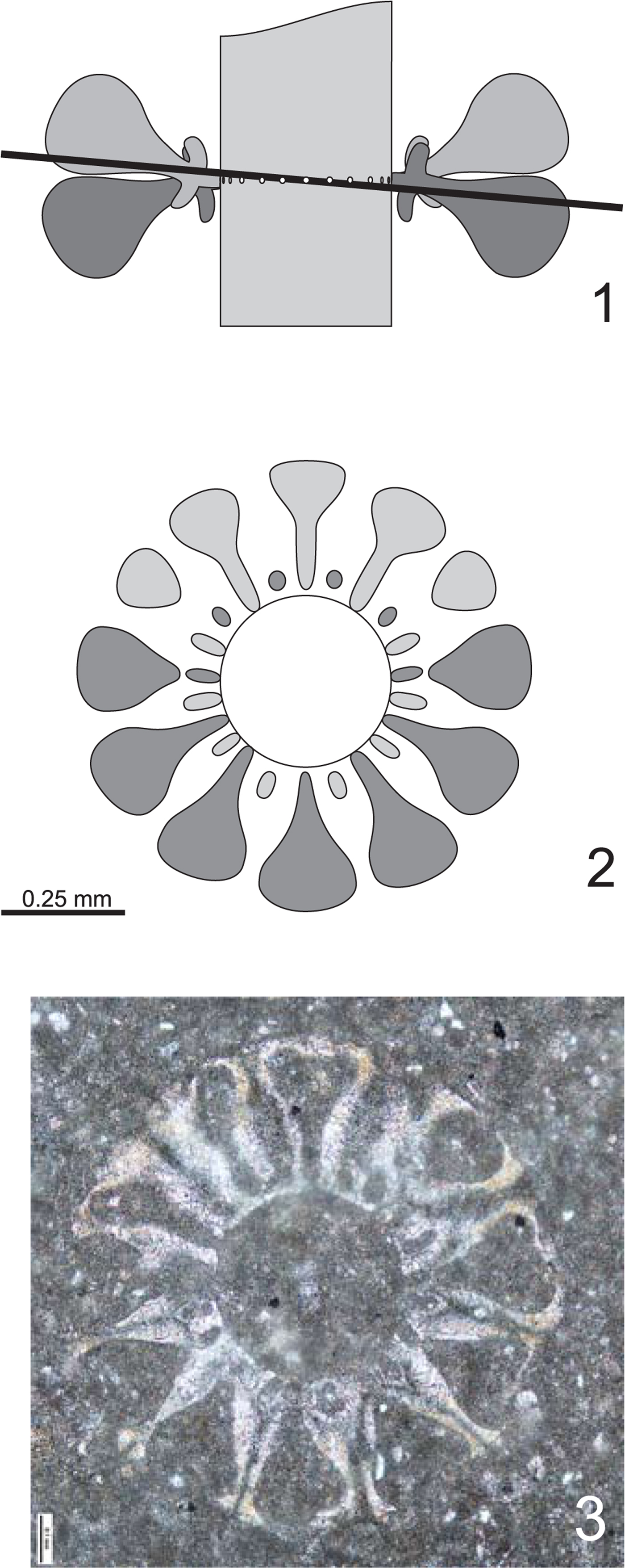
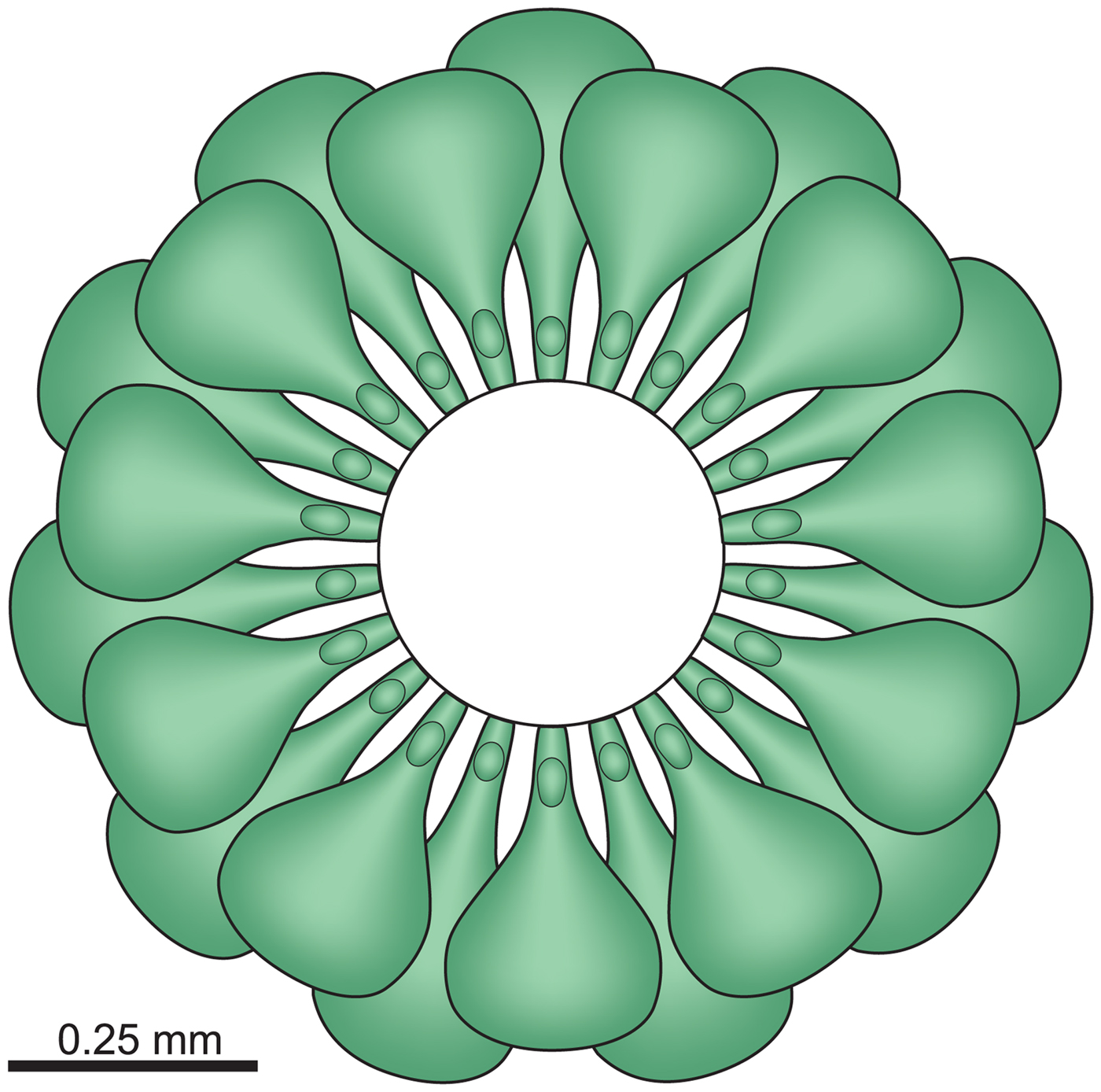
As assessed in the preceding, wprox = 16–25 and wdist ≅19–24, which means that the wdist/wprox ratio is equal to one instead of three, as in the metaspondyl model. Consequently, the two rows of 12 pores arrangement is attained by the upward and downward divergence of the ‘gametangia,’ due to their outward flaring. In this context, the proximal tuft of three pores has no influence on the distal number of laterals. Such a paradox is explained considering that two out of the three pores correspond to the short, ‘blind’ structures, visible in longitudinal section (Fig. 3.5). The corresponding new model is shown in Figures 8 and 9, where it is illustrated in subtransversal-oblique section (Fig. 8.2), in tangential section (Fig. 8.3), and in almost transversal section (Fig. 9.2).
Actinoporella campanensis (Azéma and Jaffrezo, Reference Azéma and Jaffrezo1972) n. comb. (1) Polyphysacean model in axial view with w = 22 and two rows of 11 laterals; (2) oblique section marked by oblique line in (1); note that transversally cut coronae are limited to the lower half of the section; (3) tangential section marked by vertical line in (1). (4) Same specimen as Figure 3.3, coherent with (2).
Actinoporella campanensis (Azéma and Jaffrezo, Reference Azéma and Jaffrezo1972) n. comb. (1) Polyphysacean model in axial view with w = 22 and two rows of 11 laterals; (2) subtransversal section marked by almost horizontal line in (1). Dark gray circles (inner upper part) correspond to the upper corona of fertile laterals directed downward (dark colored); light gray ellipses (inner lower part) intersect the lower corona of the fertile laterals inclined upward. Dark and light gray ellipses (inner left and right sides) represent the basal parts of fertile laterals. (3) Same specimen as Figure 3.1, coherent with (2).
Assignment at genus level
The two short, ‘blind’ structures are depicted in Figure 10. An interpretation of transversal, slightly oblique sections is given in the Systematic paleontology in the section describing Actinoporella campanensis, under Remarks (Fertile laterals and coronae). Polyphysacean coronae have been interpreted as: (1) expansions of the central stem, (2) primary laterals together with the basal part, or (3) secondary laterals (see review in Dumais and Harrison, Reference Dumais and Harrison2000). Here, coronae are considered as corresponding to secondary laterals, concurring with Valet's (Reference Valet1968, p. 62–63) interpretation.
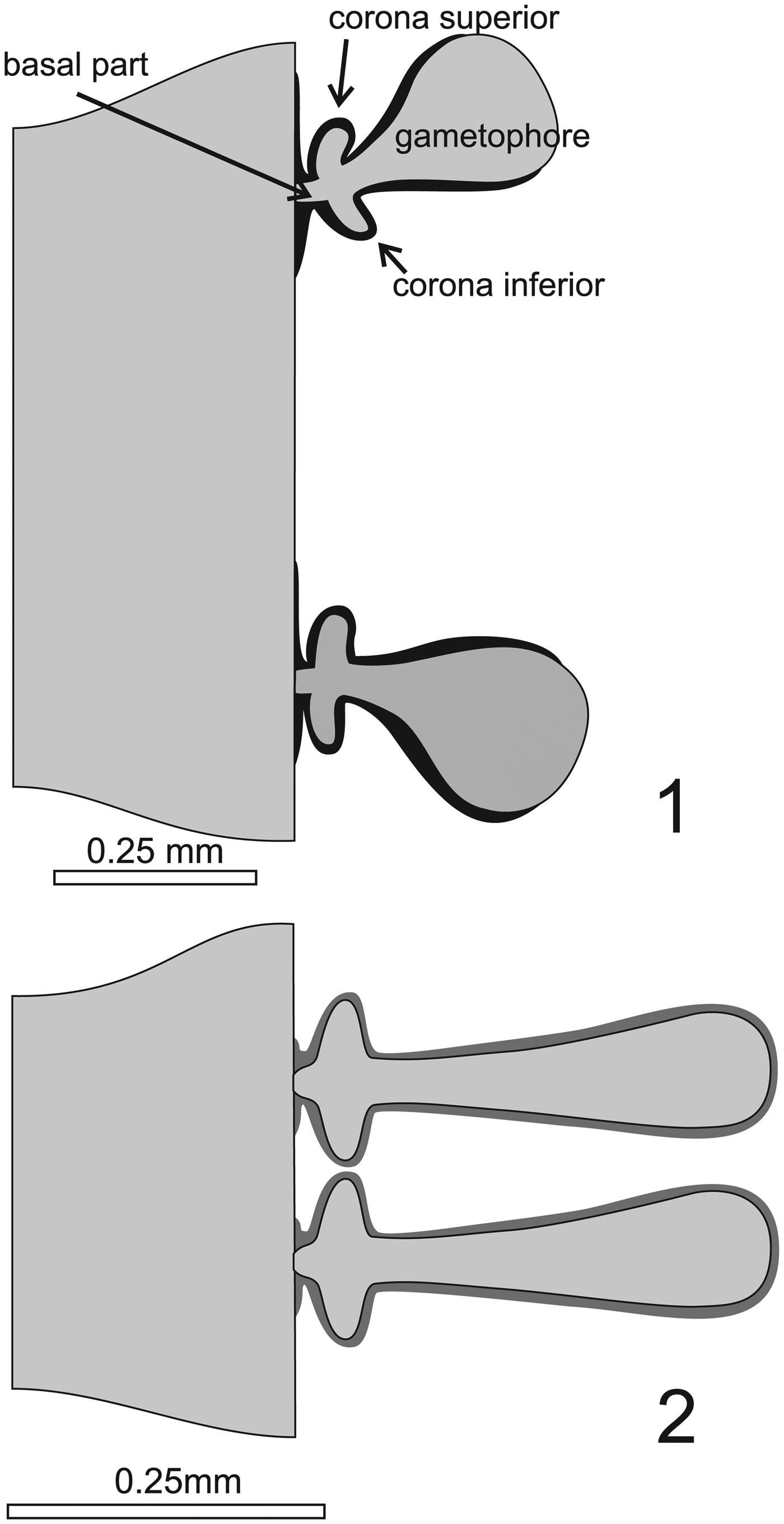
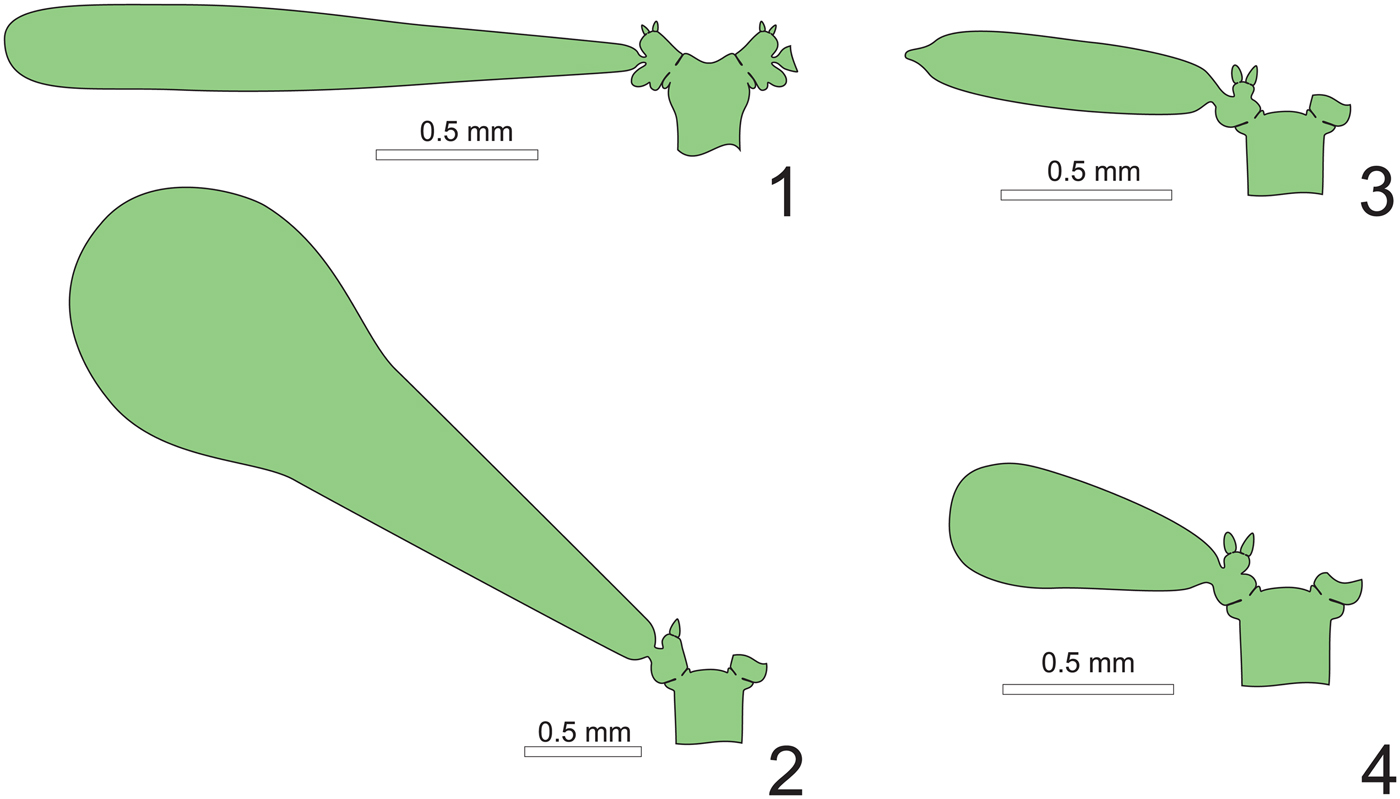
(1) Actinoporella campanensis (Azéma and Jaffrezo, Reference Azéma and Jaffrezo1972) n. comb. (2) Actinoporella podolica (Alth, Reference Alth1878). (1) Components of the fertile lateral and calcification (fibrous calcite) colored black. The basal part corresponds to the primary (first-order) lateral arising from the axis of the plant. (2) Notice the same structural elements in Actinoporella podolica. Compared with Actinoporella campanensis, laterals appear slenderer with closer whorls; calcification (aragonite) is drawn in dark gray.

From a taxonomic perspective, the occurrence of upper and lower coronae arising from a short basal lateral, and large outer pores corresponding to fertile laterals serving for reproduction (gametophores), make F. campanensis match the genus Actinoporella. Conversely, the presence of heterocline euspondyl laterals, with gametophores markedly swollen outward (pear-like), a calcareous skeleton made of fibrous calcite, and spaced whorls, make F. campanensis markedly different from Actinoporella podolica (Alth, Reference Alth1878), the type species of the genus.
Heterocline laterals
Several factors, such as the spacing of the whorls, size of laterals (p), number (w) of laterals per whorl, and calcification may condition the isocline versus heterocline arrangement. In extant genera Acetabularia and Parvocaulis, both isocline (e.g., A. acetabulum [Linnaeus, Reference Linnaeus1758] Silva, Reference Silva1952 and P. parvula [Solms-Laubach, Reference Solms-Laubach1895] Berger et al., Reference Berger, Fettweiss, Gleissberg, Liddle, Richter, Sawitzky and Zuccarello2003) and heterocline (e.g., A. peniculus and P. clavata [Yamada, Reference Yamada1934] Berger et al., Reference Berger, Fettweiss, Gleissberg, Liddle, Richter, Sawitzky and Zuccarello2003) species exist (e.g., Valet, Reference Valet1969; Berger et al., Reference Berger, Fettweiss, Gleissberg, Liddle, Richter, Sawitzky and Zuccarello2003). In our opinion, heteroclinity, taken solely, is not enough to support the ordering of taxa at the level of the genus, especially in family Polyphysaceae.
Gametophores markedly swollen outward (pear-like)
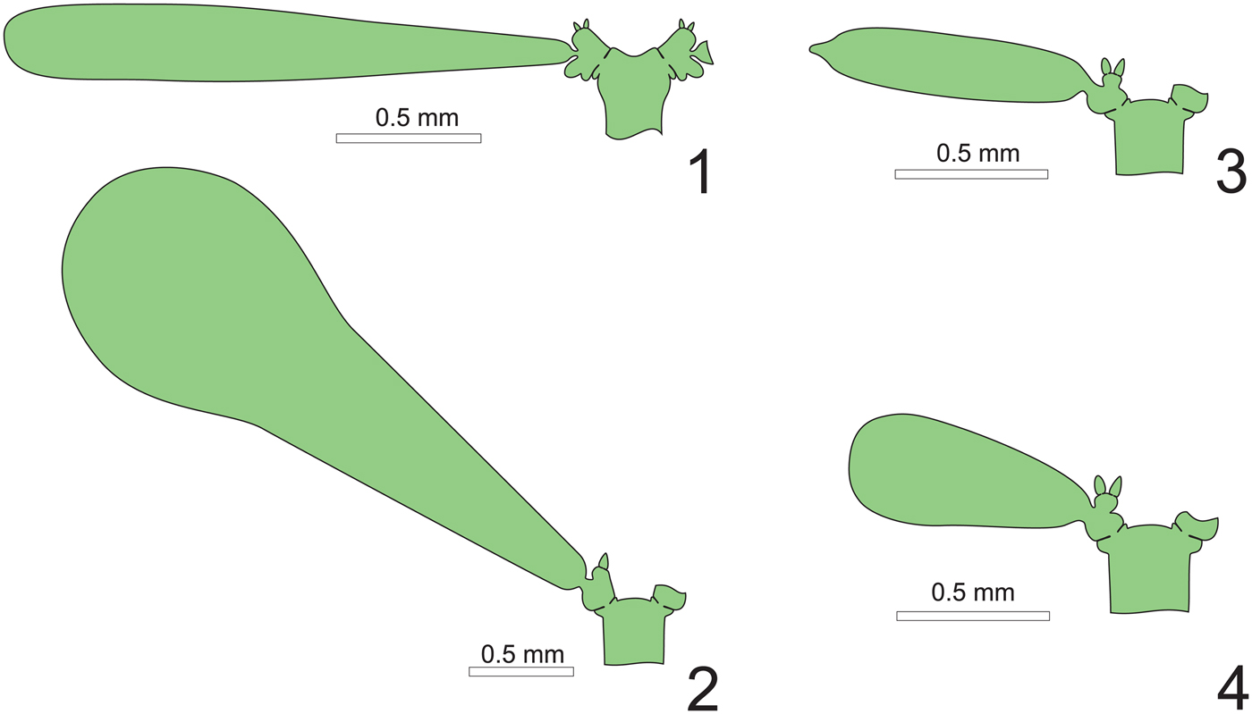
Sometimes, authors use the shape, more or less swollen, of the laterals to differentiate two genera (e.g., Suppiluliumaella from Triploporella in Barattolo, Reference Barattolo1983). Such a character, taken as an individual element of differentiation at the level of the genus, looks problematic. The shape of gametophores in extant genera Acetabularia and Parvocaulis is highly variable (Berger and Kaever, Reference Berger and Kaever1992; Berger et al., Reference Berger, Fettweiss, Gleissberg, Liddle, Richter, Sawitzky and Zuccarello2003; Fig. 11). Species such as Acetabularia acetabulum, A. crenulata Lamouroux, Reference Lamouroux1816, and A. farlowii Solms-Laubach, Reference Solms-Laubach1895 hold slender gametophores, but in Acetabularia peniculus, they are ‘vesiculiferous’ (Valet, Reference Valet1969, p. 619). The same happens in genus Parvocaulis, with Parvocaulis exigua (Solms-Laubach, Reference Solms-Laubach1895) Berger et al., Reference Berger, Fettweiss, Gleissberg, Liddle, Richter, Sawitzky and Zuccarello2003 holding elongated (acrophorous) gametophores and P. pusilla (Howe, Reference Howe1909) Berger et al., Reference Berger, Fettweiss, Gleissberg, Liddle, Richter, Sawitzky and Zuccarello2003 holding pear-shaped gametophores. The same, but intraspecific, variability is also very high in extant taxa such as Acetabularia crenulata (Berger and Kaever, Reference Berger and Kaever1992, p. 144).
Shape of gametophores in the extant genera Acetabularia and Parvocaulis. (1) Acetabularia farlowii Solms-Laubach, Reference Solms-Laubach1895; (2) Acetabularia peniculus (Brown, Reference Brown and Turner1819) Solms-Laubach, Reference Solms-Laubach1895; (3) Parvocaulis exigua (Solms-Laubach, Reference Solms-Laubach1895) Berger et al., Reference Berger, Fettweiss, Gleissberg, Liddle, Richter, Sawitzky and Zuccarello2003; (4) Parvocaulis pusilla (Howe, Reference Howe1909) Berger et al., Reference Berger, Fettweiss, Gleissberg, Liddle, Richter, Sawitzky and Zuccarello2003. Notice the absence of corona inferior in Parvocaulis and the slender to ‘pear-shaped’ morphology of the gametophores in the two genera. Drawings based on Berger and Kaever (Reference Berger and Kaever1992) and Berger et al. (Reference Berger, Fettweiss, Gleissberg, Liddle, Richter, Sawitzky and Zuccarello2003).
Calcareous skeleton made of fibrous calcite
Such a structure of the calcareous wall (fibrous calcite), even if less common than the original aragonitic structure changed into blocky calcite, also occurs in other fossil taxa. A generic, if not suprageneric, significance of the mineralogy of the calcareous skeleton is assigned by Simmons et al. (Reference Simmons, Emery and Pickard1991). But in the literature, a much broader consensus assigns a species-level rank to this character, as shown by various examples found in different fossil genera, such as Salpingoporella (S. muhelbergii [Lorenz, Reference Lorenz1902] Pia, Reference Pia and Trauth1917, the type species, originally aragonitic, and S. dinarica Radoičić in Granier and Doloffre, Reference Granier and Deloffre1993, fibrous calcite), Clypeina (C. marginoporella Michelin, Reference Michelin1840, aragonitic, and C. sulcata [Alth, Reference Alth1881] Granier and Brun, Reference Granier and Brun1991, fibrous calcite), Pseudoclypeina (P. cirici Radoičić, Reference Radoičić1975 and P. distomensis Barattolo and Carras, Reference Barattolo and Carras1990, fibrous calcite, and P. farinacciae Radoičić, Reference Radoičić1975, aragonitic), and Falsolikanella (F. campanensis, fibrous calcite, and F. danilovae Granier et al., Reference Granier, Bucur and Trabold1999, aragonitic).
Spacing of the whorls
In families Triploporellaceae and Dasycladaceae, distance between the whorls, close versus spaced, is an important character since directly related to the general aspect of the alga, reflecting the organization of the thallus. Close whorls usually occur in taxa provided with an external cortex and a strong calcareous skeleton. Close versus spaced whorls is one of the characters used to macroscopically discern Dasycladus from Batophora (Valet, Reference Valet1969, p. 578, 584). The problem arises when applied to the Polyphysaceae. In this family, most extant species are single whorled at maturity, with umbrella-like thalli, while Halicoryne is the only extant genus where comparisons between the relative distance between whorls were carried out. The h/D ratio (h = distance between whorls, D = outer diameter of the thallus) was estimated in H. spicata (Kützing, Reference Kützing1863) Solms-Laubach, Reference Solms-Laubach1895 by Valet (Reference Valet1968, pl. 20, fig. 4) and in H. wrightii Harvey, Reference Harvey1859 by Valet (Reference Valet1969, pl. 147, fig. 1). In H. spicate, the h/D ratio is relatively large compared to H. wrightii (i.e., 0.40 versus 0.20, respectively). The same parameter in Falsolikanella campanensis (Fig. 10.1) and Actinoporella podolica (Fig. 10.2) is even more emphasized (0.56 versus 0.15). As a matter of fact, Actinoporella podolica exhibits an extreme variability in size and distance between whorls, as shown by the topotype specimens illustrated by Conrad et al. (Reference Conrad, Praturlon and Radoičić1974, fig. 6b), where the h/D ratio is around 0.50, a value also found in Actinoporella campanensis.
Bearing in mind such a close examination of the taxonomic characters, in our opinion the moderately phloiophorous shape, isocline gametophores, and rather closer whorls in Actinoporella podolica, as opposed to the markedly flared out, heterocline laterals and spaced whorls in F. campanensis, are characters of specific weight only. At genus level, and for the same reason, there are no strong arguments to separate Falsolikanella from Actinoporella. Consequently, Falsolikanella campanensis is ascribed to the genus Actinoporella, and the genus Falsolikanella Granier, 1987 is dealt with as a junior synonym of Actinoporella Gümbel, Reference Gümbel1882.
Systematic paleontology
Order Dasycladales Pascher, 1931
Family Polyphysaceae Kützing, 1843
Systematics at tribe level
The family Polyphysaceae is traditionally subdivided in three tribes: Acetabularieae Decaisne, Reference Decaisne1842, Halicoryneae Valet, 1969, and Clypeineae (Elliott, Reference Elliott1968) Bassoullet et al., Reference Bassoullet, Bernier, Deloffre, Génot, Jaffrezo and Vachard1979 (Deloffre, Reference Deloffre1988).
The tribe Acetabularieae includes genera presenting a more or less calcified thallus, with a disc at top of the axis, made of reproductive chambers (definition in Deloffre, Reference Deloffre1988: “Thalle plus ou moins calcifié. Disque reproducteur au sommet de l'axe formé par des loges reproductrices”). According to Valet (Reference Valet1969, p. 578), the tribe Halicoryneae is defined as follows (translated from French): “Plants with numerous alternating sterile and fertile verticils.” The last tribe, Clypeineae, is defined by Deloffre (Reference Deloffre1988, p. 182) with the following diagnosis: “Ramifications primaires, secondaires possibles et de troisième ordre éventuelles; verticilles espacés saillants; présence éventuelle de corona; cavité axiale cylindrique” (free English translation: Primary branches, possibly secondary branches, tentatively third-order branches; corona possibly present; axial cavity cylindrical.)
Remarks
Even considering that the reproductive function of the laterals of fossil taxa is widely speculative, such a definition makes the Clypeineae tribe a catchall taxon since it aggregates cladospore (e.g., Pseudoclypeina) and choristospore lato sensu (e.g., Actinoporella and Hamulusella) genera. In extant acetabulariacean algae, fertile laterals hold further, sterile laterals arising from the corona, but no terminal second and perhaps higher-order laterals, such as in the fossil Pseudoclypeina. Consequently, genera with fertile laterals bearing terminal ramifications must be separated from genera with crown-like proximal structures included in tribe Halicoryneae. Certainly, the most remarkable character of Halicoryne is the occurrence of alternating sterile and fertile whorls, but this character is also visible in Acetabularia crenulata (Berger and Kaever, Reference Berger and Kaever1992, p. 144), resulting that the only possibly distinguishing character is the presence of coronae: upper or upper and lower coronae in tribe Acetabularieae and only more or less evident lower corona in the Clypeineae. At this point, these two tribes are herein defined as follows:
Tribe Acetabularieae: thallus cylindrical, umbelliform, or with spaced-out whorls of fertile rays. Upper corona only, or upper and lower coronae. Genera ascribed: Acetabularia, Parvocaulis, Chalmasia, Halicoryne, Orioporella, Actinoporella.
Tribe Clypeineae: thallus cylindrical, umbelliform, or with spaced-out fertile laterals. Possible presence of variously marked lower corona. Genera ascribed: Clypeina, Hamulusella.
Tribe Acetabularieae Decaisne, Reference Decaisne1842
Genus Actinoporella Gümbel, Reference Gümbel1882, emended
Type species
Actinoporella podolica (Alth, Reference Alth1878)
Emended diagnosis
Thallus simple, unbranched, bearing spaced-out whorls of short primary laterals. Second-order laterals are three in number: one upper and one lower corona, both arising from the proximal part of the primary lateral, and one large gametophore arising from the tip of the primary lateral.
Taxonomic position at higher rank
Deloffre (Reference Deloffre1988) ascribed Actinoporella to the tribe Clypeineae. In this paper, drawing on the preceding discussion showing the presence of lower and upper coronae, compliant with Génot et al. (Reference Génot, Saint-Martin and Bucur2008), the genus Actinoporella is assigned to the tribe Acetabularieae.
Remarks
The paper of Conrad et al. (Reference Conrad, Praturlon and Radoičić1974) includes a historical record of Actinoporella, a synonymy (Verticilloporella Raviv and Lorch, Reference Raviv and Lorch1970 is dealt with as a junior synonym), reproductions of key, historically important illustrations issued by Alth (Reference Alth1878) and Pia (Reference Pia1920), an emended diagnosis, similarities with the extant Polyphysaceae, and a review study of Actinoporella podolica based on topotype specimens collected at Tanutinska Gora, near Nizniow city, now in Ukraine. The emended diagnosis was as follows: “Cylindrical stalk bearing regularly spaced, disc-like whorls of branches. More or less marked outgrowths, or excrescences occur at the branches’ proximal ends. These outgrowths develop upper and/or lower coronas surrounding the stalk” (Conrad et al., Reference Conrad, Praturlon and Radoičić1974, p. 4).
Compared to the previous diagnosis, somewhat outdated terms such as ‘branches’ and ‘outgrowths’ are respectively replaced by ‘laterals’ and ‘corona,’ the major difference calling for the presence of coronae arising from and located above as well as below the primary lateral, a character considered at the level of the genus. Other characters, such as expansions (vestibules) of the axis of the plant, whorls made of iso- or heterocline laterals, dimensions, and pattern of calcification, are less important for the systematics.
In this paper, three species, A. podolica, A. campanensis n. comb., and A. kukoci are assigned to Actinoporella, but in the literature, no less than 15 other species were assigned, although sometimes with doubt, to this genus. A comprehensive review of all of them is beyond the scope of this paper. Updating a list issued by Conrad et al. (Reference Conrad, Praturlon and Radoičić1974), an alphabetical list of species assigned to Actinoporella in the literature follows. As for the genus Pseudoactinoporella Conrad, 1970, reference is made to a review issued by Bucur et al. (Reference Bucur, Rashidi and Senowbari-Daryan2012).
Actinoporella durandelgai Jaffrezo and Fourcade ex Jaffrezo in Bassoullet et al., Reference Bassoullet, Bernier, Conrad, Deloffre and Jaffrezo1978, name fide Granier and Deloffre (Reference Granier and Deloffre1993). Basionym: Clypeina durandelgai. Tithonian–Berriasian. Spaced-out whorls of laterals bearing in their lower part conspicuous proximal outgrowths evoking a corona. Transferred to Hamulusella by Barattolo (Reference Barattolo1998).
?Actinoporella geredeensis Farinacci and Radoičić, Reference Farinacci, Radoičić, Farinacci, Ager and Nicosia1991, Berriasian. Upper and lower proximal outgrowths. Laterals strongly tilted. The questionable assignment to Actinoporella is original.
Actinoporella guembeli Alth, Reference Alth1882, a junior synonym of Actinoporella podolica according to Pia (Reference Pia1920) and Conrad et al. (Reference Conrad, Praturlon and Radoičić1974).
Actinoporella fragilis (Conrad, Reference Conrad1970) Granier, Reference Granier1994, late Hauterivian–early Aptian. Laterals closely packed, strongly tilted, bearing a single, upper proximal feature suggesting a corona. In this paper, the original name, Pseudoactinoporella fragilis (Conrad, Reference Conrad1970) Conrad and Peybernés, Reference Conrad and Peybernés1976, is upheld.
Actinoporella israelensis Raviv and Lorch, Reference Raviv and Lorch1992, Callovian. A nomen nudum, close if not a synonym of Salpingoporella grudii Radoičić, Reference Radoičić1975.
Actinoporella jaffrezoi Granier, Reference Granier1994, Berriasian–Hauterivian. Tentative assignment to Praturlonella, pending a revision.
Actinoporella krymensis Maslov, Reference Maslov1958, nom. nud., Late Jurassic. Possibly a Clypeina according to Conrad et al. (Reference Conrad, Praturlon and Radoičić1974). Not properly typified.
Actinoporella kukoci Radoičić, Reference Radoičić1974, Eocene. Similar to A. podolica but usually larger, with a larger number of laterals per whorl (w value) (see also Radoičić, Reference Radoičić1995).
Actinoporella lucasi (Emberger, Reference Emberger1955) Granier, Reference Granier1992, Valanginian. Basionym: Clypeina lucasi Emberger, Reference Emberger1956. A synonym of A. podolica according to Conrad et al. (Reference Conrad, Praturlon and Radoičić1974).
Actinoporella maslovi Praturlon, Reference Praturlon1964, Late Jurassic to basal Neocomian. Transferred to Clypeina by Radoičić (Reference Radoičić1986).
Actinoporella nigra (Conrad and Peybernès, Reference Conrad and Peybernès1978) Granier, Reference Granier1994, late Hauterivian–late Barremian. Basionym: Clypeina nigra Conrad and Peybernés, Reference Conrad and Peybernès1978. The ‘excrescences’ of Granier (Reference Granier1994, pl. 3, fig. 1, arrow), next to the axis of the plant, denote solid (closed) thickenings of the dark, microgranular calcareous mantle and probably are not genuine coronae. The attribution to Actinoporella remains open.
Actinoporella podolica (Alth, Reference Alth1878) Alth, Reference Alth1882, type species of Actinoporella. Basionym: Gyroporella podolica. Tithonian–early Barremian. See Conrad et al. (Reference Conrad, Praturlon and Radoičić1974) for details.
Actinoporella sadalanensis (Elliott, Reference Elliott1977) Granier, Reference Granier1994. Hamulusella sadalanensis, the basionym, is upheld in this paper. Paleocene. Widely spaced whorls of laterals showing a short proximal portion below the connection with the axial stem. See Barattolo (Reference Barattolo1998) for a discussion.
Actinoporella? silvaeregis—Basionym: Pseudoactinoporella silvaeregis Bucur, Reference Bucur1981. Bucur (Reference Bucur1992): questionable assignment to Pseudoactinoporella. Bucur (Reference Bucur1999): questionable assignment to Falsolikanella. Granier (Reference Granier1994): questionable assignment to Actinoporella. Barremian–early Aptian. Notice: no upper and/or proximal features evoking coronae occur, but horizontal (transversal) proximal swellings of the laterals.
Actinoporella sulcata Alth, Reference Alth1882. Basionym: Clypeina sulcata. Quoted by some authors as a senior synonym of Clypeina jurassica Favre in Favre and Richard, Reference Favre and Richard1927. The transfer to Clypeina, first made by Bassoullet et al. (Reference Bassoullet, Bernier, Conrad, Deloffre and Jaffrezo1978, p. 32), is debatable. See also Conrad et al. (Reference Conrad, Praturlon and Radoičić1974).
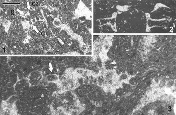
Based on the preceding analysis of taxonomic criteria (assignment at genus level), F. campanensis is revised and transferred to the genus Actinoporella, whose systematics is emended. Currently, the genus comprises three species, formally valid, A. podolica, A. kukoci, and A. campanensis. Actinoporella kukoci is the youngest species (Eocene). In Figure 12, some specimens from the type material are supplied, showing the typical characters of the genus.
Actinoporella kukoci Radoičić, Reference Radoičić1974. Type material. (1) Tangential section cutting a whorl proximally; the two rows of aligned rounded pores correspond to gametophores (arrow g) and one of two coronae (arrow c1); the pore pointed to by arrow c2 probably corresponds to the other corona; thin section n. RR2808; (2) longitudinal oblique section cutting a single whorl; the upper and lower protuberances set proximally aside each pore can be interpreted as calcification around recrystallized coronae; original illustration by Radoičić (Reference Radoičić1974, pl. 1, fig. c); (3) tangential section of a whorl; one of two superimposed pores (arrow) probably corresponding to a corona; thin section n. RR2808. Scale bar = 0.50 mm.

Actinoporella campanensis (Azéma and Jaffrezo, Reference Azéma and Jaffrezo1972) new combination
Figures 3, 4
- 1972
Likanella campanensis Azéma and Jaffrezo, p. 126, pl. 1, figs. 1–12.
- 1972
Likanella campanensis; Fourcade et al., p. 235, pl. 2, fig. 9.
- 1974
Likanella campanensis; Jaffrezo, p. 28, pl. 2, figs. 13, 14.
- 1977
Likanella campanensis; Conrad, p. 199.
- 1978
Likanella? campanensis; Barattolo, p. 9.
- 1978
Likanella campanensis; Bassoullet et al., p. 141, pl. 16, figs. 4–6, bibliographic review.
- 1978
Likanella campanensis; Radoičić, p. 189, pl. 1, 2.
- 1978a
Selliporella campanensis; Sokač and Velić, pl. 5.
- 1979
Likanella campanensis; Canérot, p. 505.
- 1979
Likanella campanensis; Peybernès, p. 735.
- 1987
Falsolikanella campanensis; Granier, p. 208, pl. 1, figs. a–d, g, h, l, m, bibliography.
- 1988
Falsolikanella campanensis; Granier, p. 52, pl. 6, 7, figs. a–c, f, i.
- 1991
Falsolikanella campanensis; Farinacci and Radoičić, p. 138, pl. 7, figs. 1, 2, 4, 5.
- 1993
Falsolikanella campanensis; Granier and Deloffre, p. 30.
- 1993a
Falsolikanella campanensis; Masse, p. 315, pl. 1, fig. 6.
- 1993b
Falsolikanella campanensis; Masse, p. 366.
- 1993
Falsolikanella campanensis; Soták and Mišik, pl. 8, figs. 3, 4.
- 1994
Falsolikanella campanensis; Schindler and Conrad, p. 75.
- 1994
Falsolikanella campanensis; Granier and Berthou, p. 115, fig. 3.
- Non 2000
Falsolikanella cf. F. campanensis; Bucur et al., p. 440, pl. 5, figs. 11–13.
- ?2014
Falsolikanella campanensis; Granier et al., p. 27, pl. 6, fig. d.
Holotype
Oblique section with three whorls (University of Lyon collection, FSL 420001), thin section n. 3486, from Puig Campana (Alicante, Spain), Berriasian, (Azéma and Jaffrezo, Reference Azéma and Jaffrezo1972, pl. 1, fig. l), here shown in Figure 3.6.
Emended diagnosis
Central stem cylindrical, bearing spaced whorls of heterocline laterals distally arranged in two rows, less frequently more than two rows. Each proximal pore corresponds to an initial short tract, corresponding to a primary lateral. Three vertically aligned elements arise from the primary lateral; the central element, arranged in distal continuity with the primary lateral, markedly flares outward, serving as fertile lateral (gametophore). Conversely, the upper and lower elements are rather short and slender, homologous to upper and lower coronae, as in Acetabularia. Calcification consists of a thin fibrous layer coating the primary laterals and the coronae. The layer extends outward, coating the gametophores for a large portion of their length. A thinner, continuous layer coats the central stem, leaving the space between whorls uncalcified.
Remarks
Topotype specimens show the calcareous skeleton to be articulated, made of a thin, yellowish, fibrous layer coating the fertile laterals and the central stem, upward and downward, but excluding the bulk of the space between adjacent whorls. The thin, calcified layer follows the outline of the central stem and fertile laterals. Despite the primary fibrous mineralization, tangential sections sometimes suggest a lateral ‘fusion’ of the proximal pores (Fig. 4.6, 4.8), misleadingly calling for two gametangia to arise from a single proximal point.
In well-preserved specimens, a darker rim, tentatively of primary origin, marks the outer side of the layer; it is particularly visible at the boundary between adjacent fertile laterals and the proximal contact between the corona and the fertile lateral (Figs. 3.6, 4.2, 4.6, 4.8). Instead, only a single layer is visible at the contact between the laterals, in the inner portion of the whorl (Fig. 3.1, 3.2). Toward the exterior, the calcareous layer distally decreases in thickness and is missing at the tip of the fertile lateral (Fig. 3.3). Along the central stem the calcareous layer also becomes thinner and thinner, coating the axis downward and upward from each whorl (Fig. 3.5), leaving the central stem mid part uncalcified, thus triggering the postmortem disarticulation of the alga.
Fertile laterals are 16 to 25, commonly 20–24 per whorl. In axial section, they arise from the tip of a short tract (e.g., Fig. 3.5) bearing three elements of different length. The longest and largest, phloiophorous and pear shaped, is set in prolongation of the first tract (Fig. 3.5). The upper element (upper corona), cylindrical and rather elongated, arises perpendicularly to the first tract and bends slightly forward, as some sections suggest (Fig. 4.1, 4.9). In transversal section (Fig. 3.1) the upper coronae appear as elliptic pores between gametophores (white arrow), but aside some equivalent pores tend to split in two circular smaller pores aligned radially (black arrow). Available material does not allow better documentation and explanation of this character. Possibly, the couple of pores shown by the black arrows correspond to corona protuberances visible on extant polyphysacean algae (Dumais and Harrison, Reference Dumais and Harrison2000, fig. 4h). The lower element (lower corona), shorter than the upper one, is gently leaning and tapered forward. Depending on whether it belongs to fertile laterals, it bends upward or downward (Figs. 3.5, 4.5). The first tract, halfway in the section from the upper and lower coronae, forms the continuation of the short primary lateral, whereas its distal phloiophorous extension corresponds to the gametophore (Fig. 10).
In each whorl, pores corresponding to the fertile laterals are roughly aligned on a single horizontal plane. Proximally, however, at level of the corona, they diverge upward and downward; thus, in the proximal tangential section, adjacent tufts of three pores look slightly twisting (Fig. 4.6, 4.7). Distally, the fertile laterals are arranged in a single plane made of two rows (Fig. 4.7, 4.9), but noticeably, more than two tilts (probably three) occur in some specimens (Fig. 3.6).
Transversal-oblique sections allow one to pursue the transition among the elements forming the fertile laterals. In this kind of section, the coronae are cut transversally and appear as circles (Fig. 3.1–3.4). In Figure 8.2, the dark-gray circles correspond to the lower corona of downward-bending gametophores, being in continuity with them, whereas the light-gray ellipses match the lower corona of those gametophores directed upward. Note that in Figure 3.3 and 3.4 (last circular pore at right), the pores occur only in correspondence with the whorl from which they are originating. This means that such upright structures are short, not forming elongated laterals. Otherwise, laterals cut transversally would have all been visible, forming circles/ellipses (e.g., model shown in Fig. 7.2; see also De Castro, Reference De Castro1997, figs. 17, 18). Figure 8.2 explicates the elements cut in subtransversal section. In the middle part of the figure, the dark-gray circles correspond to the lower corona of downward-bending fertile laterals, while the two light-gray ellipses correspond to the lower corona of upward-bending fertile laterals. The two small, dark-gray elongated ellipses correspond to basal parts of downward-bending fertile laterals.
In tangential section (e.g., Fig. 4.6, 4.8) the upper and lower coronae are ‘fused’ with the basal part forming an elliptic vertical pore (see also Fig. 8.3). Noticeably, in this kind of section, the threefold tuft (three vertically aligned pores) passes sideways to a couple of large pores (the gametophores, Fig. 4.1, 4.9) in agreement with wdist/wprox ratio equal to 1.
No trace of reproductive organs (gametangia) was observed in the large pear-shaped gametophores or in the calcareous skeleton, making that the kind of reproduction that is supposedly homologous to that of Acetabularia.
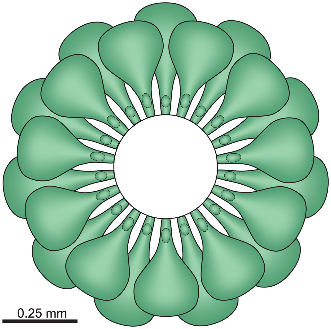
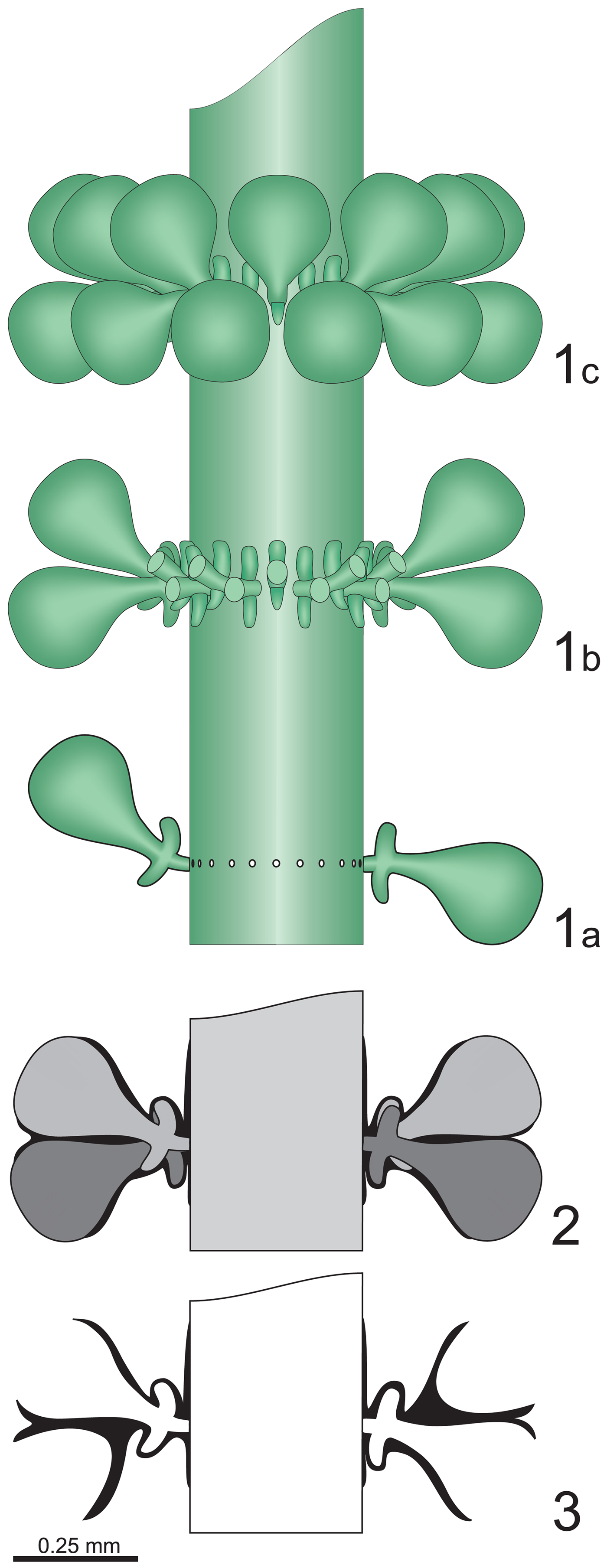
The reconstructions of the thallus (Figs. 13, 14) depict five whorls in longitudinal view/section and one in upper view. Only the top and the bottom of the thallus are not recorded, probably uncalcified. Tentatively, we assume that the shape of these uncalcified parts was like in the extant genus Halicoryne (Berger and Kaever, Reference Berger and Kaever1992). In fact, Actinoporella and Halicoryne belong to the same family, with a cylindrical thallus bearing spaced whorls of fertile laterals. Thus, in A. campanensis, the fertile laterals were probably decreasing gradually in size and inclination toward the apex. Moreover, because known specimens consist of whorls of fertile laterals, the calcified thallus most likely corresponds to the mature stage of the alga while the juvenile, sterile stages were probably not calcified and were lost during the algal growth.
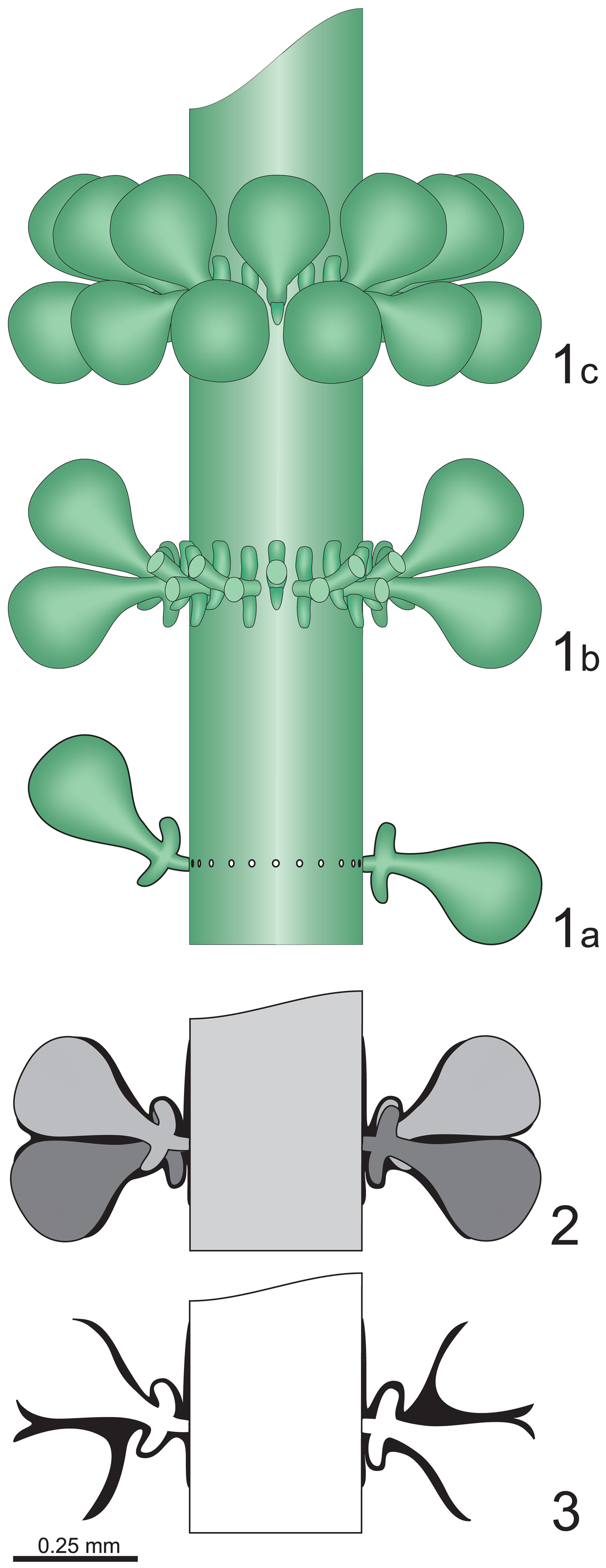
Actinoporella campanensis (Azéma and Jaffrezo, Reference Azéma and Jaffrezo1972) n. comb. Reconstruction of the alga in axial view; (1) three consecutive whorls, removing the calcification; (1a) deprived of frontal fertile laterals; (1b) frontal fertile laterals truncated to show the coronae; (1c) whorl shown in perspective view; (2) soft parts and calcareous skeleton of a whorl; (3) calcareous skeleton (black) of a whorl.
Actinoporella campanensis (Azéma and Jaffrezo, Reference Azéma and Jaffrezo1972) n. comb. Reconstruction of the alga in transversal view, removing calcification.
Systematics of species previously assigned to the genus Falsolikanella
Several species have been ascribed to the genus Falsolikanella Granier, 1987; the status is here discussed for some of them.
Pseudoactinoporella? silvaeregis (Bucur, Reference Bucur1981)
- 1981
Pseudoactinoporella silvaeregis Bucur, p. 151, pls. 1, 2.
- 1992
Pseudoactinoporella? silvaeregis; Bucur, p. 450, pl. 3.
- 1999
Falsolikanella(?) silvaeregis; Bucur, p. 58, pl. 3, fig. 8, with synonymy.
Holotype
Perpendicular–oblique section, sample 48E, thin section N.I. 1051, Carnet - Valea Poienii, Romania (Bucur, Reference Bucur1981, pl. 1, fig. 1).
Remarks
Although with doubt, the Barremian to early Aptian Pseudoactinoporella silvaeregis Bucur, Reference Bucur1981 is assigned by Bucur (Reference Bucur1999) to the genus Falsolikanella. Quoting this author, the specimen in longitudinal section illustrated by Bucur (Reference Bucur1992, p. 451, pl. 3, fig. 7; reillustrated in Bucur Reference Bucur1999, pl. 3, fig. 8) “exhibits a short globulous primary branch … splitting in two phloiophorous branches arranged in a vertical plane.” The laterals start with a short peduncle, then those verging upward (e.g., second, third, fifth, and sixth whorls on the right) exhibit a sort of upward-verging ‘triangular’ expansion. Those slightly verging downward (second whorl on the left, fourth and seventh on the right) show the similar ‘triangular’ expansion to be set downward, resulting in all laterals widening regularly outward. The lack of proximal tangential sections makes it difficult to understand whether the short proximal peduncle corresponds to a primary lateral. Therefore, in this paper, the species is, provisionally at least, left in the genus Pseudoactinoporella.
Praturlonella danilovae (Radoičić, Reference Radoičić1975)
Figure 15
- 1975
Likanella danilovae Radoičić, p. 151, pls. 1, 2.
- 1978
Praturlonella danilovae; Barattolo, p. 9, 31.
- 1999
Falsolikanella danilovae; Granier et al., p. 58, pl. 3, fig. 8, with synonymy.
Praturlonella danilovae (Radoičić, Reference Radoičić1975). (1–6) Type material; (7) Bosnia, Kljuc sheet; (1) transversal section, thin section n. RR1865; (2) oblique section, thin section n. RR1864; (3) subtransversal section, thin section n. RR1822; (4) oblique section, thin section n. RR1865; (5) tangential section, thin section n. RR1866; (6) tangential section, thin section n. RR1893; (7) longitudinal section, thin section n. RR4243. Scale bar = 0.5 mm.

Holotype
Oblique section designed by Radoičić (Reference Radoičić1975), thin section RR4243 of the Radoičić collection (Fig. 15.7), corresponding to Radoičić (Reference Radoičić1969, pl. 1, fig. 1).
Remarks
The late Hauterivian to early Barremian Likanella danilovae Radoičić, Reference Radoičić1975 is transferred by Granier et al. (Reference Granier, Bucur and Trabold1999) to the genus Falsolikanella. Quoting Granier et al. (1999, p. 177), the “thallus bears single whorls consisting of ‘bundles’ of branches, and that each bundle consists of a few secondary branches on top of a short vestibular part. Vestibules were first regarded as expansions of the main axis; they are presently considered as equivalents to primary branches.” The authors do not specify the number of secondary laterals per tuft.
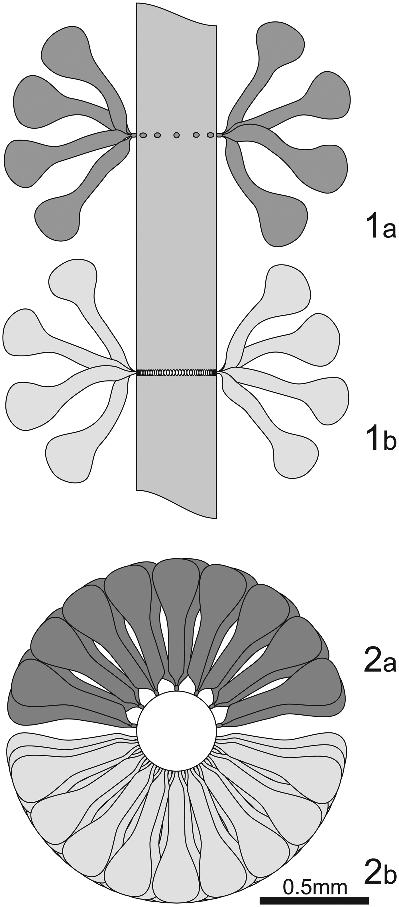
Figure 16, based on the holotype (Fig. 15.7) of P. danilovae, compares the metaspondyl model (Fig. 16.1a, 16.2a) to the heterocline model (Fig. 16.1b, 16.2b) applicable to this species. In the metaspondyl model, the distal number of laterals (wdist) is estimated to be 80, while the number of secondary laterals per tuft is supposed to be six. Also in the metaspondyl model, some 13 vestibules about 86 µ from each other are expected to be present. In the heterocline model, 80 laterals are strictly and regularly aligned in a single whorl. Sections of Likanella danilovae belonging to the type material are shown in Figure 15. Although tangential sections (Fig. 15.5, 15.6) misleadingly show an arrangement in tufts (see Fig. 6 and relative discussion), transversal and oblique sections (Fig. 15.1, 15.3, 15.4) display regularly crowded heterocline laterals. The metaspondyl arrangement of P. danilovae is therefore not proved, and in this paper, the assignment of the species to the genus Praturlonella, as proposed by Barattolo (Reference Barattolo1978), is accepted.
Organization of Likanella danilovae Radoičić in (1) longitudinal and (2) transversal view. (1a, 2a) Sensu Granier et al. (Reference Granier, Bucur and Trabold1999); (1b, 2b) sensu Barattolo (Reference Barattolo1978).

Praturlonella(?) teakolarae (Radoičić, Jurkovšek, and Jovanović, Reference Radoičić, Jurkovšek and Jovanović2011) new combination
- 2011
Clypeina teakolarae Radoičić, Jurkovšek, and Jovanović, p. 13, pl. 1–6.
- 2012
Falsolikanella? teakolarae; Sokač et al., p. 174, pl. 9–11, 18 (figs. 1–4).
Holotype
Subaxial-tangential section from Alveolinid-Nummulitid Limestone of Kras Group, thin section BJ2796-27, Bogdan Jurkovšek collection housed in the Geological Survey of Slovenia, Ljubljana. Figured in Radoičić et al. (Reference Radoičić, Jurkovšek and Jovanović2011, pl. 1, fig. 2).
Remarks
Sokač et al. (Reference Sokač, Velić, Grgasović, Ćosović and Vlahović2012) tentatively assigned the Eocene species Clypeina teakolarae Radoičić, Jurkovšek, and Jovanović, Reference Radoičić, Jurkovšek and Jovanović2011 to Falsolikanella. Like in Clypeina jurassica, the specimens assigned by Sokač et al. (Reference Sokač, Velić, Grgasović, Ćosović and Vlahović2012) to Falsolikanella? teakolarae, including the species original material, exhibit a yellowish, fibrous calcareous wall. Whorls cut longitudinally show diverging laterals forming tufts (e.g., Sokač et al., Reference Sokač, Velić, Grgasović, Ćosović and Vlahović2012, pl. 9, fig. 2, pl. 11, fig. 8). Better evidence of metaspondyl arrangement could be shown in proximal tangential sections, but unfortunately, despite the abundant and well-preserved material, none have been supplied. The ‘tuft effect’ therefore may result from high values of w in the whorl, combined with the heterocline arrangement. Calcification is frequently poor or missing close to the insertion of the laterals on the central stem. In the author's new Falsolikanella macropora new combination (see the following), for example, laterals sometimes are proximally well calcified and arranged in two rows of alternating pores (Sokač et al., Reference Sokač, Velić, Grgasović, Ćosović and Vlahović2012, pl. 13, fig. 5, second whorl from the bottom), while sometimes they are less calcified, looking like pores fused together (e.g., Sokač et al., Reference Sokač, Velić, Grgasović, Ćosović and Vlahović2012, pl. 13, fig. 7). In our opinion, the metaspondyl versus the euspondyl heterocline arrangement of Falsolikanella? teakolarae is not demonstrated. Moreover, as shown by Sokač et al. (2012, pl. 11, figs. 5, 6), laterals distally widen abruptly, while conversely, the calcareous wall becomes very thin (e.g., Sokač et al., Reference Sokač, Velić, Grgasović, Ćosović and Vlahović2012, pl. 11, fig. 6, pl. 10, fig. 5, lowest pore). In fact, it is uncertain whether the laterals are distally closed, forming a cortex such as in Clypeina, or continue, forming a swollen vesicle such as in Praturlonella or Humiella. In this paper, the species is tentatively assigned to Praturlonella, introducing Praturlonella? teakolarae n. comb., noting however that among the so far nine species originally or later, either validly or not, assigned to the Tithonian–Eocene genus Praturlonella, none displays the aforementioned special pattern of calcification typical of Clypeina jurassica.
Humiella(?) macropora (Sokač et al., Reference Sokač, Velić, Grgasović, Ćosović and Vlahović2012) new combination
- 2012
Falsolikanella? macropora Sokač et al., p. 177, 180, pl. 12, figs. 1, 2, 3a, 4, 5, 6a, 7–9, pl. 13–17, 18, figs. 5–7.
- 2012
Praturlonella salernitana; Sokač et al., pl. 6–8, 12, fig. 3b.
Holotype
Oblique section labeled RAK–40A/11 northern foothills of Mt. Biokovo, near the village of Kozica (Croatia), thin section RAK–40A, figured in Sokač et al. (2012, pl. 13, fig. 5).
Remarks
Sokač et al. (Reference Sokač, Velić, Grgasović, Ćosović and Vlahović2012) tentatively assigned to Falsolikanella their new Eocene species Falsolikanella? macropora. As proposed by these authors (Sokač et al., Reference Sokač, Velić, Grgasović, Ćosović and Vlahović2012, p. 178), the occurrence of phloiophorous laterals differentiated into a stem and a clearly swollen outer part is consistent with a cladospore reproductive function. The fact that the inflated part is markedly elongated (Sokač et al., Reference Sokač, Velić, Grgasović, Ćosović and Vlahović2012, e.g., pl. 14, fig. 3) and distally calcified (Sokač et al., Reference Sokač, Velić, Grgasović, Ćosović and Vlahović2012, e.g., pl. 17, figs. 2, 5) apparently supports the hypothesis put forward by these authors, assuming the alga is metaspondyl, with six to eight tufts per whorl and each tuft made of five to seven laterals. However, transversal sections cut at the level of a whorl (Sokač et al., Reference Sokač, Velić, Grgasović, Ćosović and Vlahović2012, e.g., pl. 17, fig. 10) should display the arrangement in tufts, while conversely, a regular, radiated distribution all around the whorl is visible. Moreover, the size of the alga varies considerably, proportionally one to four (Sokač et al., Reference Sokač, Velić, Grgasović, Ćosović and Vlahović2012, compare pl. 17, figs. 5, 6). In the Dasycladales, the number of pores per whorl proportionally increases with size. In F. macropora, small specimens show the pores cut initially to be roughly arranged on a single line and distally diverging in two rows (Sokač et al., Reference Sokač, Velić, Grgasović, Ćosović and Vlahović2012, e.g., pl. 17, fig. 6). Therefore, the alga must be considered euspondyl. As to the forms assigned by Sokač et al. (Reference Sokač, Velić, Grgasović, Ćosović and Vlahović2012) to Clypeina lucana and Praturlonella salernitana, in our opinion they are small specimens of a taxon to be assigned, at first glance, to Falsolikanella? macropora. In this paper, the heterocline arrangement of the laterals is thought to be due to their high number per whorl, combined with their vesiculiferous shape, and therefore secondary in importance compared to the euspondyl arrangement, specifically the reproductive function of the laterals. In this paper, F. macropora is therefore tentatively assigned to the genus Humiella Sokač and Velić, Reference Sokač and Velić1981.
Conclusions
In 1972, Azéma and Jaffrezo described Likanella campanensis, a new species from the early Cretaceous (upper Berriasian or basal Valanginian) of the eastern Betic Cordillera, southern Spain. The diagnosis and reconstruction referred to a cylindrical axis bearing twin whorls of all equal, pyriform (vesiculiferous), and diverging laterals. A few years later, L. campanensis was implicitly transferred by Sokač and Velić (Reference Sokač and Velić1978b) to the genus Selliporella Sartoni and Crescenti, Reference Sartoni and Crescenti1963 emend. Barattolo, De Castro, and Radoičić, 1988. Although not formally emended, Selliporella is newly described and assigned to the tribe Diploporeae. Later, Granier (Reference Granier1987) supplied an alternative interpretation of L. campanensis, calling for the whorls to be simple, made of clusters (tufts) of three, all similar first-order fertile laterals, lined up along the axis of the thallus and arising from a proximal vestibule. Separately, the author noticed the special pattern of calcification, comparable to that of Clypeina jurassica. Based on L. campanensis as type species, the genus Falsolikanella was introduced. Our observations largely agree with those made by Granier (Reference Granier1987). We acknowledge the presence of three laterals (‘pores,’ ‘branches,’ ‘laterals,’ ‘ramifications,’ and ‘rays’ are corresponding terms often used in the specialized literature dealing with the fossil record) aligned axially, close to the central stem. The three laterals originate from a single, short proximal lateral, in this paper interpreted as the basal, first-order origin of a tuft of second-order laterals, not a vestibule. As shown by Granier (Reference Granier1987), the tufts are transversally very close to each other but not completely aligned in a single horizontal row, shifting upward and downward by lateral compression. Due to the vertical arrangement of the three second-order laterals forming a tuft, each of the close-set laterals counted in transversal section at whorl level corresponds to a single tuft, thus enabling estimation of the number of proximal, first-order laterals per whorl to a value of wprox = 16–25.
The second-order, pear-shaped laterals are distally arranged in two rows. Their heterocline arrangement makes the assessment of the number of pear-shaped laterals more complicated in thin section. It is however possible to guess the total, distal number of laterals pertaining to a single whorl by doubling the distal number of large distal laterals obtained in each row from transversal sections. In addition, a trigonometric method applied to oblique sections is proposed (see Supplemental Data), resulting in a value of wdist = 19–24. Therefore, the wdist/wprox ratio is one, showing that the presence of tufts of three second-order laterals has no influence on the distal number of laterals. Such a paradox is explained considering that two out of the three pores primally found correspond to short, ‘blind’ structures, coronae, likewise found in the extant genus Acetabularia. From this clarification, F. campanensis is transferred from family Diploporaceae to family Polyphysaceae. The genus Actinoporella, whose systematics is emended, currently comprises three species, formally valid, A. podolica, A. kukoci, and A. campanensis. A discussion on the species previously assigned to the genus Falsolikanella is supplied, particularly on Falsolikanella danilovae, whose transfer to the genus Praturlonella is confirmed.
Acknowledgments
The authors thank E. Robert, UCBL-FSL (Collection de l'Université Lyon 1), for the loan of the type specimens of Likanella campanensis. We are grateful for the comments and suggestions received from S. Lo Duca and from reviewers B. Granier, J.-P Masse, and F. Schlagintweit. A sincere thank you also goes to N. Scafetta, Dipartimento di Scienze della Terra dell'Ambiente e delle Risorse, Università di Napoli Federico II, and L. Carbone, Dipartimento di Matematica, Università di Napoli Federico II, for their useful suggestions and remarks concerning the mathematical part (Supplemental Data). We thank H. Gollop for the English review.
Accessibility of supplemental data
Data available from the Dryad Digital Repository: https://doi.org/10.5061/dryad.664h2vf.