Introduction
Carotid body tumour, also known as carotid body paraganglioma, is a neoplasm arising from the chief cells in the carotid body paraganglia, a chemoreceptor situated at the bifurcation of the common carotid artery. Most carotid body tumours are benign, slow-growing,Reference Somasundar, Krouse, Hotsetter, Vaughan and Covey1 and tend to double in size approximately every four years, compressing or invading the surrounding neurovascular structures.
Malignancy is said to occur in at least 5 per cent of carotid body tumours.Reference Knight, Gonzalez, Rary and Rush2 A diagnosis of malignant carotid body tumour is very difficult to make, partly due to the aggressive local growth pattern of benign tumours, but also because there are no histological features which distinguish benign from malignant tumours. Rather, the only distinguishing feature is that malignant tumours metastasise, whereas benign tumours do not.Reference Lee, Barich, Karnell, Robinson, Zhen and Gantz3, Reference Luna-Ortiz, Rascon-Ortiz, Villavicencio-Valencia, Granados-Garcia and Herrera-Gomez4
Our recent clinical experience with malignant carotid body tumour has led us to conclude that some form of regional node dissection should accompany surgical excision of such tumours. This paper describes two case studies and summarises the literature in support of our view.
Case one
A 39-year-old woman presented with a one-year history of a slowly enlarging left neck lump.
Fine needle aspiration biopsy was insufficient for diagnosis. Computed tomography (CT) and ultrasound scans were consistent with a diagnosis of carotid body tumour. Magnetic resonance imaging (MRI) showed the tumour extending to within 1 cm of the skull base, from below the carotid bifurcation. Angiography confirmed that the tumour was encasing the internal and external carotid arteries.
Surgical resection included the internal carotid artery, with saphenous vein interposition graft reconstruction. While the vein graft was being harvested, deep cervical lymph nodes from levels II and III were removed for histological analysis. The hypoglossal and vagal cranial nerves were preserved.
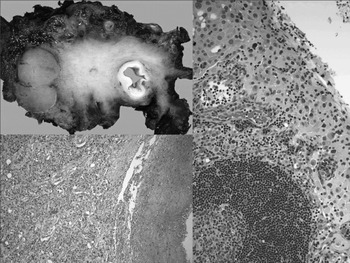
Histological examination showed a carotid body paraganglioma measuring 40 × 25 × 20 mm with an infiltrative border that was present at the surgical margin in places. Metastatic tumour was found in an upper deep cervical lymph node (Figure 1).
Histopathology of case one. The macroscopic specimen (top left) shows the carotid artery and the tumour, with an obvious infiltrative margin of the primary tumour extending towards a lymph node. Photomicrographs show: at bottom left, the primary tumour with a nested pattern in proximity to the wall of the carotid artery (H&E; original magnification ×100); and at right, a deposit of carotid body tumour in one of the regional lymph nodes (H&E; original magnification ×200).

An octreotide scan was negative for residual local disease and distant metastases.
Post-operative radiotherapy to a total dose of 60 Gy was given in 30 fractions over six weeks.
At two years' follow up, the patient was well with no residual symptoms or cranial nerve weakness, and clinically clear of recurrent disease.
Case two
A 27-year-old woman presented with an eight-month history of a left upper neck mass which had doubled in size. It was associated with some pharyngeal irritation and numbness of the left cheek.
Computed tomography scans were suggestive of a carotid body tumour. An MRI angiogram supported this diagnosis, and showed that the lesion was encasing the carotid vessels.
At surgery, the tumour measured 6 × 3 cm. The internal carotid artery (ICA), vagus nerve, sympathetic trunk and superior laryngeal nerves were involved, and were therefore sacrificed. A saphenous vein interposition graft was used for reconstruction of the ICA. Based on our previous case, a selective neck dissection was also performed.
Post-operatively, the patient had a vocal fold palsy, Horner's syndrome and reduced function of the left hypoglossal nerve. She underwent a Gore-tex thyroplasty to medialise the paralysed vocal fold, and a left upper eyelid ptosis repair.
Histological examination of the surgical specimen showed a sclerotic and invasive paraganglioma measuring 35 × 30 × 22 mm, with microscopic tumour at the superior and inferior resection margins. There was also metastatic involvement of one of the level II lymph nodes.
Post-operative radiotherapy of 50 Gy over five weeks was given to the ipsilateral neck.
Eighteen months after surgery, the patient was well with no clinical or radiological evidence of disease.
Discussion
Benign carotid body tumours cannot be differentiated reliably from malignant tumours on histological grounds, despite attempts to establish histological criteria for malignancy.Reference Lee, Barich, Karnell, Robinson, Zhen and Gantz3, Reference Luna-Ortiz, Rascon-Ortiz, Villavicencio-Valencia, Granados-Garcia and Herrera-Gomez4 Typical features of malignancy (such as nuclear pleomorphism, mitoses, necrosis and local tissue infiltration) are not uniformly present in malignant carotid body tumours, yet may be found in benign tumours. Our two cases both required carotid artery resection, and as such were not typical of carotid body tumour excision procedures. It is possible that the locally invasive nature of these two lesions increased the risk of regional metastases, but this is not apparent from other authors' experience.
Zbaren and LehmannReference Zbaren and Lehmann5 studied 106 cases of malignant carotid body tumour published between 1893 and 1980, and found that the only histological determinant of malignancy was the presence of metastases. Several other authorsReference Gaylis and Mieny6–Reference Cooper, Slevin, Johnson and Evans8 have reached the same conclusion. Gaylis and MienyReference Gaylis and Mieny6 found a malignancy rate of 30 per cent among 23 carotid body tumour cases (two of them diagnosed by lymph node biopsy prior to carotid body tumour excision); however, they were unable to identify the malignant tumours on histological assessment of the primary tumour alone.
The incidence of either regional or distant metastasis in carotid body tumour cases is usually less than 10 per cent,Reference Ling, Weinrach and Eskandari7 and is normally confined to regional nodes.Reference Lee, Barich, Karnell, Robinson, Zhen and Gantz3
One clinical seriesReference Gaylis and Mieny6 reported two cases in which malignancy was diagnosed six and eight years after carotid body tumour excision, due to late appearance of nodal metastases. At the other end of the spectrum, a carotid body tumour may not be clinically apparent, and may only be suspected when metastatic carotid body tumour is found in a lymph node.Reference Zbaren and Lehmann5, Reference Cooper, Slevin, Johnson and Evans8 Generally, however, nodal metastases are occult at the time of surgery, so that a diagnosis of malignant carotid body tumour can only be made by directed, regional cervical lymph node sampling.
The consensus nowadays is that carotid body tumours are benign until or unless metastases (to non-neuroendocrine tissue) are found.Reference Somasundar, Krouse, Hotsetter, Vaughan and Covey1 This has led some to suggest that carotid body tumour should be managed by excision and ipsilateral selective neck dissection.Reference Liapis, Evangelidakis, Papavassiliou, Kakisis, Gougoulakis and Polyzos9 Netterville et al. Reference Netterville, Reilly, Robertson, Reiber, Armstrong and Childs10 and Pacheco-OjedaReference Pacheco-Ojeda11 both reported performing a limited, selective neck dissection, in order not only to optimise tumour exposure but also to sample lymph nodes for occult metastases. Zbaren and LehmannReference Zbaren and Lehmann5 have advocated Universal Selective Neck Dissection. The US National Cancer Data Base Report on Malignant Paragangliomas of the Head and Neck actually recommends that lymphadenectomy should be undertaken in conjunction with primary tumour excision.Reference Lee, Barich, Karnell, Robinson, Zhen and Gantz3
Selective neck dissection adds very little morbidity to the primary carotid body tumour resection. It is obviously a critical means of detecting regional metastases, and acts effectively as a form of sentinel node biopsy. Early detection of regional metastatic disease is especially important given that patients can live for long periods with disseminated disease but control of loco-regional disease can be difficult.
Data are emerging regarding a favourable effect of adjuvant radiotherapy in cases of metastatic regional disease. Adjuvant radiotherapy probably has a survival benefit, although further studies are needed.Reference Lee, Barich, Karnell, Robinson, Zhen and Gantz3
• A reliable diagnosis of malignant carotid body tumour can only be made in the presence of metastatic disease, as the histological features of the primary tumour do not correlate with clinical behaviour
• This report describes two cases of malignant carotid body tumour in which regional nodal biopsy, undertaken at the time of primary tumour excision, revealed unsuspected metastatic disease
• Sentinal node dissection ensures that occult metastases are not only detected but also appropriately managed
Despite the number of authors recommending regional lymph node sampling in association with carotid body tumour excision, this approach does not seem to have been adopted widely. Perhaps it is because such recommendations have been presented within a large clinical series report or a broad literature review. We believe that a focussed paper, such as the present report, may help raise awareness of the appropriateness of node dissection at the same time as carotid body tumour resection.
Conclusion
In the absence of other reliable clinical, histological or radiological methods of detecting malignancy in a timely manner, lymph node sampling at the time of tumour removal provides the optimal means of diagnosing a malignant carotid body tumour. A selective neck dissection ensures that occult metastases are not only detected but also appropriately managed, thereby safeguarding against a late diagnosis of malignancy and the subsequent need to reoperate on a scarred and previously treated neck. We advocate the consideration of ipsilateral, selective neck dissection in all surgical cases.