The catheter ablation of anteroseptal accessory pathways can be challenging due to their proximity to the atrioventricular node, resulting in lower success rates and higher incidence of atrioventricular blocks compared to other accessory pathways. Rarely, oblique and/or deep routes can make ablation even harder, requiring alternative access routes for better catheter contact and ablation success. Non-coronary aortic cusp is one of such alternative routes for successful ablation in rare cases, but it also involves risks like atrioventricular block and potential damage to the coronary arteries or aortic valves. The data about its electrophysiological characteristics, safety, and efficacy are limited, especially in the paediatric population. Here, we present a case of Wolff–Parkinson–White syndrome with an oblique and deep-seated anteroseptal accessory pathway in which ablation was successfully performed from non-coronary aortic cusp. Reference Letsas, Efremidis and Vlachos1–Reference Kiliçaslan, Uz, Işilak and Tokatli4
Case presentation
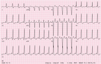
A 10-year-old male patient was admitted to a paediatrician with a complaint of palpitations. Wolff–Parkinson–White syndrome was detected in his ECG, and sotalol treatment was started. He was referred to our clinic. His 12-channel ECG showed manifest preexcitation with a delta wave positive in I, II, negative in V1, and isoelectric/positive in aVF leads, suggesting an anteroseptal localisation (modified Arruda algorithm) (Fig 1).
12-lead surface ECG recorded on admission showing marked delta waves consistent with an anteroseptal manifest AP.

A septal paradoxical bulging movement of the left ventricular septal region was observed on echocardiographic evaluation, causing dyssynchrony and left ventricle systolic dysfunction (left ventricle ejection fraction was 34% with the modified Simpson’s method). The dysynchrony measurements made with M-mode echocardiography revealed a septal-to-posterior wall motion delay of 300 ms. In 12-lead Holter ECG recordings, a persistent manifest accessory pathway was detected. Wolff–Parkinson–White accessory pathway did not disappear during the exercise test despite the high heart rate. Electrophysiological study and ablation was scheduled for the patient. The procedure was conducted under general anaesthesia. EnSite Precision® Mapping System (St. Jude Medical Inc., St Paul, MN, USA) was used with limited fluoroscopy.
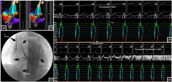
An accessory pathway risk assessment was performed at baseline electrophysiological study (EPS) and found a low risk of sudden cardiac death (APERP was 330 ms and no pre-excitation during atrial fibrillation). The accessory pathway was unresponsive to intravenous adenosine administration. Supraventricular tachycardia was induced with basal programmed atrial stimulations. During supraventricular tachycardia (cycle length 270 ms), the earliest atrial activation was found between the right anteroseptal and anterior regions (40 ms earlier than the CS proximal signal) forward, and the earliest ventriculoatrial interval was 34 ms). When the rhythm was sinus, HIS bundle signal could also be seen, so cryoablation was applied with an 8-mm tip catheter. After five lesions, supraventricular tachycardia could not be induced, but ventricular preexcitation was still existing. This time an 8F 4-mm tip irrigated radiofrequency (RF) catheter was used from the jugular vein route to apply limited deep lesions on the anterior side of the oblique accessory pathway (five lesions, each with 20–25 W 30°C and 90 ohms). After these lesions, there was no ventricular preexcitation on surface ECG, and no supraventricular tachycardia could be induced. However, ventricular preexcitation again recurred at the 15th minute of the waiting period. After insufficient femoral and jugular cryo and RF application, aortic cusp mapping was started near the epicardial area. Aortic cusps were mapped in detail using the retrograde aortic approach (Fig 2, A–C). Earliest location during delta mapping was within non-coronary aortic cusp (and found the earliest signal −62 ms ahead, with a clear QS pattern as a unipolar signal) (Fig 2, D and E). The 8-mm tip cryoablation catheter was chosen due to its proximity to HIS, and ventricular preexcitation disappeared at −80 degrees in the first second. After each three cryo lesions for 240 seconds and with careful P-R interval watch for prolongation, there was no ventricular preexcitation on surface ECG, also checked under adenosine (without any preexcitation), and VA conduction was found decremental and dissociated at 430 ms. No supraventricular tachycardia could be induced by programmed atrial stimulations under basal and isoproterenol infusion. The procedure was completed successfully and without complication. The total procedure time was 200 minutes, and the total fluoroscopy time was 6.3 min. The patient is still under follow-up for 1 year, without any complaints, with a normal 12-lead ECG and normal ventricular functions on echocardiography (LV EF %65) with no dyssynchrony.
Three-dimensional anatomy of the right atrium is shown in A and B . A ; transjugular and B ; aortic noncoronary cusp were mapped with EnSite Precision® Mapping System and fluoromapping. ( C ) Fluoroscopic image, Current catheter positions are shown while preexcitation disappeared. Intracardiac and surface 12-lead electrograms are shown in D and E . ( D) The earliest place was within the NCC and found the earliest signal -62 ms ahead, in the retro aortic route. ( E ) The cryoablation was chosen due to its proximity to HIS, and ventricular preexcitation disappeared at -80 degrees in the first second. CS - coronary sinus; RF - radiofrequency; RV - right ventricle.

Discussion
EPS and ablation are recommended as first-line therapy for children beyond 5 years of age with Wolff–Parkinson–White syndrome. However, anteroseptal and mid-septal accessory pathways remain as challenging targets. Accessory pathways (AP) may cross the central fibrous trigone or involve the right atrial appendage; they can be confused with anteroseptal accessory pathways, and sometimes conservative energy delivery in these regions can be seen. Failed cryoablation attempt or recurrence after cryoablation has been reported. Reference Tanıdır, Özyılmaz, Ünsal, Güzeltaş and Ergül2,Reference Kiliçaslan, Uz, Işilak and Tokatli4 In patients with successful cryo or RF ablation, unusual locations with deep epicardial locations (especially aortic non-coronary cusp) may be present in possible recurrence. In our patient, although AP was successfully eliminated by both cryo and RF via the right-sided femoral and jugular pathways, recurrence occurred in a short time. And as it is known, the anteroseptal parahisian pathways are endocardial. Although we eliminated the ventricular retrograde pathway in this patient, the continuation of antegrade conduction and ablation of the non-coronary aortic cusp suggests that it may also be epicardial.
Alternative methods can be used to increase the success of ablation in this difficult locations, and one of these ways is ablation from the non-coronary cusp. Aortic cusp ablation is uncommonly performed in paediatric age group. In childhood, the non-coronary aortic cusp approach is rarely preferred because of possible damage to the coronary arteries, aortic valve leaflets, and compact AV node. However, recent experience in the adult population has demonstrated that non-coronary aortic cusp ablation can be performed safely and effectively. Nguyen Reference Nguyen, Ceresnak and Janson3 et al. reported the aortic cusp procedure in various arrhythmia substrates results from 5 EPS centres in 13 young patients (<21 years). 100% success with the procedure, no complications or valve damage were observed. It was emphasised that careful visualisation of the aortic valve and coronary arteries during ablation is vital for the safety of the procedure.
In patients with Wolff–Parkinson–White syndrome, the ventricles are electrically and mechanically pre-excited through an accessory pathway. The eccentric ventricular activation via accessory pathways results in an asynchronous spread of ventricular depolarisation. This dyssynchronous left ventricle motion could cause inefficient cardiac performance by reducing the ventricular filling time and producing mitral regurgitation. Echocardiographic observations displayed an abnormal interventricular septal motion in patients with Wolff–Parkinson–White syndrome. Significant improvement in LV function has been reported after successful catheter ablation of the accessory pathways. Reference Köşger, Sevinç Şengül, Kafali, Uçar and Ergul5,Reference Haraguchi, Kondo and Ishii6 It has been observed that dyssynchrony-associated cardiomyopathy may develop even without tachycardia, and cardiac functions may improve after ablation.
Conclusion
In patients with challenging anteroseptal APs, the non-coronary aortic cusp should be considered an alternative and safe route when the ablation is not possible on the right atrial side.
Financial support
This research received no specific grant from any funding agency, commercial, or not-for-profit sectors.
Conflict of interest
None.
Ethical standards
The authors assert that all procedures contributing to this work comply with the ethical standards of the relevant national guidelines on human experimentation and with the Helsinki Declaration of 1975, as revised in 2008, and has been approved by the institutional committees.