Alzheimer disease (AD) is the most common form of dementia with a high prevalence rate among the ageing population, and recent estimates suggest that it affects about 35.6 million individuals worldwide (Ref. Reference Li1). Clinical symptoms are characterised by loss of selective cognitive functions, particularly memory loss that begins early in the disease process (Refs Reference Lacor2, Reference Selkoe3, Reference Citron4). These traits are accompanied by neuropathological features observed in postmortem AD brains, which include loss of neurons and synapses in cortical and subcortical regions, extracellular plaques of aggregated beta-amyloid protein (Aβ), deposits of amyloid within the cerebral vasculature and intracellular neurofibrillary tangles that contain hyperphosphorylated tau protein (Refs Reference Lacor2, Reference Selkoe3, Reference Wenk5). Evidence from many laboratories suggests that the accumulation of fibrillar forms of amyloid-beta protein (Aβ) has a central role in the pathogenesis of AD (Ref. Reference Glenner and Wong6). Aβ peptides are normally generated from beta-amyloid precursor protein (APP) through proteolytic cleavage by β- and γ-secretases (Refs Reference Gralle and Ferreira7, Reference Selkoe8, Reference Selkoe9). However, an increasing body of work indicates that small Aβ oligomers, also known as Aβ-derived diffusible ligands (ADDLs), are potent central nervous system (CNS) neurotoxins (Refs Reference Selkoe3, Reference Lambert10). This new view of AD pathogenesis postulates that unlike the insoluble fibrils, ADDLs are diffusible molecules that attach with specificity to particular synapses, acting as pathogenic ligands (Refs Reference Selkoe3, Reference Gong11, Reference Walsh12). The attack on synapses results in severe disruption of synaptic function and plasticity, including impairment of long-term potentiation (LTP) and facilitation of long-term depression (LTD), which are classic paradigms for memory-related synaptic plasticity (Refs Reference Gong11, Reference Lambert13). The pathological relevance of ADDLs has been substantiated by their disease-specific accumulation in transgenic mouse AD models (Ref. Reference Lesné14) and by the accumulation of structurally equivalent oligomers in the human brain and cerebrospinal fluid (CSF) (Refs Reference Gong11, Reference Georganopoulou15). There is great interest in understanding the mechanisms whereby soluble Aβ oligomers affect synaptic functions involved in learning and memory with the anticipation that such knowledge may provide significant insights into AD pathogenesis and potentially lead to better strategies for the prevention and treatment of AD. There is considerable evidence that specificity of Aβ binding at neuronal surfaces and synapses requires membrane proteins, and several candidate receptors that may have an important role in expression of Aβ neurotoxicity have been postulated as potential targets for Aβ (Ref. Reference Lambert10). Aβ oligomers can interact with α7-type nicotinic acetylcholine receptor (α7 nAchRs) (Refs Reference Dineley16, Reference Snyder17, Reference Wang18), trigger endocytosis of glutamate receptor subunits (Refs Reference Lacor2, Reference Zhao19) and the insulin receptor (IR) (Refs Reference De Felice20, Reference Xie21), and possess the ability to alter trafficking of metabotropic glutamate receptors 5 (Refs Reference Renner22, Reference Wang23). The focus of this article is to review the interactions between soluble Aβ oligomers and specific neuronal receptors in the context of Aβ-mediated synaptic disruption and neurotoxicity.
Moieties of Aβ
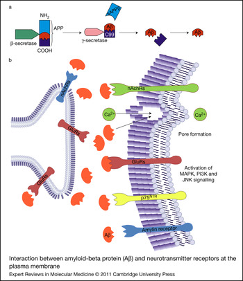
Aβ is generated by the sequential action of β- and γ-secretase enzymes on the parent transmembrane APP. β-Secretase cleaves APP at the Aβ N-terminus, resulting in a soluble form of APP (sAPPβ) and a 99-residue C-terminal fragment. This C-terminal fragment undergoes processing by γ-secretase within the transmembrane region of APP, resulting in the formation of Aβ peptides of various lengths (39–43 amino-acid residues) (Ref. Reference Gralle and Ferreira7). The two most common primary isoforms generated are Aβ40 and Aβ42: the shorter form is typically produced by cleavage that occurs in the endoplasmic reticulum, whereas the longer form is produced by cleavage in the trans-Golgi network. The Aβ40 form is the more predominant isoform produced by neurons and other cells, and accounts for over 70% of the total Aβ produced (Refs Reference Iwatsubo24, Reference Fraering25), whereas Aβ42, which accounts for only 10–20% of total Aβ, is the major protein component of the Aβ neuritic plaques (Refs Reference Iwatsubo24, Reference Fraering25). Aβ42 is more hydrophobic and has a greater propensity to form aggregates into fibrils and also greater neuronal toxicity in tissue culture models than Aβ40, implying that Aβ42 is a more important moiety in AD plaque formation and pathogenesis (Ref. Reference Kuperstein26). Aβ monomers are known to aggregate into higher molecular weight oligomers and fibrils. Aβ oligomers and fibrils can interact with neuronal membrane proteins, which may induce change or loss of protein function (Ref. Reference Verdier, Zarandi and Penke27). Increases in either total Aβ levels or the relative concentrations of Aβ40 and Aβ42, where the former is more concentrated in cerebrovascular plaques and the latter in neuritic plaques, have been suggested in the pathogenesis of both familial and sporadic AD, thus making it an important biomarker for this disease (Ref. Reference Kuperstein26).
Glutamate receptors
The extent of synaptic degeneration, including impairment in synaptic plasticity and loss of synapses in the brain, provides one of the best morphological correlates of cognitive impairment in clinical AD (Refs Reference Sze28, Reference Terry29). Increasing evidence suggests that involvement of soluble Aβ oligomers in degenerative synaptic changes in vulnerable brain regions such as the hippocampus leads to cognitive impairments in learning and memory (Refs Reference Lambert13, Reference Shankar30, Reference Townsend31, Reference Ondrejcak32). LTP facilitates synaptic plasticity by strengthening communication across a synapse, and is essential for hippocampal-dependent learning and memory processes (Refs Reference Lynch33, Reference Martin and Morris34). Specific neuronal receptors involved in mediating LTP and synaptic plasticity processing are therefore likely candidates for the binding of Aβ oligomers. Glutamatergic neurotransmission has an important role in the synaptic transmission and neuronal plasticity processes associated with learning and memory (Ref. Reference Lee35). Impairment in glutamatergic neurotransmission signalling has been proposed to be a crucial factor in AD pathology (Refs Reference Lee35, Reference Bennett36). Glutamate receptors consist of two classes, ionotropic (cation-specific ion channels) and metabotropic (G-protein-coupled), and members of both classes are implicated as neuronal receptors in Aβ-oligomer-mediated neurotoxicity.
NMDA ionotropic glutamate receptors
N-methyl-d-aspartate receptors (NMDARs) constitute a major class of glutamate receptors and have key roles in glutamatergic transmission and synaptic plasticity in the CNS (Refs Reference Snyder17, Reference Yamin37, Reference Texido38, Reference Rammes39, Reference Bordji, Becerril-Ortega and Buisson40). These receptors are critically important for induction of LTP and synaptic strengthening (Refs Reference Derkach41, Reference Lau and Zukin42, Reference Paoletti and Neyton43). A number of studies have shown that Aβ can affect the function of NMDARs and abolish induction of NMDAR-dependent LTP at the neuronal plasma membrane (Refs Reference Lambert13, Reference Wang44). Aβ binds to GluN1 and GluN2B subunits of NMDAR at the postsynaptic terminals of hippocampal neurons in AD brains (Ref. Reference Lacor2). Aβ oligomers are capable of regulating the surface expression and endocytosis of NMDA-type glutamate receptors by binding to α7 nAchRs (Ref. Reference Snyder17) and are also involved in dysregulation of NMDA signalling pathways (Refs Reference Roselli45, Reference Shankar46). Partial colocalisation between Aβ oligomers and the GluN2B subunit of NMDAR has been observed in hippocampal slices, although the maximum colocalisation was less than 50% (Ref. Reference Deshpande47). Furthermore, Aβ oligomers were found to coimmunoprecipitate with NMDAR subunits (Ref. Reference Renner22). However, it is unclear whether Aβ directly binds to NMDA receptor subunits. The GluN2B subunit of NMDAR has been implicated in regulating the action of Aβ oligomers by increased intracellular Ca2+ into dendritic spines (Ref. Reference Shankar46), resulting in reduced dendritic spine and synaptic density that led to early synaptic dysfunction (Refs Reference Shankar46, Reference Rönicke48). Interestingly, relatively low doses of antagonists selective for the GluN2B subunit of NMDAR have been reported to reverse Aβ-induced synaptic disruption (Refs Reference Lacor2, Reference Shankar46, Reference Rönicke48). In addition, Aβ has been shown to form clusters at the postsynaptic membrane, reducing levels of scaffolding proteins such as postsynaptic density-95 (PSD-95), which have an important role in synaptic plasticity and stabilisation of glutamate receptors located at excitatory synapses (Refs Reference Lacor2, Reference Roselli45). Moreover, Aβ disrupts axonal transport initiated by NMDAR-dependent mechanisms and mediated by the enzyme glycogen synthase kinase 3β (GSK-3β) (Ref. Reference Decker49). Additionally, Aβ, through an NMDAR-dependent mechanism, stimulates excessive generation of reactive oxygen species (ROS) (Ref. Reference De Felice50) and induces mitochondrial damage that leads to neuronal death (Refs Reference De Felice50, Reference Alberdi51).
Furthermore, STEP61, a member of the STEP (striatal-enriched phosphatase) family of brain-enriched Tyr-specific and cytoplasmic phosphatases (Ref. Reference Pelkey52), is associated with regulating the NMDA subclass of glutamate receptor trafficking. Studies have shown that Aβ activates and increases the levels of STEP61, which leads to a decrease in NMDA receptor GluN2B subunit activity and consequent endocytosis of GluN1/GluN2B NMDA receptors (Ref. Reference Venkitaramani53). Moreover, recent studies have revealed elevated levels and activity of STEP61 in the transgenic AD mouse model and in human AD brains (Ref. Reference Kurup54), and reducing STEP61 levels and activity through small-molecule inhibitors might have a therapeutic benefit in AD (Refs Reference Kurup54, Reference Zhang55). Interestingly, genetic reduction of STEP in AD mouse models reverses both cognitive deficits and loss of glutamate receptors from synaptosomal membranes (Ref. Reference Zhang55).
mGluR5 metabotropic glutamate receptor
The GluR5 subtype of metabotropic glutamate receptors has important regulatory roles in excitatory synaptic transmission at hippocampal synapses, modulation of LTP and potentiation of NMDAR-mediated Ca2+ influx (Refs Reference Renner22, Reference Wang23, Reference Zieminska and Lazarewicz56). A recent study identified mGluR5 as a novel neuronal receptor target for Aβ using single-particle tracking of quantum-dot-labelled Aβ oligomers on hippocampal neurons and examining their interactions with mGluR5 receptors. Membrane-bound Aβ oligomers accumulated at extrasynaptic synapses, where they progressively aggregated to form large nonmobile clusters through lateral diffusion. Pathological clusters of Aβ formed complexes with mGluR5 receptors, leading to decreased mobility of mGluR5 and causing their aberrant accumulation at the postsynaptic membrane. This was followed by Ca2+ dysregulation, synaptic disruption and loss of NMDARs, suggesting that their role in early synaptic impairment is induced by Aβ (Ref. Reference Renner22). Interestingly, mouse hippocampal neuronal cultures from mGluR5-knockout mice revealed an approximately 80% decrease in Aβ binding to the neuronal surface and a loss of NMDARs from the cell surface (Ref. Reference Renner22). In another study, Aβ42 was shown to decrease the frequency of spontaneous excitatory glutamatergic postsynaptic events in the identified cholinergic diagonal band of Broca neurons (Ref. Reference Chin57). This Aβ42-mediated reduction in the glutamate neurotransmission effect was expressed through activation of presynaptic mGluRs. However, the precise receptor subtype of mGluRs (group I/II) was not identified in this study (Ref. Reference Chin57). This study indicates that Aβ42 is capable of modulating transmitter release, but the functional consequences of such modulation of synaptic activity under physiological or pathophysiological conditions are at present unknown.
mGluR signalling is downregulated and desensitised in the frontal cortex of AD patients and has been shown to correlate with AD-related neuropathological changes (Ref. Reference Albasanz58). Moreover, mGluR5 shares a functional relationship with NMDAR, in particular because it relates to development of NMDA-dependent LTP and learning (Refs Reference Bortolotto59, Reference Jia60). Furthermore, chronic activation of mGluR5 increases NMDA-dependent Aβ neurotoxicity, whereas its inhibition shows neuroprotective effects against Aβ excitotoxic processes (Refs Reference Zieminska and Lazarewicz56, Reference Bruno61, Reference Karanian62). Thus, there is persuasive experimental evidence that Aβ oligomers are able to influence glutamatergic transmission by NMDAR and mGluR5 receptors. As a result, selective glutamate receptor antagonists have become promising targets for the treatment of AD. Memantine (a nonselective NMDAR antagonist) is currently being used in routine clinical practice for the treatment of patients with moderate to advanced AD (Ref. Reference Winblad63). However, there have been a few reports regarding the lack of efficacy of memantine in the treatment of patients with mild to moderate AD (Ref. Reference Schneider64). Some of the reasons for the lack of effect could be the lack of reliable biomarkers for the disease and heterogeneity of patients (variable response to memantine treatment).
Acetylcholine receptors
Nicotinic acetylcholine receptors
nAchRs are found throughout the nervous system and exist as a diverse family of ligand-gated ion channels consisting of different pentameric combinations of similar but genetically distinct subunits derived from at least 17 genes (Ref. Reference Lindstrom65). In the mammalian brain, the most abundant forms are the functional nAchRs assembled as either heteromers containing α4β2 subunits or homomers containing α7 subunits (Ref. Reference Lindstrom65). Several studies have shown that an important characteristic of most patients with clinical AD is the prominent loss of cholinergic neurons in the basal forebrain (Refs Reference Arneric, Holladay and Williams66, Reference D'Andrea and Nagele67) and the selective loss of different nAchR subunits in AD brains (Refs Reference Gotti68, Reference Martin-Ruiz69, Reference Sabbagh70).
Several studies have reported Aβ interactions with nAchRs, in particular with the α7 subtype, which is known to bind soluble Aβ with high affinity (Refs Reference Wang18, Reference Wang71, Reference Liu, Kawai and Berg72, Reference Buckingham73), leading to the speculation that Aβ might be internalised by complex formation with α7 nAchR (Ref. Reference Nagele74). This notion is supported by immunohistochemical studies on postmortem brain tissue from patients with sporadic AD, which reveal a high level of colocalisation of α7 nAchR and Aβ in individual cortical neurons and neuritic plaques, suggesting that Aβ could be tightly associated with nAchR (Ref. Reference Nagele74). These authors observed similar findings under in vitro conditions, where the addition of soluble Aβ to the culture medium of neuroblastoma cells transfected with α7 nAchR resulted in more Aβ internalisation than in nontransfected control cells (Ref. Reference Nagele74). In addition, a triple transgenic mouse model of AD showed age-dependent loss of α7 nAchRs, which was preceded by intracellular Aβ accumulation in specific brain regions compared with the age-matched control group (Ref. Reference Oddo75).
In contrast to the above observations, other studies have been unable to confirm the high-affinity binding of Aβ to α7 nAchRs (Ref. Reference Small76). Additionally, there are data indicating a variety of effects following Aβ exposure to nAchRs, suggesting that Aβ modulation of α7 nAchR function is more complex than originally reported. For example, picomolar concentrations of Aβ have been observed to act as an agonist at α7 nAchRs (Ref. Reference Fodero77) and mediate activation of the ERK2–mitogen-activated protein kinase (MAPK) signalling cascade (Ref. Reference Dineley16, Reference Dineley78). However, nanomolar concentrations of Aβ cause functional antagonism of both human and rat homomeric α7 nAchRs, which might be due to inhibition of presynaptic nAchRs, resulting in changes in presynaptic Ca2+ levels (Refs Reference Lee and Wang79, Reference Pettit, Shao and Yakel80, Reference Tozaki81). Additionally, Aβ regulations of presynaptic nAchRs (α7 and α4β2 subunits) have also been observed (Ref. Reference Chin57). Soluble Aβ42 is capable of modulating postsynaptic transmission by activating presynaptic α4β2 nAchRs in septo-hippocampal neurons of the basal forebrain, an important pathway for learning and memory (Ref. Reference Chin57). Another study identified Tyr188 residues in α7 nAchRs as one of the key components in the agonist-binding domain for the activation of presynaptic α7 nAChRs by Aβ (Ref. Reference Tong82).
These different findings might depend on a number of important factors such as the nAchR subtype being activated, as well as different forms of Aβ preparation and varying concentrations of Aβ (physiological concentrations can activate and higher concentrations can desensitise or inactivate nAchRs) (Ref. Reference Dineley78). Furthermore, contradictory findings have been reported from in vivo animal work, where transgenic AD mice deficient in α7 nAchR expression exhibited enhanced Aβ oligomer accumulation and exacerbated cognitive decline in one study (Ref. Reference Hernandez83), but amelioration of AD pathology was reported in another study (Ref. Reference Dziewczapolski84). Moreover, some studies suggest that α7 nAchR agonists could be beneficial for treatment of AD in animal models (Refs Reference Mazurov, Hauser and Miller85, Reference McLean86). However, one study revealed that activation of α7 nAchRs with a selective agonist of choline in the presence of Aβ could result in impairment of synaptic plasticity, whereas blocking α7 nAchRs by a selective methyllycaconitine antagonist protected against Aβ-induced impairments of hippocampal LTP in rats (Ref. Reference Li1).
Interestingly, most α7 nAchRs are formed as homomeric pentamers, but a novel interaction between nAchR α7 and β2 subunits, which coassemble to form a functional α7β2 heteromeric nAchR subtype, has been reported. Functional α7β2 nAchRs have an important role in learning and memory and in cholinergic neuronal functions (Ref. Reference Liu87). Therefore, selective, high-affinity interactions between pathologically relevant concentrations of soluble Aβ oligomers and α7β2 nAchRs could contribute to disruption in cholinergic signalling and impairment in learning and memory (Ref. Reference Liu87). In conclusion, there is considerable evidence to suggest that interaction of Aβ with different subtypes of nAchRs has a significant role in AD pathogenesis, and hence these receptors could serve as potentially useful therapeutic targets in this condition. However, the precise nature of such interplay requires further study.
M1 muscarinic acetylcholine receptors
Aβ has also been suggested to exert its neurotoxic effects on the cholinergic system by interfering with the function of G-protein-coupled muscarinic acetylcholine receptors (mAchRs), a family of five receptor subtypes (M1–M5) (Refs Reference Levey88, Reference Wei89). The densities of M1 mAchRs located postsynaptically are found to be relatively unaltered; however, there is some evidence for impaired coupling of the mAChRs to heterotrimeric guanine nucleotide-binding (G) proteins and second messengers (Refs Reference Ferrari-DiLeo, Mash and Flynn90, Reference Tsang91, Reference Warpman, Alafuzoff and Nordberg92). The postsynaptic M1 mAchR is the most abundant subtype in the brain regions of the cortex and hippocampus (Refs Reference Levey88, Reference Wei89, Reference Flynn93) – areas that are most affected in AD. M1 mAchRs also have an important role in hippocampus-dependent memory and cognition, and in particular short-term memory (Ref. Reference Anagnostaras94), which is impaired in AD (Refs Reference Levey95, Reference Thathiah and De Strooper96). Moreover, activation of M1 AchRs stimulates nonamyloidogenic sAPPα (amyloid precursor protein) processing in in vitro and in vivo studies, supporting a role for mAchR activation in promoting increased α-secretase activity and thus preventing the formation of Aβ by the amyloidogenic pathway (Refs Reference Buxbaum97, Reference Nitsch98). In keeping with this observation, deletion of M1 mAchR in a transgenic mouse model of AD leads to increased levels of pathogenic Aβ peptides and amyloid plaque deposition in the brain (Ref. Reference Davis99). These findings suggest that M1 AchRs are more associated with nonamyloidogenic production of APP and reduction in Aβ plaque load rather than as a neurotransmitter receptor target for Aβ interactions. Therefore, considerable effort has been directed towards developing selective agonists for M1 mAChR that might be capable of restoring the cognitive deficits in patients with AD.
AF267B, a selective M1 mAchR agonist, reduces amyloid and tau-related pathologies, and attenuates impairments in hippocampus-dependent learning and memory in an AD mouse model (Ref. Reference Caccamo100). However, other results demonstrate that AF267B equally or more potently activates M3 mAchRs compared with M1 mAchRs (Ref. Reference Jones101). Therefore, a lack of drugs that are able to distinguish among subtypes of mAchRs makes it difficult to assess the precise role of M1 mAchRs in AD. Although it has been difficult to synthesise fully specific M1 mAchR agonists, a novel allosteric agonist of the M1 mAchR, TBPB [1-(1′-(2-methylbenzyl)-1,4′-bipiperidin-4-yl)-1H-benzo[d]imidazol-2(3H)-one], was recently shown to be highly selective for mAchR in rodents (Ref. Reference Jones101). Additionally, TBPB was also shown to potentiate NMDAR-mediated currents in hippocampal pyramidal cells and could have an important role in enhancing NMDAR-mediated LTP (Ref. Reference Shinoe102). Use of M1 mAchR selective agonists would avoid the need for enhancing synaptic acetylcholine levels through administration of acetylcholinesterase inhibitors, which results in activation of all subtypes of mAchR, including M2 and M4 that are known to inhibit sAPPα release and enhance Aβ generation (Refs Reference Davis99, Reference Farber103, Reference Fisher104). Clinical trials for newer generations of M1-specific agonists are currently under investigation. Activation of other mAchR subtypes (e.g. M2) are associated with significant peripheral side effects and therefore are not suitable for clinical use (Ref. Reference Bymaster105).
G-protein-coupled receptors, diabetes and AD
Insulin and insulin growth factor (IGF) receptors
Many studies have focused on the role of insulin signalling in the aetiology and pathogenesis of AD (Refs Reference Craft106, Reference Hoyer107, Reference Perry108, Reference McClean109, Reference Chakravarthy110). Insulin-resistant type 2 diabetes mellitus (T2DM) has been identified as a risk factor for AD (Refs Reference Craft106, Reference Perry108), and CNS insulin signalling is important for many aspects of neuronal function, including neuronal repair, synaptic plasticity and memory formation (Refs Reference Gispen and Biessels111, Reference Zhao112, Reference Zhao and Alkon113), all of which potentially link insulin resistance to AD dementia (Ref. Reference Craft106). Dysregulation of the insulin signalling pathway has been observed in the brains of patients with AD, with markedly downregulated expression of IRs, and insulin receptor substrate (IRS)-1 and IRS-2, which correlate with severity of neurodegenerative changes (Refs Reference Zhao114, Reference Moloney115). Furthermore, Aβ oligomers interact with neuronal IRs to cause impairment of the expression and function of these receptors (Refs Reference De Felice20, Reference Zhao114). Such impairments mimic the Aβ-oligomer-induced inhibition of LTP and could represent an important early mechanism underlying memory impairment and other pathological features of AD. Most significantly, this loss of surface IRs, oxidative stress and synaptic disruption induced by Aβ were shown to be completely prevented by insulin and insulin-sensitising drug (e.g. rosiglitazone) treatments (Refs Reference Townsend, Mehta and Selkoe116, Reference Lee117, Reference Risner118, Reference Pedersen119). Another promising intervention is the use of the incretin hormone glucagon-like peptide 1 (GLP-1), which facilitates insulin release and has very similar growth-factor-like properties as insulin, as a potential treatment for neurodegenerative diseases (Refs Reference Perry108, Reference McClean109, Reference Lovshin and Drucker120). In vivo administration of GLP-1 and its stable analogues (Val8 and liraglutide) in rodents prevented Aβ-induced memory impairment, synapse loss, synaptic disruption and reduced Aβ plaque load (Refs Reference McClean109, Reference Gengler121). Therefore, GLP-1 and its analogues such as Val8 and liraglutide are promising new treatments for AD. However, clinical trials to validate such neuroprotective effects in humans are essential.
Impaired IGF 1 receptor (IGF-1R) signalling has also been linked to Aβ pathology in the brains of patients with AD (Refs Reference Moloney115, Reference Steen122). Circulating IGF-1, a physiologically relevant neuroprotective hormone, has been shown to facilitate CSF clearance of Aβ by favouring the passage of Aβ carrier proteins such as albumin, transthyretin (Ttr) or apolipoprotein J (apo J) through the choroid plexus epithelium (Refs Reference Carro123, Reference Carro124). Impairment of brain insulin and IGF-1 receptors in primary neuronal cultures and immortal cell line models contributes to increased Aβ aggregation and subsequent synaptic loss (Ref. Reference Zhao125). Additionally, impaired IGF-1–insulin signalling in a mouse model of AD leads to hyperphosphorylated tau and cognitive impairment (Ref. Reference Zhao125), whereas systemic IGF-1 treatment ameliorates amyloidosis and loss of cognitive function.
Insulin-degrading enzyme (IDE) is another potential explanatory link between insulin and AD. In addition to insulin, IDE binds to IGF-1 and IGF-2, amylin and Aβ (Refs Reference Qiu and Folstein126, Reference Qiu127). IDE is one of the major proteases responsible for degradation of Aβ (Ref. Reference Qiu and Folstein126), which might be competitively inhibited by insulin (Ref. Reference Qiu127), resulting in decreased Aβ degradation (Ref. Reference Qiu127). By contrast, Aβ peptide has been shown to reduce insulin binding and receptor autophosphorylation, suggesting that Aβ could be a direct competitive inhibitor of insulin binding to IR and attenuate its action (Ref. Reference Xie21).
These findings suggest that Aβ could affect neuronal IR–IGF-1R signalling in AD; however, little is known about the potential role of Aβ in insulin signalling in vivo. Therefore, molecular mechanisms whereby Aβ impairs neuronal insulin signalling in vivo deserve further investigation.
Amylin receptor
Human amylin (HA) (also known as islet amyloid polypeptide, diabetes-associated peptide) is a 37-residue amyloidogenic peptide first isolated from protein deposits within the pancreatic islets of Langerhans of patients with type 2 diabetes (Refs Reference Cooper128, Reference Arispe, Pollard and Rojas129). Distribution of amylin and its receptors is fairly widespread in the brain. Perhaps the most widely investigated function of amylin relates to its integrative role in the development and operation of neural circuits involved in the regulation of food intake, fluid balance and energy homeostasis (Refs Reference Woods130, Reference Lutz131). Recent findings indicate that amylin might act as a neurotrophic factor for the development of brainstem pathways involved in the control of food intake (Refs Reference Woods130, Reference Lutz131). However, its role in cognitive functions has not been completely understood in spite of localisation of this peptide in areas related to memory and learning.
Interestingly, HA, which acts through a G-protein-coupled receptor (amylin receptor) composed of the calcitonin receptor (CTR) and a receptor-associated membrane protein (RAMP), possesses amyloidogenic properties and a profile of neurotoxicity that is strikingly similar to that of Aβ (Refs Reference May, Boggs and Fuson132, Reference Tucker133, Reference Lim134, Reference Lim135). Accordingly, treatment of rat cholinergic basal forebrain neuronal cultures with an amylin receptor antagonist, AC187, attenuates Aβ42- and amylin-induced neurotoxicity by blocking caspase activation (Refs Reference Jhamandas and MacTavish136, Reference Jhamandas137). Furthermore, acute applications of nanomolar concentrations of Aβ in primary cultures of human fetal neurons (HFNs) result in the activation of a suite of potassium conductances, which can be blocked by exposure to another amylin receptor antagonist AC253 (Ref. Reference Jhamandas138). In addition, Aβ-induced toxicity mediated by caspase-dependent and caspase-independent pathways in HFNs can be attenuated with pretreatment of cultures with AC253 or through downregulation of amylin receptor gene expression with small interfering RNA (Ref. Reference Jhamandas138). Moreover, in transgenic mice that overexpress APP (TgCRND8), amylin receptor expression in the brain was upregulated in an age-dependent manner within specific brain regions that demonstrate increased amyloid burden (Ref. Reference Jhamandas138). The precise nature of how amylin receptor antagonists AC187 and AC253 block the neurotoxic effects of Aβ is unclear, as is the case for many other putative receptors that have been postulated as targets for the action of Aβ. However, the ability of an amylin receptor antagonist to block Aβ response at a cellular level is particularly interesting, and suggests a direct interaction between Aβ and amylin receptors located on the postsynaptic membrane. Amylin-receptor-based compounds, originally developed for treatment of T2DM, may therefore also represent a new therapeutic avenue for treating AD.
Receptor for advanced glycation end products (RAGE)
RAGE is a multiligand transmembrane receptor of the immunoglobulin superfamily (Refs Reference Bierhaus139, Reference Schmidt140). Because RAGE was first reported as a cell surface binding site for Aβ on neurons, microglia and astrocytes, its function in AD neuropathology has attracted much attention (Refs Reference Schmidt140, Reference Yan141, Reference Schmidt142, Reference Yan143). RAGE has been suggested to mediate Aβ-induced disruption of cellular homeostasis, synaptic disruption and neuronal cell death (Refs Reference Maczurek, Shanmugam and Munch144, Reference Sousa145, Reference Origlia146, Reference Han, Kim and Mook-Jung147). Furthermore, significantly increased expression of RAGE in murine AD models and human AD patients positively correlates to the severity of AD pathology and supports RAGE–Aβ interactions as key features in the pathogenesis of AD (Refs Reference Yan141, Reference Jeynes and Provias148, Reference Deane149). A study by Origlia showed that nanomolar concentrations of Aβ oligomers impair LTP, leading to synaptic disruption in entorhinal cortex through neuronal RAGE-mediated activation of the p38 MAPK signalling pathway (Ref. Reference Origlia150). Another study by the same author proposes a pivotal role for microglial RAGE as the main receptor for mediating Aβ-induced synaptic depression and LTP impairments in the entorhinal cortex (Ref. Reference Origlia146). The latter showed that LTP impairment evoked by Aβ could be restored by suppression of microglial RAGE using microglial-targeting dominant-negative RAGE (Ref. Reference Origlia146). In addition, chronic treatment with soluble RAGE (sRAGE) restores Aβ-induced LTP and impaired learning in mAPP and mPS1 transgenic mice (Refs Reference Deane149, Reference Chen151), signifying the therapeutic efficacy of sRAGE in AD. Moreover, RAGE controls the entry of Aβ into the blood–brain barrier (BBB), by binding soluble Aβ in a dose-dependent manner at nanomolar concentrations and mediating Aβ transcytosis across the BBB in a time- and temperature-dependent manner (Refs Reference Deane149, Reference Chen151, Reference Deane, Wu and Zlokovic152, Reference Mackic153, Reference Martel154). Interestingly, RAGE-mediated transport of Aβ across the BBB was blocked by treatment with sRAGE or RAGE-specific antibody and was absent in RAGE-deficient mice (Ref. Reference Deane149). sRAGE, which binds ligands of the receptor, but does not enter the CNS, because it lacks the cellular signalling domain, acts as a decoy receptor (Refs Reference Zhang155, Reference Raucci156). Formation of sRAGE–Aβ complexes blocks the entry of Aβ into the brain, resulting in decreased Aβ deposits in the brain (Ref. Reference Deane149). The fact that sRAGE levels decrease with the progress of disease severity in the brains of AD patients indicates that restoring circulating sRAGE to normal levels could represent a potential strategy for slowing down disease progression (Ref. Reference Emanuele157). Two potential therapeutic approaches are being developed, based on sRAGE and those that can inhibit activation of RAGE cellular signalling (RAGE antagonist), as a treatment for AD. Recently, safety studies (including Phase I and Phase IIa trials) have been completed for RAGE antagonist PF-04494700, an agent that was shown to be effective in lowering Aβ load in transgenic mice that develop Aβ plaques. In addition, development of sRAGE fusion protein for use as anti-RAGE therapy is also under investigation (Ref. 158).
p75 neurotrophin receptor (p75NTR)
The low-affinity nerve growth factor receptor [pan neurotrophin receptor p75 (p75NTR)], a transmembrane glycoprotein, is a member of the tumour necrosis factor receptor superfamily, which binds all neurotrophins with similar affinity but different kinetics (Refs Reference Coulson159, Reference Bothwell160, Reference Chao and Hempstead161). Its function in neurons remains elusive, but it may act as a coreceptor for the tropomysin-receptor kinase A (TrkA), the high-affinity neurotrophin receptor by increasing its affinity for nerve growth factor (Refs Reference Ibanez162, Reference Ip163). In addition, p75NTR mediates a range of neurobiological functions, including cell fate (death or survival), axon guidance and modulation of neurite outgrowth (Refs Reference Barker164, Reference Nykjaer165, Reference Wang166).
Reports on the levels of p75NTR expression in the brains of AD patients are controversial (Ref. Reference Chakravarthy110). However, a common observation from these studies is a universal downward shift of TrkA expression with increased, reduced or unchanged expression of p75NTR levels (Refs Reference Hu167, Reference Ginsberg168, Reference Counts and Mufson169). Moreover, higher expression of Aβ1–42 and p75NTR in hippocampal cells of older triple transgenic AD mice was found compared with levels in age-matched wild-type mice (Ref. Reference Chakravarthy110). These results are relevant to another study that reported increased p75NTR expression in hippocampal CA1 and CA2 neurons in human AD brains (Ref. Reference Hu167). Aβ1–40 aggregates of undetermined structure bind to and immuonoprecipitate with p75NTR (Ref. Reference Yaar170), suggesting that Aβ could be directly responsible for activating p75NTR-mediated cell death in AD (Ref. Reference Yaar170). Additionally, fluorescence resonance energy transfer analysis showed that neurotoxic Aβ oligomers can directly interact with the p75NTR extracellular domain (Ref. Reference Knowles171). Accumulating evidence indicates that p75NTR mediates Aβ-induced degeneration of cholinergic neurons in vitro and in vivo (Ref. Reference Sotthibundhu172) in human neuroblastoma cells (Ref. Reference Perini173), PC12 cells (Ref. Reference Rabizadeh174) and NIH 3T3 cells (Ref. Reference Yaar170), an effect requiring the extracellular domain of p75NTR (Ref. Reference Perini173). These findings are consistent with the finding that the extracellular domain of p75NTR mediates or enables the ability of oligomeric Aβ to induce neuron death and neuritic dystrophy under in vitro and in vivo conditions, which requires the presence of intact, wild-type p75NTR (Ref. Reference Knowles171). Deletion of p75NTR exon III significantly reduces the loss of basal forebrain cholinergic neurons and neuritic dystrophy in a chronic transgenic model of Aβ overexpression (Ref. Reference Knowles171). Moreover, it has been shown that Aβ toxicity mediated by p75NTR occurs through Jun kinase (JNK) and p38, Gi/o proteins, nuclear factor κB (NFκB), and MAPKs and p53 activity signalling pathways in different in vitro cell line models (Refs Reference Coulson159, Reference Costantini175).
Nevertheless, it should be acknowledged that conflicting data also exist for the role of p75NTR in the context of Aβ neurotoxicity and AD pathogenesis. For example, reduced levels of p75NTR in the basal forebrain neurons correlate with impairment of cognitive function (Ref. Reference Mufson176), and p75NTR expression in an APP and PS1 mouse model of AD does not correlate with increased cell death (Ref. Reference Jaffar177). Moreover, p75NTR is shown to be neuroprotective against Aβ-induced neurotoxicity in the sympathetic nervous system in a mouse model of AD (Ref. Reference Bengoechea178). Additionally, although deletion of the p75NTR gene leads to reduced soluble Aβ, the total Aβ levels, specifically insoluble fractions of this peptide, were significantly increased in p75NTR -knockout AD mice and suggest that p75NTR has a protective role in mitigating against Aβ plaque formation and deposition in AD (Ref. Reference Wang179). These investigators also showed that the extracellular domain of p75NTR has an important role in inhibiting Aβ aggregation and facilitating its clearance under in vitro and in vivo conditions (Ref. Reference Wang179). These data, along with other previous studies on p75NTR-mediated Aβ neurotoxicity, suggest that p75NTR represents a valid therapeutic target for the treatment of AD, and the recombinant extracellular domain of p75NTR may be a desirable antiamyloid therapeutic target on account of its ability to inhibit Aβ aggregation and facilitate clearance of the peptide by blocking its interaction with p75NTR.
Current status of Aβ-specific drug targets and future directions
Currently marketed drugs for the treatment of AD only provide symptomatic relief. Unfortunately, the development of effective treatments has been a challenging endeavour, but the pursuit of novel disease-modifying therapeutics for this condition is an object of intense investigation. Investigational drugs presently being tested in clinical trials are mostly based on the decade-old ‘amyloid hypothesis’ (Ref. Reference Hardy and Selkoe180). On the basis of hypothesis, numerous drug discovery efforts that target β-amyloid have been investigated, which includes Aβ vaccines (Ref. Reference Morgan181), humanised Aβ monoclonal antibodies (Ref. Reference Rinne182) and secretase inhibitors of β-amyloid production (Ref. Reference Tomita183). However, the failure of these amyloid-specific drugs, which specifically targeted critical steps within the framework of the amyloid hypothesis, has been disappointing, due in part to associated side effects. There are several reasons for the failure of amyloid-hypothesis-based clinical drug trial results. For example, many highly potent inhibitors of γ-secretase have been shown to interfere with Notch signalling, which is involved in many cell differentiation events that occur from embryogenesis into late adulthood (Ref. Reference Augelli-Szafran184). Therefore, selective inhibition of γ-secretase-mediated cleavage of APP without affecting the proteolysis of the Notch receptor is important in developing practical therapies for AD. Moreover, ‘housekeeping’ mechanisms underlying Aβ function in the brain have not been completely understood. Therefore, more directed antibodies at specific Aβ epitopes or passive immunisation with Aβ antibodies may serve to improve vaccine-related therapeutics in AD. Additionally, the therapeutic effects of anti-Aβ drugs may have been suboptimal in terms of efficacy and safety, in part because of their use in patients with advanced AD or the insufficient duration of clinical trials to accurately assess benefit in a disease, whose pathogenesis begins many years before symptoms are observed.
One approach to address the lack of clinical efficacy of the Aβ clinical trials is to consider other molecular pathways that may work independently or in concert with amyloid peptides. One such key approach would be to reverse soluble Aβ-oligomer-induced synaptic failure and halt a critical step in disease progression. Neurotransmitter receptors within the CNS discussed in this review are potentially important therapeutic targets in AD because they are involved in important synaptic functions and their levels are known to be decreased in the AD brain. Moreover, Aβ could be directly involved in altering the behaviour of neuronal receptors and modulation of ion channels linked to these receptors, thereby providing new avenues for therapeutic drug design in AD. Soluble Aβ can also form pores or de novo channels within the membrane that permit unregulated Ca2+ influx into cells and disruption of homeostasis. These molecular mechanisms could be important determinants of neurotoxicity attributed to soluble oligomeric Aβ (summarised in Fig. 1) and might also be amenable to therapeutic intervention. The diversity of potential receptor targets to explain the deleterious effects of Aβ certainly poses challenges, but at the same time provides opportunities for modulating the function of this protein at several targets. Moreover, it is entirely possible, and indeed likely, that several of these putative Aβ targets may be coupled at the membrane level or downstream signal transduction pathways. There are certainly precedents for linkage and cooperativity between a particular neurotransmitter or ligand and a diverse set of ionotropic and G-protein-coupled receptors. An in-depth understanding of how these neurotransmitter receptors influence synaptic plasticity and memory processes and an elucidation of mechanisms whereby soluble Aβ oligomers cause neuronal receptor dysfunction will likely serve as important steps in the development of novel therapeutic strategies for AD.
Interaction between amyloid-beta protein (Aβ) and neurotransmitter receptors at the plasma membrane. The interaction results in activation of several intracellular signalling pathways. (a) In the amyloidogenic pathway, sequential cleavage of amyloid precursor protein (APP) by β-secretase and γ-secretase generates the neurotoxic Aβ peptide. Interaction of Aβ with a variety of pre- and postsynaptically located neuronal receptors within the lipid membrane leads to activation of ion channels. Some examples of receptor targets illustrated are glutamate receptors (GluRs; NMDA and metabotropic mGluRs), p75 neurotrophin (p75NTR) receptors, nicotinic acetylcholine receptors (nAchRs) and amylin receptors. (b) Alternatively, Aβ can also cause the formation of pores or de novo channels within the membrane that permit uncontrolled Ca2+ influx, disruption of Ca2+ homeostasis and consequent activation of apoptotic pathways. Aβ activation of postsynaptic receptors results in the triggering of intracellular signal transduction pathways that include mitogen-activated protein kinase, PI3K and Jun kinase. Aβ might also facilitate endocytosis of N-methyl-d-aspartate receptors. At the presynaptic level, Aβ modulation of transmitter release has been best studied in the context of interactions with nAchRs and mGluRs.

Acknowledgements
Research contributions of the authors were supported by the Canadian Institutes of Health Research (MOP 93601), the Canada Research Chairs program and the University Hospital Foundation. We thank the peer reviewers for review of this manuscript.