Case report
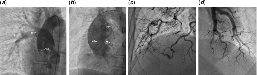
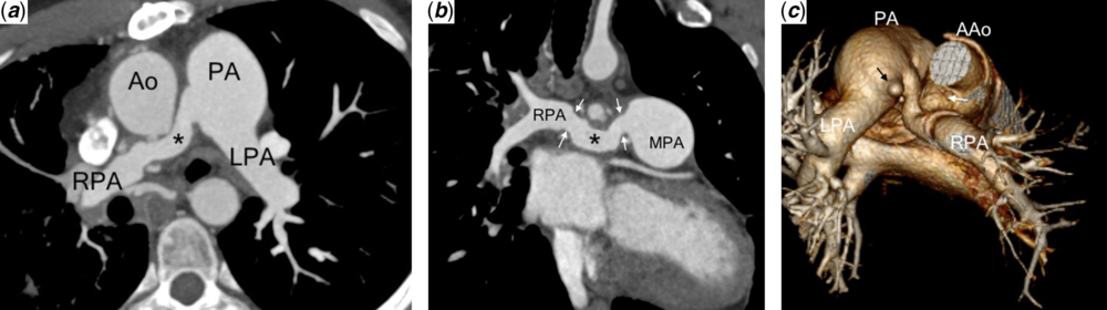
A 43-year-old male patient, who was 9 years old, was evaluated for dyspnoea during moderate exercise, the New York Heart Association (NYHA) Class was II. Upon physical examination, a cardiac murmur was found; an echocardiography was performed and found a patent ductus arteriosus (PDA) 5 mm in minor diameter and high probability of pulmonary hypertension; the confluence of the pulmonary arteries is not described in the report, so it was decided to close the PDA surgically, and after surgery, there was a slight improvement in symptoms, and the patient did not return to their respective controls. At the age of 34, the patient returned to the cardiology consultation, his NYHA class was III, he presented sporadic episodes of haemoptysis, and he received treatment as a pulmonary infection with remission of haemoptysis and without variation of the functional class. At the age of 43, he presented another episode of haemoptysis, chest pain of moderate intensity, palpitations, and dyspnoea at the rest. Echocardiography showed normal-sized cardiac chambers, left ventricle with concentric remodelling, normal biventricular systolic function, and the anomalous origin of the right pulmonary artery (AORPA) of the ascending aorta. The computed tomography (CT) scan confirmed the presence of AORPA and bronchiectasis in the right lung. The heart catheterisation (Fig 1) found that the left pulmonary artery (LPA) pressure was 50/25 (mean 33) mmHg and the right pulmonary artery (RPA) pressure was 120/60 (mean 80) mmHg, which were equal to systemic pressure (aorta); the pulmonary vascular resistance (PVR) was 9.5 WU/m2, and the PVR/systemic vascular resistance (SVR) index was 0.42. A vasoreactivity test with 100% oxygen was performed without significant change in pressures, the LPA pressure of 50/20 (mean 30) mmHg, the RPA pressure of 120/60 (mean 80) mmHg, and the systemic pressure of 120/60 (mean 80) mmHg, but with a decrease in the PVR of 6.1 WU/m2 and the PVR/SVR index of 0.34. The coronary arteries did not have any significant angiographic lesions on coronary angiography. With these data, a multidisciplinary team discussion was held prior to the decision to proceed to surgical correction. The surgery consists of reimplantation of the RPA to the main pulmonary artery (MPA), with a dacron tube of 16 mm in diameter and a creation of an 8 mm interatrial communication, for the risk of postoperative pulmonary hypertension and low cardiac output. One month after surgery, under treatment with sildenafil, the symptoms improved, the NYHA class was II, and the first control of heart catheterisation was performed, which reported: the pulmonary pressure of 119/49 (mean 77) mmHg, the PVR of 16.7 WU/m2, the systemic pressure of 99/56 (mean 75) mmHg, and the SVR of 8.4 WU/m2. In the vasoreactivity test with 100% oxygen, a significant decrease in pulmonary pressure and resistance was observed up to 68/19 mmHg (mean 41) and the PVR was 6.1 WU/m2 with no decrease in cardiac output. Twenty months after surgery, another heart catheterisation was performed and found that the pulmonary pressure was 47/17 (mean 31) mmHg, the PVR was 4.7 WU/m2, the systemic pressure was 104/60 (mean 77) mmHg, the SVR was 16.5 WU/m2, and the PVR/SVR index was 0.28; there was no difference in pressure between RPA and MPA. Control CT (Fig 2) reported patency of the implanted RPA and no stenosis at the anastomotic site. The clinical evolution was favourable; currently, the patient continues with medical treatment with a sildenafil of 25 mg three times a day, and the NYHA class is I.
Cardiac catheterisation: (a, b) Angiography in ascending aorta shows AORPA from AAo posterior wall (white arrow). (c) Magnified pulmonary angiography of RPA shows tortuous distal arteries with a small amount of monopedial arteries and slow washing of medium contrast. (d) Magnified pulmonary angiography of LPA shows heterogeneous capillary blush with moderate amount of monopedial arteries. (AAo = proximal ascending aorta; RPA: right pulmonary artery.)

Multislice tomography: (a) Axial view: shows RPA implantation toward the MPA, which is dilated. (b) Coronal view: reimplanted RPA, with permeable dacron tube graft (asterisk) without evidence of stricture in its path and in its sites of anastomosis (white arrows). (c) Volumetric reconstruction: tube path is lower than the aortic remainder of the previous site of implantation of the right branch (white arrow). The LPA is of normal calibre. Presence of PDA pulmonary end (black arrow). (MPA: main trunk of the pulmonary artery; PDA: patent ductus arteriosus.)

Discussion
Anomalous origin of a branch pulmonary artery from the aorta is a rare malformation, accounting for 0.12% of all congenital heart defects. This type of cardiac malformation was described first by Fraentzel in 1868.Reference Wang, Song and Cheng1,Reference Shakibi, Rastan and Nazarian2 AORPA has a reported mortality rate as high as 70% among those patients who did not undergo surgical repair within 1 year of life; the main cause of the high mortality rate was pulmonary hypertension and irreversible pulmonary vascular disease, because the pulmonary artery of abnormal origin receives high pressure from the systemic circulation, and as the pulmonary resistance decreases after birth, the flow through the anomalous pulmonary artery increases, leading to pulmonary artery hypertension (PAH) and congestive heart failure and subsequently irreversible pulmonary vascular disease.Reference Liu, Juan and Chen3,Reference Prifti, Crucean and Bonacchi4 The present case is important, because it is very infrequent, and is the oldest adult, reported in the literature, who has undergone surgical correction with favourable evolution. The AORPA is frequently associated with other cardiac malformations and is rarely presented as an isolated anomaly. AORPA was most commonly associated with patients with PDA, whereas anomalous origin of the left pulmonary artery (AOLPA) was most commonly associated with patients with aortic arch anomalies.Reference Shakibi, Rastan and Nazarian2,Reference Higeno, Inamura and Kayatani5,Reference Bockeria, Makhachev and Tkh6
These patients may have a different clinical presentation; usually, most neonates and infants are asymptomatic, but they may develop a progression of symptoms, such as exercise intolerance, dyspnoea, chest pain, haemoptysis, and recurrent pulmonary infections, throughout infancy and adolescence.Reference Bockeria, Makhachev and Tkh6,Reference Pankaj, Sachin and Shyam7 These symptoms depend on the anatomy of the affected lung and the types of the blood vessels providing its perfusion.Reference Liu, Juan and Chen3,Reference Pankaj, Sachin and Shyam7,Reference Raymond, Pedretti and Privitera8 The heart catheterisation is a very important procedure and is the gold standard for diagnosis of PAH and should be indicated preoperatively in all patients.Reference Pankaj, Sachin and Shyam7,Reference Raymond, Pedretti and Privitera8 The acute vasoreactivity testing (AVT) is a particularly important component of the heart catheterisation in congenital heart disease (CHD) with PAH, as it informs prognosis and can guide the correct treatment. According to current guidelines, assessment of reversibility is limited to hemodynamic variables: those in favour of reversible PAH-CHD are a left-to-right shunt and a PVR index is <4 Woods units. Surgical correction is contraindicated when the net shunt is directed right-to-left and is discouraged when the PVR index is >8 Woods units. When the PVR index is between 4 and 8 Woods units, “individual patient evaluation in tertiary centres” is advised.Reference Van der Feen, Bartelds, de Boer and Berger9 These recommendations, however, are predominantly based on expert opinion and are hardly supported by data. In fact, in PAH-CHD, no prospective studies have yet identified reliable hemodynamic cutoffs that predict the reversal of pulmonary vascular disease and normalisation of hemodynamic after cardiac correction. Acute pulmonary vasodilator tests (AVT) are performed during heart catheterisation to test the effect of short-acting pulmonary vasodilators on PVR, pulmonary artery pressure (PAP), and shunting. Although the use of AVTs to estimate reversibility prior to corrective surgery is widespread in current clinical practice, no haemodynamic cutoffs have shown sufficient accuracy in predicting reversal after shunt correction.Reference Van der Feen, Bartelds, de Boer and Berger9,Reference Galie, Humbert and Vachiery10 The haemodynamic change that defines a positive response to AVT in PAH associated with a shunt defect (CHD) should be considered as a >20% fall in PVR and PVR/SVR with the respective final values of <6 indexed Wood units (iWU) and <0.3. Nevertheless, operative safety in PAH-CHD with a shunt cannot be, and an element of clinical subjectivity is inevitable.Reference Apitz, Hansmann and Schranz11 In the present case, the AVT was considered as “reactivity,” and give to this patient the possibility of surgical correction with favourable evolution. Different surgical techniques have been employed. The direct anastomosis of the anomalous pulmonary artery from the aorta remains the technique of choice.Reference Liu, Juan and Chen3,Reference Xie, Gao and Wu12 The most frequent complication is stenosis at the level of the anastomosis and pulmonary hypertension.Reference Prifti, Crucean and Bonacchi4,Reference Xie, Gao and Wu12,Reference Garg, Talwar, Kothari, Saxena, Juneja, Choudhary and Airan13 In our case, there were no complications during the medium-term follow-up, and the last heart catheterisation showed a decrease in vascular pulmonary disease, which is consistent with the notable improvement in symptoms and functional class, despite the fact that the diagnosis was made very late. The factors that determine this evolution are still unknown due to the low incidence and intervention in such cases.
Conclusion
The interval between diagnosis and correction of anomalous origin of one pulmonary artery from the ascending aorta should be as short as possible to prevent pulmonary hypertension and congestive heart failure, because survival rate is around 30% among those patients who did not undergo surgical repair within 1 year of life.Reference Liu, Juan and Chen3 Some authors suggest that surgical correction should be performed during the first three months of life. We report the case of an older patient from all series undergoing AORPA corrective surgery with a favourable evolution in the medium term.
Acknowledgements
None.
Financial support
This research received no specific grant from any funding agency, commercial or not-for-profit sectors.
Conflicts of interest
None.