Coronary arterial aneurysms caused by Kawasaki disease may often develop obstructive lesions. This may lead to myocardial ischaemia or sudden death, which can occur from the early phase to even more than 20 years after the onset of the disease.1 Patients with aneurysms due to Kawasaki disease, therefore, should receive follow-up examinations throughout their lives. Although, X-ray coronary angiography is considered to be the gold standard for the detection of coronary arterial lesions, it is a very invasive, hazardous, and expensive procedure. Furthermore, coronary angiography cannot detect the thickness of the vessel wall without using intravascular ultrasound, which is a more risky and expensive procedure. A non-invasive technique that can visualize coronary arterial lesions and the vessel wall is desirable.
Cross-sectional echocardiography can detect coronary dilated lesions non-invasively. The technique does not, however, reveal obstructive lesions and aneurysms located on peripheral coronary arteries. Moreover, it becomes incredibly difficult to visualize the coronary arteries as children grow. Multi detector computed tomography is a less invasive examination compared to X-ray coronary angiography. The disadvantage of this technique is that it entails exposure to a large amount of ionizing radiation, and is performed using contrast medium and breath-holding, which makes it impossible to use in infants and young children. As magnetic resonance coronary angiography has developed remarkably over the past few years, we hypothesized that it would be possible to use the technique to perform non-contrast enhanced, free-breathing examinations even in young children with Kawasaki disease. In this study, therefore, we evaluated, retrospectively, the efficiency of magnetic resonance coronary angiography in detecting coronary arterial lesions in 106 patients with Kawasaki disease, ranging from infants to adults.
Subjects
Since July 2000, when requested, we have included magnetic resonance coronary angiography in the routine follow-up examinations of coronary arterial lesions due to Kawasaki disease. Since June 2003 to June 2005, the magnetic resonance coronary angiography examination included both bright blood and black blood imaging, and these techniques have now been used in 106 patients. Their age ranged from 4 months to 33 years, with a median of 13.0 years. Among these patients, 15 visited our hospital and asked us to perform magnetic resonance coronary angiography, and the other 91 were treated in the acute phase and/or followed up at our hospitals (Japan Red-Cross Medical Center and Tokyo Postal Services Agency Hospital). There were four patients who failed to complete the study. One of them was excluded because of a metallic artifact (zipper) in her clothing, and the others because of snoring and irregular respiration due to nasal obstruction while sleeping. One patient was a restless sleeper, and another woke-up before finishing the procedure. Our success rate of completing the examination, therefore, was 96%. The last three patients were re-examined almost two weeks later, and the final results were good enough to be included in this study.
Methods
Cross-sectional echocardiography
This was performed on a Hewlett Packard SONOS 5500 using 8 to 12 megaherz transducers in all patients within 7 days before magnetic resonance coronary angiography. The diameter of the aorta, the main trunks of the left and the right coronary artery, and the maximal diameter of aneurysms were measured. Intimal thickness, seen as diffuse layers with low echodensity, and thrombus, seen as vague masses on the aneurysmal wall were also observed.
Conventional X-ray coronary angiography
We have followed up 70 patients with large aneurysms and/or obstructive lesions using conventional X-ray coronary angiography, and their angiograms were compared with the magnetic resonance coronary angiographic images. The duration between the two examinations ranged from 1 day to 12 years. In 18 patients, the last X-ray coronary angiography was performed within a 6-month interval before or after the magnetic resonance coronary angiography. Among the 18, 12 patients with grid images taken in the same direction of each injection underwent the last magnetic resonance coronary angiography examination, within 6 months before X-ray angiography.
Magnetic resonance coronary angiography procedure
Magnetic resonance coronary angiographic studies were performed on a commercial 1.5 T Gyroscan Intera Master R.9 (Philips Medical Systems, Best, The Netherlands) equipped with cardiac software, a fast gradient system (maximum gradient: 30 milli-metre, maximum slew rate: 150 milli-Tesra/metre), and a synergy cardiac coil. A two-element flex-M coil was used for 35 children less than 7 years old, because of their body size. Magnetic resonance coronary angiography was performed using the steady-state free precession (SSFP) sequence, or “bright blood imaging” and navigator-echo proton density weighted black blood imaging, or “black blood imaging”, simultaneously. Images were acquired during free respiration using a navigator echo2 to monitor the diaphragm motion during free respiration. For respiratory gating and prospective tracking of the position of the three-dimensional volume, we used a two-dimensional selective navigator localized at the dome of the right hemidiaphragm. The gating window was set to 5 millimetres, and real-time motion correction used a constant correction factor of 0.6.
A flow-insensitive T2-prepulse to enhance the contrast without contrast agents was followed by a localized anterior saturation pre-pulse, a navigator pulse, a spectrally selective fat saturation pulse, and then by a three-dimensional segmented k-space gradient echo sequence comprising an echo time of 2.3 milliseconds, a repetition time of 4.6 milliseconds, and a radial scan technique. This sequence design was used for both 3 point plan scan and whole heart imaging, with 8 phase-encoding steps per cardiac cycle, thus producing bright blood imaging. The parallel imaging technique was used with a SENSE factor 2. Data were acquired along the major axis of the artery. Flow-compensating gradients were not used. Twenty slices, which were 3-millimetres thick, interpolated to 1.5 millimetres, were acquired with a 300 to 360 millimetre field-of-view, and were reconstructed with a matrix of 512 by 360, giving an in plane voxel size of 0.59 by 0.59 millimetres. A three-point plan scan was used until November 2003. From then, we used whole heart imaging in 67 of the 106 patients.
Black blood imaging using triple inversion recovery pulses was also performed, using the sequence of an echo time of 26 milliseconds, a repetition time of 1600 milliseconds for 2 beats, and a linear scan technique. Data were acquired along the major axis of the artery. Ten slices, which were 3-millimetres thick and a −1.5 millimetre slice gap, were acquired with a 220 by 360 millimetre field-of-view, and were reconstructed with a 512 by 385 matrix, giving an in plane voxel size of 0.7 by 0.7 millimetres.
For all magnetic resonance coronary arterial images, data were acquired with electrocardiographic gating during mid diastole.
The procedure was performed without the use of sedation, and with free breathing in patients over 8 years old. Infants and young children less than 7 years were provided with sodium trichloroethyl phosphate syrup, at a dose of 0.8 to 1.0 millilitres per kilogram, so that they could sleep during the examination. If the syrup was not effective, we administered thiopental sodium, at 2 to 5 milligrams per kilogram, by intravenous infusion. The scanning protocols were accomplished within approximately 40 minutes in each patient.
The difference of the arterial diameter between X-ray angiography and magnetic resonance coronary angiography was analyzed in the 12 patients in whom routine X-ray angiography was performed with grid imaging within 6-months after the magnetic resonance coronary arteriography, since dilated lesions often disappear or the diameter decreases very rapidly. On X-ray angiograms, the arterial size on the image at the end-diastolic phase was measured based on the grid images. The severity of localized stenoses was evaluated also in the 18 patients in whom X-ray coronary angiography was performed within a 6-month interval before or after the magnetic resonance coronary angiography. The obstructive lesions, thrombus, and/or intimal thickness were evaluated in 70 patients by X-ray angiography.
The magnetic resonance coronary arterial investigators were blinded to the X-ray angiography, but were able to view the findings of echocardiography from the electronic medical records.
Coronary arterial lesions due to Kawasaki disease have been classified by the Japanese Circulation Society Joint Research Group.3 Small aneurysms or dilations are lesions with localized findings of dilation with a luminal diameter 4 millimetres or less, and in older children, 5 years and over, lesions with a luminal diameter less than 1.5 times that of the normal coronary artery. When the internal lumen is larger, the lesion is diagnosed as an aneurysm. Obstructive lesions are classified into three types based on the X-ray angiographic findings: localized stenoses, occlusions that are 100% obstructed, and segmental stenosis.4 Segmental stenosis4 is defined as the appearance of new vessels that have a different morphology and pathological causes of recanalization. We have reported them as braid-like lesions. These being multiple tortuous vessels located in the lumen of the native coronary artery, bridging lesions represented by connecting vessels surrounding the occluded aneurysms, and pericoronary arterial communication, which are intracoronary collateral arteries.4
Written informed consent was obtained from all participants before X-ray angiography and magnetic resonance examinations at each time, and this study was approved by the Committee on Clinical Investigations of our hospital.
Statistics
The diameter of the coronary artery and aneurysms were measured by two independent observers at separate times, and correlative statistics by linear regression and Bland-Altman plots were used for the relation between the measurement of X-ray angiography and the both methods of magnetic coronary angiography. The inter-observer variability were analyzed by Bland-Altman plots. We considered differences significant at a p value less than 0.05.
Results
On bright blood imaging, aneurysms were seen in 62 patients, dilations in 17 patients, occlusion in 13 patients, localized stenosis in 20 patients, recanalized vessels in 10 patients, irregularity of the arterial wall in 6 patients, and no involvement of the coronary artery in 22 patients. Complete regression of the aneurysms was proven in 12 of these 22 patients.
Echocardiography versus magnetic resonance coronary angiography
Dilated lesions.
In 62 of the 106 patients, 69 aneurysms were observed on echocardiography, and an additional 28 aneurysms were visualized by bright blood imaging (Fig. 1). Of the 28, 23 were located at the periphery, where they were invisible by echocardiography, and the other 5 aneurysms were occluded and could not be evaluated by echocardiography.
In 20 instances, slight dilation was detected on echocardiography, but only 17 by bright blood imaging, and 19 by black blood imaging. One of the 20 dilations was misdiagnosed by echocardiography, proving to be recanalized tortuous vessels on bright blood imaging and X-ray angiography. On black blood imaging it also appeared like a dilated lesion. The other 2 slight dilations on echocardiography were diagnosed as normal sized coronary arteries by bright blood imaging and X-ray angiography, but looked slightly dilated not only on echocardiography but also on black blood imaging.

Figure 1. Aneurysms on bright blood imaging: 5-year-old male. A: Right and left coronary arteries. B: Left circumflex artery. C: Whole heart imaging. Black arrows point to a right coronary artery which has two small aneurysms. Red arrowheads point at a large aneurysm on the left anterior descending artery. White arrows point to a left anterior descending artery. Yellow arrows point to a small aneurysm at the bifurcation of the left coronary artery.
Magnetic resonance coronary angiography versus X-ray angiography with grid within a 6-month interval
The diameters of the coronary artery in the 12 patients were measured on the X-ray angiography and the magnetic resonance coronary angiography. The correlation coefficient between the arterial diameter measured by X-ray angiography and bright blood imaging at 27 points was 0.978, and between X-ray angiography and black blood imaging at 23 points was 0.929. On the Bland-Altman plots, the mean difference between X-ray angiography and bright blood imaging was 0.0, and the 95% confidence interval was from −1.4 to +1.5. The mean difference between X-ray angiography and black blood imaging was −0.6, and the 95% confidence interval was −3.7 to +2.5. The bigger aneurysms tended to show larger differences, and the size of arterial diameter on X-ray angiography correspond better on bright blood than on black blood imaging.(Fig. 2) As for the inter-observer variability for measurement of the arterial diameter on Bland-Altman plots, the mean difference between the two observers was 0.02, and the 95% confidence interval ranged from −0.54 to +0.59.

Figure 2. Difference of the size of arterial diameter between X-ray angiography and magnetic resonance coronary angiography.
X-ray angiography versus magnetic resonance coronary angiography, within a 6-month interval
Localized stenosis.
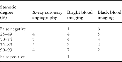
There were 18 localized stenoses in the 54 coronary arterial branches of the 9 patients among 18 in whom X-ray angiography was performed within 6 months before or after magnetic resonance coronary angiography. One false negative was a stenosis of 50% narrowing at the inlet of a large aneurysm, which was not detected by either bright or black blood imaging. One 90% stenosis on bright blood imaging, which was located at the inlet portion of a large aneurysm, was a false positive, not being seen on black blood imaging or X-ray angiography. Generally, with bright blood imaging, the degree of stenosis tends to appear more severe, and 3 cases of 75–90% narrowing of localized stenosis on X-ray angiography were detected as more than 90% localized stenosis on bright blood imaging (Table 1). With black blood imaging, the vague shadow in the lumen makes the stenoses appear to be of a lesser degree, or even non-existent, and 6 localized stenoses were not detected. The sensitivity of detecting localized stenosis based on X-ray angiography was 94.4% with bright blood imaging, and 66.7% with black blood imaging, with a specificity of 97.2% and 100%, respectively, and with a negative predictive value of 97.2% and 85.7%, respectively (Table 2).
Table 1. Detection of obstructive lesions

Table 2. Estimation of stenotic degree

X-ray angiography versus magnetic resonance coronary angiography
Occlusion.
X-ray angiography detected 17 occlusions in 13 of the 70 patients. The findings of occlusions on magnetic resonance coronary angiography were separated into 3 types. The first type included 8 aneurysms with an extremely high signal in bright blood imaging, and no blood flow detected by black blood imaging (Fig. 3). On bright blood imaging, the mean ratio of the 8 occluded aneurysmal signals to the signal at an adjacent segment of the intact coronary artery (mean: 2.1, standard deviation: 0.3) was significantly higher (p value less than 0.0001) than the ratio of the 8 non-occluded aneurysmal signals, which were selected randomly as controls, to the signal of the adjacent intact coronary artery (mean: 1.1, standard deviation: 0.1).

Figure 3. Occlusion: 8-year-old male after aortocoronary bypass surgery. A: X-ray coronary angiography: the left anterior descending artery is connected to the left internal thoracic artery. An arrowhead shows calcification of the occluded aneurysm. B: Bright blood imaging; a: occluded aneurysm with a high signal, b: the signal was measured adjacent to the intact coronary artery. C: Black blood imaging: The aneurysm had no lumen and showed a signal higher than that of blood. D: Calcification on X-ray coronary angiography. Yellow arrowheads point at a black lines of calcification.
The second type was an abruptly discontinued coronary artery, which was observed in another 8 occlusions (Fig. 4). Differing from the occlusion finding on X-ray angiography, the distal arteries of these two types of occlusion were visualized with a very low signal by magnetic resonance coronary angiography. The third type was a vessel undetected by magnetic resonance coronary angiography, which was an occlusion of an obtuse marginal branch of the circumflex artery (data not shown). It could not be visualized by either magnetic resonance coronary angiography or X-ray angiography.

Figure 4. Occlusion: 20-year-old male. A: X-ray coronary angiography: The left anterior descending artery is occluded (arrow). B: Occlusion classified as an abruptly disconnected coronary artery (arrow). A collateral artery (yellow arrow) connects to the distal artery.
One false negative was an occlusion of the second branch of the circumflex artery, which was not detected on magnetic resonance coronary angiography but was confirmed on X-ray angiography. In another patient after aortocoronary bypass surgery, a false positive was seen in an aneurysm with localized stenosis on the left anterior descending artery, which was connected to a graft of the left internal thoracic artery. The native coronary arterial flow through the stenosis went against the retrograde stream from the bypass graft. These opposing flows in the aneurysm made it appear to be occluded on magnetic resonance coronary angiography. The sensitivity was 94.2%, the specificity was 99.5%, and negative predictive value was 99.5% (Table 1).
Recanalized vessels.
We detected 10 recanalized vessels with X-ray angiography, and 8 of them showed excellent agreement with the findings of bright blood imaging (Figs 5A, B). These 8 lesions were braid-like lesions (Fig. 5A). With black blood imaging, the recanalized vessel walls appeared like white strings in the dilated lumen (Fig. 5C) but one of the braid-like lesions on black blood imaging did not show fine white strings in the lumen of the dilated artery, and it was misdiagnosed as a dilatation. Another braid-like lesion was a tiny obtuse marginal branch of the left circumflex artery that could not be detected by magnetic resonance coronary angiography.

Figure 5. Recanalized vessels: 20-year-old male. A: X-ray coronary angiography: The tortuous vessels are considered to be recanalized vessels. B: Bright blood imaging: The vessels on bright blood imaging show excellent agreement with the findings of X-ray coronary angiography. C: Black blood imaging: The walls of the new vessels in the dilated lumen appear like white strings (arrows).
One of the 2 recanalized vessels of a bridging lesion was observed as larger vessels with bright blood imaging and was misdiagnosed as arteries with irregular walls. The sensitivity, therefore, was 80% on bright blood imaging, and 70% on black blood imaging, and the specificity was 100% on both methods of magnetic resonance coronary angiography (Table 1).
Thrombus and/or intimal thickening, calcification.
On X-ray angiography, thrombus was seen in only 5 lesions as irregular negative shadows in the aneurysmal lumen. On echocardiography, thrombus was seen as vague massive masses in the aneurysmal lumen and diffuse layers, with low echo density on the aneurysmal wall were seen in 10 lesions. With bright blood imaging, thrombus was seen in 9 lesions and with black blood imaging, thrombus and/or intimal lesions were seen in 38 lesions. On black blood imaging, thrombus was seen as an eccentric mass on the aneurysmal wall, with an irregular edge facing the aneurysmal lumen with heterogeneous signal intensity. The masses appeared as light and dark shades of gray on black blood imaging. On the other hand, the uniformly thick gray layer with homogenous signal intensity covering the inside of almost the whole aneurysmal wall was considered to be intimal thickness. In the large aneurysms with a thick intima, the diameter of the aneurysm corresponded between X-ray angiography and bright blood imaging. These two methods detected the blood flow in the lumen. On the other hand, the results of echocardiography and black blood imaging agreed and detected not only the lumen but also the diameter of the aneurysm including the intima and/or thrombus (Fig. 6).

Figure 6. 10-year-old male. A thrombus in an aneurysm on the left anterior descending artery cannot be visualized on X-ray coronary angiography (A). However the size of the aneurysm is different from that observed on two-dimensional echocardiography (B). Therefore, magnetic resonance coronary angiography was performed. The maximal size of the aneurysm is 9.2 millimetres on X-ray coronary angiography (A) and 9.1 millimetres on bright blood imaging (C), but is 18.1millimetres on two-dimensional echocardiography (B) and 18.2 millimetres on cross-sectional black blood imaging (D). The size of the black blood flow inside the aneurysm is 9.3millimetres (D). Red arrowheads point at a thrombus
Calcification was seen in 11 lesions on X-ray angiography, 9 of which were detected as circles on the aneurysmal wall and 2 as tiny dots. All of the 9 circle shaped calcifications were detected as a black line on both bright and black blood imaging (Fig. 3) but 2 tiny calcifications were not detected.
Discussion
Generally, patients with regressed aneurysms due to Kawasaki disease are considered to be cured, so follow-up examinations with X-ray angiography are discontinued.5 Some recent studies, however, detected localized stenoses even after the aneurysms had regressed.1, 6 Serial evaluation is necessary, therefore, not only for the patients with large coronary aneurysms, who have a high risk of thrombotic occlusion of the aneurysm and/or progressing localized stenosis, but also for those with regressed aneurysms. It is almost impossible to recall patients with regressed aneurysms and to perform invasive X-ray angiography to screen for coronary arterial lesions when the patients do not show symptoms of ischaemic heart disease.
X-ray angiography is essential, nevertheless, in very young children in the convalescent phase of Kawasaki disease but often entails some other risks, such as thrombotic occlusion of the femoral artery due to puncture of a post-inflammatory vessel in the state of hypercoagulability, or worsened anaemia of Kawasaki disease because of bleeding from the punctured artery. The aneurysms decrease in size rapidly soon after forming. Hence, evaluation of the size of aneurysms, which is necessary for risk stratification and therapeutic management, must be conducted as soon as possible.1 All these aspects show the need for a non-invasive, easy and simple method to detect coronary arterial lesions.
Some investigators have already reported that magnetic resonance coronary angiography showed a high sensitivity and high accuracy in detecting coronary arterial lesions.7, 8 Moreover, the development of magnetic resonance coronary angiography has enabled examination of patients without breath holding or the use of contrast medium. Because of this, we thought it was now possible to perform magnetic resonance coronary angiography in infants and young children.
In this study, magnetic resonance coronary angiography could be performed easily even in the early phase of the disease. As expected, the dilated lesions were detected better with magnetic resonance coronary angiography than with echocardiography. A couple of slight dilations on echocardiography and on black blood imaging were detected as normal coronary arteries on bright blood imaging and X-ray angiography. After the aneurysms had decreased in size, slight dilation was often observed with echocardiography over a long period, and this difference was also found between echocardiography and X-ray angiography.9 This suggests that echocardiography and black blood imaging may show the lumen with slight intimal proliferation. Among comparative studies of coronary arterial size in Kawasaki disease between magnetic coronary angiography and X-ray coronary angiography, Greil et al8 reported excellent agreement between the two techniques, such as a mean difference of 0.4 millimetres and standard deviation 0.6 millimetres.
They also reported that arterial size on magnetic resonance coronary angiography measurements was slightly larger than that seen with X-ray angiography. They performed bright blood imaging. Our results with bright blood imaging showed a mean difference of 0.0, this being −0.6 with black blood imaging. In our study, as the aneurysms enlarged, the difference between the two measurements increased. As we mentioned before, this may have been due to differences in intimal thickness. As for coronary arterial walls observed by black blood imaging, Fayad et al10 proposed that large eccentric plaques with a heterogeneous signal intensity might possibly be due to differences in tissue composition. Remarkable intimal thickening, and/or organized thrombus of the arterial walls in aneurysms which had decreased in size, were detected by black blood imaging. This study is the first to show the thick intima in aneurysms caused by Kawasaki disease with magnetic resonance coronary angiography. The thickened intima observed by black blood imaging may predict future coronary atherosclerotic changes. Besides, another important aspect of following-up aneurysms is to observe fresh formation of thrombus. In a large aneurysm, thrombus is sometimes formed very rapidly and frequently even after successful thrombolytic therapy.11 With black blood imaging, thrombus was detected more clearly, which may suggest that this technique is effective in evaluating intravascular thrombolytic therapy.
Evaluation of localized stenosis on magnetic resonance coronary angiography is difficult because the stenoses are often located at the inlet or outlet of a large aneurysm, and they are covered by the aneurysm, like the one false negative and one false positive finding in our study. Our radiologists did not know the findings of X-ray arteriography, and they tried to evaluate these stenoses not only by the long axis views, but also by a series of cross-sectional views at these sites. After we started to employ whole heart imaging, observations from all directions become possible.
Hence, they detect localized stenoses fairly well with bright blood imaging. It is more difficult to estimate the degree of stenoses by black blood imaging. Recently, we are using spiral black blood imaging12, 13 at these sites, and trying to detect localized stenosis more accurately.
The images of occlusion obtained with magnetic resonance coronary angiography were often different from those of X-ray angiography. We still do not know why some of the occluded aneurysms showed an extremely high signal on bright blood imaging. The duration of the occlusion is not clear, because most of the patients had no signs or symptoms of myocardial infarction. At least, there was no significant difference in the duration from the onset to the magnetic resonance coronary angiography between the group of occlusions with an extremely high signal and the group of abruptly disconnected arteries. Our one false negative case was an occlusion of a tiny side-branch from a left circumflex artery and it seems to be one of the limitations of this study.
There are limitations in the use of magnetic resonance angiography. With black blood imaging, occlusion is detected clearly. Turbulent flow in a huge aneurysm makes a thrombus-like shadow, and sometimes it is difficult to distinguish turbulent flow from thrombus. Moreover, the side-branches are not always detectable by magnetic resonance coronary angiography. With the recent rapid technical progress, we expect further improvements. For instance, spiral black blood imaging can distinguish the arterial walls from the lumen precisely,12, 13 and a software tool, like Soap-Bubble,14 can even detect side-branches. These techniques can prevent false negatives of localized stenosis and occlusions. Extremely tortuous micro-vessels of recanalization are often shown as larger vessels by bright blood imaging. By observing the magnetic resonance coronary angiography image carefully, these surplus vessels make it easy to suspect the presence of recanalized vessels.6
We expect that knowing the limitations and the unique pattern of images on magnetic resonance coronary angiography will improve the rates of evaluating coronary arterial lesions due to Kawasaki disease.
In conclusion, therefore, magnetic resonance coronary angiography was proven to be a useful method for evaluating all types of coronary arterial lesions due to Kawasaki disease, for all states of the disease from the early to the late phase in patients of all ages. The use of both bright and black blood imaging is desirable for evaluating coronary arterial lesions due to Kawasaki disease. Magnetic resonance coronary angiography can reduce the number of diagnostic catheterizations. Until magnetic resonance-guided coronary artery interventions become standardized, however, magnetic resonance coronary angiography will not replace interventional catheterizations.
Acknowledgements
We thank Professor Kouichi Nakamura, Jichi Medical University for helpful comments on the statistical analysis of the present data, and Ms. B Levene for English correction. This study was supported by a research grant from Advanced Medical Science and Treatment of the Postal Services Agency.