

Introduction
Shoulder pain is one of the most common musculoskeletal symptoms, responsible for 16% of all musculoskeletal complaints.Reference Andrews1 The incidence of shoulder pain ranges from 6.6 to 25 cases per 1000 patients, with a peak incidence in those aged 45–64 years.Reference Andrews1 The incidence falls after the age of 65 years, which has been attributed to the fact that older people can often tolerate discomfort better due to a diminished activity level.Reference O'Reilly and Bernstein2 However, when specifically asked, older people report shoulder problems to be a common cause of disability and pain, affecting at least 25% of hospital and community populations.Reference Chard and Hazleman3
The shoulder is a complex structure that accounts for three different joints: the glenohumeral joint, the acromioclavicular joint (ACJ) and the scapulothoracic joint. The rotator cuff consists of the supraspinatus, infraspinatus, teres minor and subscapularis muscles. There are active and passive stabilizers that maintain joint congruency and allow full range of motion. In order to identify shoulder problems, it is essential to perform a detailed physical examination that should include asking about the characteristics of the pain. In most of the cases when examining older patients, a simple X-ray of the shoulder could give enough information to make the diagnosis when combined with the physical examination.
In this review, we describe the main groups of pathologies that usually affect older people and the surgical approach to each of them, focusing on the specific characteristics in this age group, such as bone quality and general health status.
Search methods
A protocol was established, and a search of published studies in English-language sources for shoulder pathology and treatment was performed. Systematic reviews and mostly studies of individuals over 60 years old were included. MEDLINE was searched via PubMed.
Traumatic pathology
Fractures of the proximal humerus
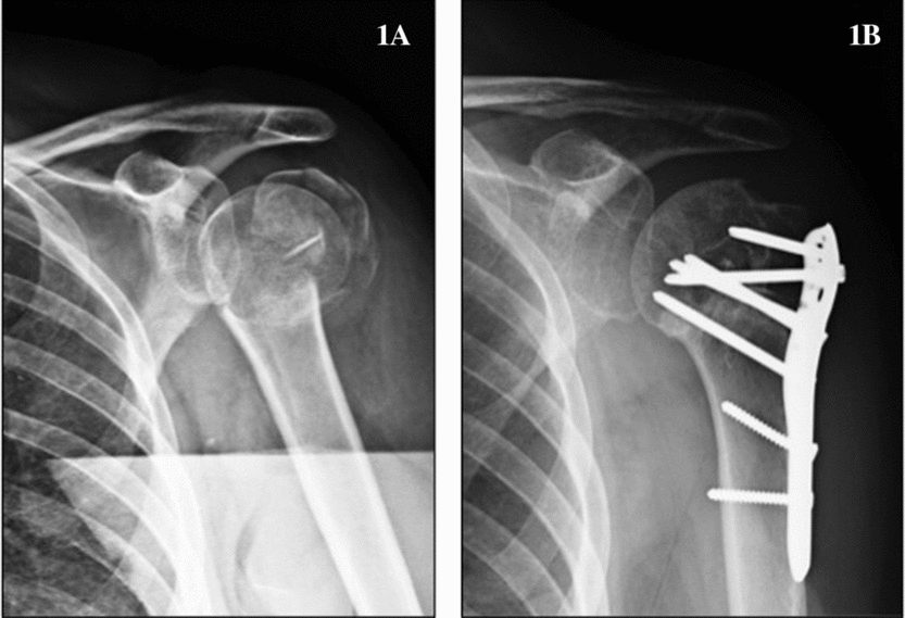
Proximal humeral fractures (Fig. 1) are the third most common fracture in older people, behind hip fractures and extra-articular distal radius fractures. They usually happen as a consequence of a fall from standing height. Patients that present with proximal humeral fractures are generally fitter than those who sustain a proximal femoral fracture, but less fit than those with distal radius fractures.Reference Kelsey, Browner, Seeley, Nevitt and Cummings4 They have been attributed to recurrent falls and the high prevalence of osteoporosis in this population, especially in post-menopausal women. Calvo et al.,Reference Calvo, de Miguel, de la Cruz and López-Martín5 in a study of 5147 women (mean age 72.6 ± 7.5 years) who had sustained 5268 fractures, found that 17.5% of them were conservatively treated proximal humeral fractures. Court-Brown et al. also found that the highest age-specific incidence occurs in women between 80 and 89 years old.Reference Court-Brown, Garg and McQueen6
A, antero-posterior plain X-ray of the shoulder showing a four-part fracture of the proximal humerus; B, the radiological result after open reduction and plate fixation

The most frequently used classification for proximal humeral fractures is Neer's classification.Reference Neer7 It is based on analysing the displacement (more than 1 cm or angulation greater than 45 deg) of the four main fragments of the proximal humerus: head, greater tuberosity, lesser tuberosity and humeral shaft.
Court-Brown et al., in their series of 1027 consecutive proximal humeral fractures, found that 49% were minimally displaced and this is a constant finding in the literature.Reference Bell, Leung and Spratt8 For these minimally displaced fractures, conservative treatment is generally advocated. This group would include one part fractures involving humeral neck, one part greater tuberosity or lesser tuberosity fractures and impacted two part fractures of the surgical neck with minimal angulation of the humeral head.Reference Neer7 Some authors also recommend conservative management in two, three or four part fractures with less severe varus or valgus angulation of the humeral head to the shaft (<30 deg of the normal 130 deg head-shaft angulation), but with residual cortical contact to the shaft.Reference Court-Brown and McQueen9 The main disadvantage of this is that there is a higher risk of non-union and the presence of mal-union is unavoidable. This could lead to pain and discomfort and a decrease in functional capacity, especially regarding self-care or daily living activities, even at 6 months after the fracture.Reference Calvo, de Miguel, de la Cruz and López-Martín5 However, operative risks, specifically infection, are avoided.
When looking at displaced fractures, there is still controversy on whether to approach them surgically or conservatively in older patients.Reference Hanson, Neidenbach, de Boer and Stengel10 As previously mentioned, in this population group, functional expectations are moderate. Moreover, co-morbidities such as diabetes mellitus, osteoporosis, rheumatoid arthritis, immunocompromise, steroid medication and concurrent neoplasm have been associated with a poor outcome and increased risk of complications.Reference Court-Brown, Garg and McQueen11 Most authors agree that the following fracture types may benefit from a surgical approach, always depending on the patient's characteristics in each case: two-part fractures involving the tuberosities with more than 1 cm of displacement, fractures with a displaced fragment involving the articular fragment with one of the tuberosities, unstable two-part surgical neck fractures with metaphyseal comminution, two- or three-part fractures with more than 30 deg of varus or varus deformity, three- or four-part fracture-dislocations and head-split fractures.Reference Murray, Amin, White and Robinson12 Foruria et al. found that some fracture patterns were associated with a worse prognosis when managed non-surgically.Reference Foruria, de Gracia, Larson, Munuera and Sanchez-Sotelo13 These included fractures with posteromedial impaction, fractures of the greater tuberosity when it was displaced medially overlapping with the posterior articular surface and lateral impaction fractures. Operative treatment includes several options, such as minimally invasive techniques, intramedullary nailing, open reduction and internal fixation (ORIF), hemiarthroplasty (HA) and more recently, reverse shoulder arthroplasty (RSA).Reference Murray, Amin, White and Robinson14 Locking plate technologyReference Bell, Leung and Spratt15 and augmentation with calcium phosphate or sulphate cement, iliac crest and intramedullary fibular bone grafts have expanded the indications for ORIF in proximal humeral fractures with osteoporotic bone.Reference Namdari, Voleti and Mehta16
Minimally invasive surgery has less risk of infection and requires less soft tissue detachment, but provides a less stable fixation and has a potential risk of neurovascular injury. Percutaneous fixation has yielded good results in two-part fractures and has also been recommended in three-part fractures in older patients, in whom an incomplete reduction can achieve satisfactory clinical results, avoiding the risks of a more aggressive surgical approach.Reference Blonna, Rossi, Fantino, Maiello, Assom and Castoldi17 On the other hand, ORIF provides a more anatomical reduction and more stable fixation, but increases the risk of infection and avascular necrosis due to soft tissue manipulation. Olerud et al. Reference Olerud, Ahrengart, Ponzer, Saving and Tidermark18 investigated three-part fractures in older people, comparing surgical treatment with locking plates to conservative treatment. At 2-year follow-up, functional outcome and quality of life score (HRQoL) indicated better results for surgical treatment. However, there were 13% of the patients with severe complications that required re-intervention and 17% required a minor second surgical intervention as well. In both groups there was a negative effect in both the upper limb functionality score and the HRQoL. Intramedullary nailing provides stable fixation in osteoporotic bone and preserves the periosteal blood supply. The entry point of the nail is close to the rotator cuff and may cause stiffness and dysfunction, leading to a high rate of re-interventions in order to remove the metalwork. It has been recommended for older patients with displaced two-part surgical neck fractures.Reference Murray, Amin, White and Robinson12 Court-Brown et al., however, advocate for conservative treatment in this type of fracture in patients over 70 years old.Reference Court-Brown, Garg and McQueen11
Regarding three- and four-part fractures in older patients, surgical options include a range from conservative management to HA or RSA. A recent study performed in four-part fractures included 55 patients with a mean age of 77 years, and reported a significant advantage in quality of life in favour of HA compared with non-operative treatment, in terms of pain control rather than differences in mobility.Reference Olerud, Ahrengart, Ponzer, Saving and Tidermark19 In the context of a fracture, HA is technically demanding surgery that requires restoring the anatomical relationship between the head and the tuberosities in order to achieve good functional results.Reference Boileau, Krishnan, Tinsi, Walch, Coste and Molé20 Cemented stems are generally recommended due to poor bone quality. In spite of new implant designs that promote bony contact, healing of the tuberosities remains a major concern. Moreover, the functionality of the rotator cuff in this population group often compromises the outcome of HA in proximal humeral fractures.
RSA has started being used for the treatment of proximal humeral fractures in the older population. This is a type of prosthesis that is generally used for the treatment of rotator cuff arthropathy. Its design provides a joint lateralization and does not need an intact rotator cuff in order to maintain shoulder function. When we compare HA with RSA in acute proximal fractures in older patients, RSA appears to produce functionally superior results to HA at 5 years post-operatively, although more long-term studies are needed to corroborate these findings.Reference Stanbury and Voloshin21, Reference Boyle, Youn, Frampton and Ball22
Shoulder instability in older patients
Anterior dislocation of the shoulder in patients aged over 60 generally occurs as a consequence of a fall from standing height and has an incidence of 11–16%.Reference Levy, Pritsch and Rath23 The main difference to anterior dislocations in the younger patient is the lower risk of recurrence, which is approximately 30%, and the higher incidence of lesions of the rotator cuff, that has been reported to range from 61 to 76%.Reference Gumina and Postacchini24 The mechanism of the injury consists of an anterior subluxation of the head, which causes an anterior force. It results in stretching of the anterior capsule and subscapularis tendon and tearing of the posterior rotator cuff or supraspinatus tendon, which is generally degenerated and weak in this population group.Reference McLaughlin and MacLellan25 Full thickness tears are more frequently observed in patients that have sustained more than one episode and they are large to massive tears that often affect the subscapularis tendon.Reference Gumina and Postacchini24 There is also a higher risk of damage to the axillary nerve or the brachial plexus, which has been reported to be 9%, with a mean recovery period of 3–12 months.Reference Gumina and Postacchini26 Rotator cuff tears (RCTs) (Fig. 2) might be under-diagnosed and should be suspected in chronic shoulder pain in older patients after a dislocation. They can often be mistaken for nerve palsies.Reference Murthi and Ramirez27
Arthroscopic image showing a severely retracted rotator cuff tear with exposure of the joint and the long head of the biceps tendon
The need for surgery is still controversial. Rapariz et al. Reference Rapariz, Martin-Martin, Pareja-Bezares and Ortega-Klein28 reported a series of 60 patients aged over 60 years when they suffered the first episode. Of these, only four required reconstructive surgery to maintain stability. Similarly, Gumina et al. published a study of 108 patients, of whom only 16 patients underwent surgery. When surgery is needed, two different problems should be addressed: on the one hand, the anterior capsulolabral complex, including injuries to the glenoid rim, and on the other hand, RCTs, which play a major role in instability of the shoulder due to muscular imbalance.Reference Shin, Yun, Kim and Yoo29 Some authors believe that one single episode could be related to previous massive RCT (failure of posterior stabilization mechanism), while recurrent instability is also caused by failure of the anterior stabilization mechanism.Reference Levy, Pritsch and Rath30 In the case of a single episode, it is generally advocated to suture the RCT. If recurrent instability develops, then repair of the anterior capsulolabral complex is also recommended. Levy et al. reported good results with open anterior capsular shift with or without anterior labral repair and rotator cuff repair.Reference Levy, Pritsch and Rath30
Regarding associated fractures, the most frequently observed is greater tuberosity fracture. It has been considered a good prognostic sign as it means that the rotator cuff is intact. Compression fractures of the humeral head (Hill-Sachs) or glenoid fractures are usually large due to osteoporosis in this population group. When they cause chronic instability, they may require a surgical approach that in older patients will generally consist of performing an arthroplasty.Reference Sonnabend31
Chronic dislocations are not uncommon in older patients. They should be suspected when there is a loss of motion, and an overall restriction in internal rotation (anterior chronic dislocations) or external rotation (posterior chronic dislocation). Closed reduction under general anaesthesia can be performed, but it is not recommended when there is a fracture of more than 20% of the humeral head or the dislocation is older than 4 weeks, due to a high risk of iatrogenic fracture. In the case of a bony defect or persistent instability, shoulder arthroplasty provides pain relief and improved range of motion.Reference Sperling, Pring, Antuna and Cofield32
Degenerative pathology
Impingement syndrome, rotator cuff tendinopathy and rotator cuff tears
Impingement syndrome represents a spectrum of entities that includes subacromial bursitis, rotator cuff tendinopathy and RCT. The aetiology of these different pathologies has been described as a combination of two mechanisms.Reference Rees33 Some authors support the fact that there is an ‘extrinsic’ compromise of the subacromial space from the anterior acromion, coracoacromial ligament (CAL), and ACJ. Bigliani et al. described three different types of acromion morphology that could be directly responsible for this compromise and represent the basis of current surgical practice.Reference Bigliani, Ticker, Flatow, Soslowsky and Mow34 Other authors support the ‘intrinsic’ theory, which believes that tendon degeneration is due to age-related changes and vascular compromise, with or without a traumatic onset.Reference Chang35
NeerReference Neer36 described the three stages of impingement disease: the first stage is characterized by oedema and hemorrhage, the second stage would include fibrosis and tendinitis, and the third stage is characterized by the presence of bone spurs and tendon rupture. Supraspinatus tendinitis is one of the most frequent causes of shoulder pain at any age. It is characterized by tenderness in the superior or superolateral aspect of the shoulder and often irradiates down the arm. It normally worsens at night, impairing normal sleep. It can also affect the long head of the biceps tendon (LHBT), in which case the pain is localized in the front of the shoulder. Pain is exacerbated on resisted supination and flexion of the arm with the elbow in extension. Along with tendinitis is often found bursitis of the subacromial, subdeltoid or subcoracoid bursa. In the case of a RCT there is a lack of active motion when compared with the passive range. Weakness can also be appreciated in forward elevation, abduction, external rotation or internal rotation depending on the muscle affected.
Conservative management of subacromial syndrome includes several options. Although physical therapy is advocated in the first phases, they are encountered typically in younger patients under 40 years old.Reference Chang35 In more advanced phases or when severe pain is present, physical therapy can even worsen the symptoms. In these cases, subacromial injections of corticosteroid and/or local anaesthetic are the initial approach before considering a surgical procedure.Reference Min, St Pierre, Ryan, Marchant, Wilson and Arrington37 Some authors have determined that patient response to the first injection predicts need for future surgery.Reference Cummins, Sasso and Nicholson38
Numerous types of injections have been described.Reference Van der Sande, Rinkel, Gebremariam, Hay, Koes and Huisstede39 Corticosteroid injections are widely used and their benefit has been attributed to their anti-inflammatory effect. Cummins et al.Reference Cummins, Sasso and Nicholson38 studied 100 patients with a diagnosis of subacromial syndrome with a non-surgical protocol of physical therapy and cortisone injections. At 2 years follow-up, 79% of the patients had not needed surgery and presented with markedly improved function and decreased pain. However, the use of corticosteroids has limitations due to the potential side-effects. Some authors have demonstrated that they can be deleterious to the rotator cuff tendons causing degeneration or rupture, as well as intra-articular cartilage damage or even systemic effects such as osteoporosis. Moreover, their use in people with diabetes can cause variations in blood sugar levels that should be monitored. Recently, Min et al.Reference Min, St Pierre, Ryan, Marchant, Wilson and Arrington37 in a prospective randomized controlled trial have demonstrated that non-steroidal anti-inflammatory (NSAID) injections can provide equivalent pain relief and improvement in function to corticosteroid injections, avoiding their risks. Therefore, depending on the concomitant pathology of the patient, one or the other could be chosen in order to minimize potential systemic side-effects.
Surgical treatment can be recommended after failure of a conservative management protocol. Subacromial decompression with anterior acromioplasty was first described by Neer in 1972 as an open procedure.Reference Neer40 However, with the advances in shoulder arthroscopy, open acromioplasty is now reserved for exceptional cases. Arthroscopic subacromial decompression consists of bursectomy, with or without anterior acromioplasty and coracoacromial ligament resection. The rationale for acromioplasty is based on the ‘extrinsic’ theory mentioned before, which supports the compression of the rotator cuff in the coracoacromial arch.Reference Neer36 Inflammation of the bursa is a constant finding in cases of impingement, and bursectomy is a simple arthroscopic procedure that has been generalized and provides good results. Acromioplasty and resection of the coracoacromial ligament, however, are still controversial in cases of degenerative rotator cuff because, according to those supporting the ‘intrinsic theory’, they could possibly contribute to proximal migration of the humeral head.Reference Donigan and Wolf41 Donigan et al.Reference Donigan and Wolf41 in a recent systematic review concluded that bursectomy alone could be as effective as bursectomy and acromioplasty performed together.
The incidence of RCTs increases with age. Magnetic resonance imaging (MRI) studies have shown full-thickness tears in 28% of those aged over 60 years, and in patients aged over 70 the prevalence of asymptomatic full-thickness RCTs is about 38% as diagnosed by ultrasound scan.Reference Tempelhof, Rupp and Seil42, Reference Sher, Uribe, Posada, Murphy and Zlatkin43 When a rotator cuff is present, a surgical repair is the preferred treatment. It can be performed openly or arthroscopically. In massive tears, there is a still a great controversy when treating older patients on whether conservative treatment should be advocated or not. Relative contraindications for rotator cuff repair could be advanced fatty infiltration of rotator cuff muscles, definite loss of tendons, or proximal humeral migration.Reference Downie and Miller44 Age of the patient, level of activity, extent of disability and co-morbidities should be considered before surgical management is decided. In cases of massive tears, in which surgical management is not considered, conservative management consists of NSAID medications, injections and gentle physical therapy focusing on strengthening of the remaining rotator cuff tendons and the deltoid muscle.
Many studies have been recently published on rotator cuff repair in patients over 60 and 70 years of age. De Carvalho et al.Reference De Carvalho, Puri and Calder45 reviewed 104 cases of openly repaired RCTs in a group of patients with a mean age of 77 years and reported a high satisfaction rate of 92.7%, as well as pain relief and high-level function. However, a recent systematic review concluded that, although there could be a benefit of a surgical approach, the heterogeneity of the treatments does not allow generalization.Reference Downie and Miller44
Numerous groups have tried to find prognostic factors that could be helpful in the decision-making process. Robinson et al. found that age appears to be an important factor associated with incidence of re-tear in patients over 70 years old, as well as male gender.Reference Robinson, Wilson, Dalal, Parker, Norburn and Roy46 Lam and Mok reported poorer outcomes in women, those with higher American Society of Anesthesiologist (ASA) grade and longer duration of symptoms.Reference Lam and Mok47 The size of the tear, the degree of retraction and the presence of fatty degeneration have also been associated with lower healing rate and less satisfactory clinical results.Reference Tashjian, Hollins and Kim48, Reference Djahangiri, Cozzolino and Zanetti49
Rotator cuff arthropathy and long head of the biceps tendon pathology
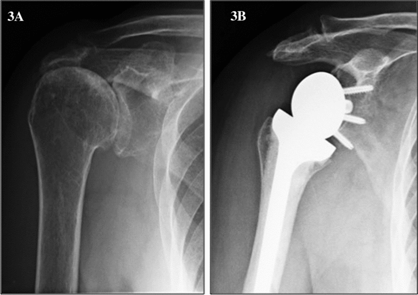
The end stage of rotator cuff pathology is rotator cuff arthropathy (Fig. 3). It is considered a spectrum of pathology that starts with rotator cuff insufficiency, which causes proximal migration of the humeral head.Reference Neer, Craig and Fukuda50 The acromiohumeral distance decreases, exacerbating impingement syndrome symptoms and causing specific arthritic changes to the glenohumeral joint and to the acromion. Biomechanical studies have demonstrated the importance of shoulder force-couples between the rotator cuff and the deltoid in order to maintain congruency of the humeral head within the glenoid.Reference Hsu, Luo, Cofield and An51 However, information is lacking regarding the natural progression of RCTs to cuff tear arthropathy.
A, antero-posterior plain X-ray of the shoulder showing a rotator cuff arthropathy with acromial acetabulatization and proximal migration of the head; B, X-ray of a rotator cuff-deficient shoulder after reverse total shoulder replacement
Clinical findings include joint effusion and pain, often worse at night and with activity, and loss of motion. Large effusion might be present because fluid is free to communicate between glenohumeral joint and subacromial bursa. Massive tear of the rotator cuff is present and causes weakness and marked atrophy of the musculature. Typical radiological findings are proximal migration of the humeral head, presence of osteophytes, narrowing of the joint, rounding of the greater tuberosity of the humerus, acetabularization of the undersurface of the acromion and superior glenoid wear with osteopenia.Reference Neer, Craig and Fukuda50, Reference Hamada, Yamanaka, Uchiyama, Mikasa and Mikasa52
Non-surgical management is the first line of treatment in most patients.Reference Nam, Maak, Raphael, Kepler, Cross and Warren53 Traditionally, surgical management of RCT arthropathy has been disappointing because of a high rate of complications and poor patient satisfaction. Surgical options include arthroscopic lavage with tenotomy or tenodesis of LHBT if it is still present, humeral tuberoplasty and shoulder replacement.
Lesions of the LHBT in this context are a potential cause of shoulder pain and dysfunction.Reference Duff and Campbell54 The spectrum of pathology varies from simple tendinitis to partial rupture, delamination, medial subluxation that causes damage to the subscapularis tendon or intra-articular entrapment due to hypertrophy (hourglass biceps). In the case of a spontaneous rupture, no further intervention is needed. Boileau et al. published the results of isolated biceps tenotomy or tenodesis in patients with massive RCTs that were not candidates for rotator cuff repair.Reference Boileau, Baqué, Valerio, Ahrens, Chuinard and Trojani55 They reported effectiveness in terms of pain relief and increased range of motion in those patients with restriction due to painful arc. However, the authors do not recommend this technique in patients with pseudoparalysis of the shoulder (forward elevation of less than 90 deg) and established rotator cuff arthropathy. In these cases, shoulder arthroplasty should be advocated.
Candidates for joint arthroplasty in this context are patients that present with intractable pain, a functional deltoid and an intact coracoacromial arch.Reference Smith, Guyver and Bunker56 There are two different categories of arthroplasty that have been used for treatment of rotator cuff arthropathy: hemiarthroplasty (HA) and reverse total shoulder arthroplasty (RSA). HA was traditionally recommended for patients under 70 years old with active elevation of more than 90 deg and has provided moderate clinical results.Reference DiGiovanni, Marra, Park and Bigliani57, Reference Goldberg, Bell, Kim, Bak, Levine and Bigliani58 Sánchez-Sotelo et al. reported an improvement in shoulder active elevation from 72 to 91 deg and a decrease in the mean pain score, with an overall satisfactory result in 67% of the cases at 5 years follow-up.Reference Sanchez-Sotelo, Cofield and Rowland59 However, they described anterosuperior instability in seven of the 30 patients and could establish a correlation between incidence of instability and prior subacromial decompression. Goldberg et al. in a more recent study did not find statistical differences in mean active forward elevation and mean external rotation gain when comparing the group of patients aged 70 or over with those less than 70 years.Reference Goldberg, Bell, Kim, Bak, Levine and Bigliani58
RSA was first designed in 1985. It consists of a reverse ball-and-socket design that relies on the deltoid muscle function to restore range of motion and compensate for rotator cuff insufficiency.Reference Boileau, Watkinson, Hatzidakis and Balg60 The semiconstrained design prevents proximal migration that was frequently seen with the use of HA. It has gained increasing popularity and presently remains the preferred treatment for rotator cuff arthropathy.Reference Smith, Guyver and Bunker56, Reference Drake, O'Connor and Edwards61
Young et al. recently performed a study comparing the results of HA versus RSA for the treatment of rotator cuff arthropathy in two comparable groups of 102 patients. They found better functional results with RSA, although the authors suggest that long-term studies are necessary to confirm these findings.Reference Young, Zhu, Walker and Poon62 The survival rate with replacement of the prosthesis and glenoid loosening as end-points have been reported as 91 and 84%, respectively, at 120 months, demonstrating better results for those cases of massive RCT.Reference Guery, Favard, Sirveaux, Oudet, Mole and Walch63 There were two breaks in the survival curve, one around 3 years post-operatively, reflecting early loosening of the prosthesis and the other occurred around 6 years post-operatively and was related to progressive deterioration of the functional result. These authors recommend the use of RSA in patients over 70 years old with very low functional demands.
Acromioclavicular joint arthritis
The ACJ is a diarthrodial joint that is surrounded by a capsule and contains a synovial lining and an intra-articular disc that degenerates with age, typically after the fourth decade. This degeneration is a frequent cause of pain and disability. Physical examination and plain X-ray can often be enough to diagnose ACJ arthritis, as well as response to local anaesthetic injections. The role of MRI as a diagnostic tool has been controversial. Shubin Stein et al. demonstrated that reactive bone oedema in the ACJ is a more reliable predictor of symptomatic ACJ pathology than degenerative changes seen on MRI.Reference Shubin Stein, Ahmad, Pfaff, Bigliani and Levine64 Conservative treatment is firstly advocated for ACJ osteoarthritis. In refractory cases, an arthroscopic excision of the joint can be performed during subacromial decompression, providing satisfactory results.Reference Levine, Soong, Ahmad, Blaine and Bigliani65 Care should be taken to avoid damage to the acromioclavicular or coracoclavicular ligaments causing secondary instability of the joint. The most frequent complication of this surgery is incomplete resection or re-growing of the osteophytes.
Glenohumeral osteoarthritis
Glenohumeral osteoarthritis of the shoulder is less common than knee or hip osteoarthritis. The causes of primary osteoarthritis are unknown, although it can be secondary to trauma, instability or previous surgery.Reference Vecchio, Kavanagh, Hazleman and King66 Clinically it is characterized by pain, decreased motion and loss of function.Reference Andrews1 Radiological studies will reveal joint space narrowing, subchondral sclerosis and osteophytes about the glenoid and the humeral head, typically inferiorly.Reference Walch, Badet, Boulahia and Khoury67 There is usually a posterior humeral head dislocation that causes posterior glenoid wear and a severe contracture of the anterior soft tissues, including the anterior capsule and subscapularis tendon.Reference Walch, Badet, Boulahia and Khoury67 This patho-anatomy is responsible for loss of external rotation.
Total shoulder arthroplasty has provided successful and reliable results for the treatment of osteoarthritis.Reference Ricchetti, Abboud, Kuntz, Ramsey, Glaser and Williams68 However, it is a technically demanding surgical procedure with moderate estimated blood loss, and thus specific characteristics of older patients should be taken into account in each case. Firstly, bone quality can be compromised due to osteopenia and bony wear, which means that a cemented stem might be used with the associated anaesthetic risks. Secondly, due to soft tissue imbalance, the stability of the implant can be difficult to achieve and the functionality of the rotator cuff can also determine a poor functional outcome. Lastly, functional demands and expectations should be individually assessed before advising the patients to undergo the procedure.
Foruria et al. reported a series of 50 total shoulder arthroplasty (TSA) performed in 44 patients aged over 80 years old. The most frequent co-morbidities were high blood pressure, myocardial infarction and atrial dysfunction, with 27 of the 44 patients ASA grade III or IV. Although 80% of the patients had a satisfactory or excellent outcome, the authors admit that non-fatal medical or surgical complications are common and there is necessity for an intensive care unit for immediate post-operative care.Reference Foruria, Sperling, Ankem, Oh and Cofield69 Richetti et al. found that the group of older patients required a longer period of in-patient care before return home and were more likely to require a blood transfusion.Reference Ricchetti, Abboud, Kuntz, Ramsey, Glaser and Williams68
There has been a special interest in reporting outcomes of TSA when considering specific co-morbidities. For example, Sperling et al. published their results on TSA in patients with rheumatoid arthritis.Reference Sperling, Cofield, Schleck and Harmsen70 In patients with inflammatory diseases and very destructive shoulder osteoarthritis, the results of TSA are less predictable due to insufficiency of the rotator cuff. Some authors recommend the use of RSA in this group of patients.Reference Hattrup, Sanchez-Sotelo, Sperling and Cofield71 Regarding Parkinson's disease, which is most common in the older population, TSA appears to be successful in terms of pain relief, but functional results have been reported to be poor, particularly in patients over 65 years.Reference Kryzak, Sperling, Schleck and Cofield72 A specific rehabilitation programme and dedicated medical management of the disease have been recommended.
The prevalence of osteoporosis in the older population has become a general concern when thinking of performing an orthopaedic surgical procedure. A recent study of 230 patients diagnosed with shoulder osteoarthritis with a mean age of 68 years revealed that 75% were unaware of suffering from osteoporosis.Reference Pervaiz, Cabezas, Downes, Santoni and Frankle73 The authors recommend that in addition to normal pre-operative planning, a shoulder computerized tomography scan should be obtained to screen patients for metabolic bone disease, in order to diminish the potential risk of lack of stability of the humeral stem, or the glenoid component compromising the survivorship of the implant.Reference Pervaiz, Cabezas, Downes, Santoni and Frankle73
Conclusion
Shoulder surgery in the older population can provide excellent results in selected patients, particularly in terms of pain relief more than functional recovery. Given the higher risk of anaesthetic and surgical complications, an established protocol of conservative treatment should be considered in the first place, when the situation allows. Co-morbidities such as osteoporosis should be identified and appropriately treated in order to prevent shoulder fractures and their complications.
Conflicts of interest
The authors declared no conflicts of interest.