Introduction
Juvenile nasopharyngeal angiofibroma is the commonest benign tumour of the nasopharynx in adolescent males. It is associated with a high risk of severe intra-operative haemorrhage, particularly in advanced stages.
Our university hospital, one of the oldest in the country, is well known (for the past seven decades) as a tertiary referral centre for juvenile nasopharyngeal angiofibroma, serving the surrounding states and Nepal. Our contribution has been declared as the largest in the world, as cited in the British reference book Scott-Brown's Otolaryngology (in the Rhinology section, fifth edition).Reference Chew, Kerr and Groves 1 The entire spectrum and evolution of juvenile nasopharyngeal angiofibroma surgery can be appreciated if one considers the cases that have presented at our centre, since the early 1960s to the current day in which pure endoscopic resection is practised.
The majority of our patients have a low socioeconomic status with no insurance, and hence the treatment is governed by the existing resources with minimal government support. Thus, the practical management options that are feasible here may not necessarily be the latest techniques adopted by the Western world. As a classical example, open surgery for juvenile nasopharyngeal angiofibroma at our centre costs less than 5000 INR (90 USD), while a single sitting of pre-operative selective embolisation (before the endoscopic procedure) costs no less than 950 USD. As the average monthly income of a poor Indian patient is less than 50 USD, the treatment options for such patients are limited. This may be the case for most other centres dealing with juvenile nasopharyngeal angiofibroma across the Third World. Recently established endoscopic surgery may not be practical in many centres, including ours, as it is not suitable for extensive disease, while the incorporation of selective pre-operative embolisation adds to the cost of treatment, which will be unaffordable for the majority of patients at such centres. Furthermore, although improvement in transnasal endoscopic skull base techniques has redefined management of juvenile nasopharyngeal angiofibroma, the limits of endoscopic resection still need to be fully defined.Reference Kamel 2 Under these (practical and theoretical) circumstances, we opt for a pure endoscopic approach only in selected patients, while the majority undergo open procedures.
Lateral extension of juvenile nasopharyngeal angiofibroma, particularly in the lateral part of the pterygopalatine fossa, infratemporal fossa and the cheek (staged as at least IIC (Radkowski et al.Reference Radkowski, McGill, Healy, Ohlms and Jones 3 ) or IIIA (Mishra et al.Reference Mishra, Shukla, Bhatia and Pant 4 )), presents a challenge regarding the selection of a minimal access approach that is the least morbid. Open facial or sublabial approaches with anterior osteotomy cause cosmetic scarring, and may also hamper the facial growth of these adolescent patients or even disrupt dentition. The selection of transpalatal approaches with endoscopic assistance, in our experience, not only prevents the above complications but also avoids blind tumour manipulation, especially at the pterygopalatine fossa, as is sometimes apparent after complete exposure of the tumour. Hence, the reported advantage of open anterior approaches, of tumour stalk visualisation, is not a limiting factor in our modified inferior approaches when assisted with endoscopy. The early practice of open external carotid ligation and/or Gelfoam® embolisation was found to be less effective, while hypotensive anaesthesia can be more practicable at most centres.
This paper aims to defend our various modifications of transpalatal techniques performed with endoscopic assistance under hypotensive anaesthesia (without pre-operative embolisation). These would seem to be more practical for managing advanced lateral extensions of juvenile nasopharyngeal angiofibroma at most worldwide centres with restricted resources. This paper in no way undermines the importance of pure endoscopic techniques, but aims to provide a firm platform for comparing our modified inferior open approaches with the other available options, especially for the lesser equipped centres.
Materials and methods
This prospective study is based on 63 patients with juvenile nasopharyngeal angiofibroma, from May 2012 to May 2015, who underwent transpalatal, transpalatal-circumaxillary (transpterygopalatine) or transpalatal-circumaxillary-sublabial approaches for surgical management of extensive lateral extension involving the pterygopalatine foramina, pterygopalatine fossa, infratemporal fossa, cheek and even the temporal fossa. The diagnosis of juvenile nasopharyngeal angiofibroma was suspected clinically but confirmed radiologically in every case pre-operatively. Of the 63 patients, 10 had previously undergone a transpalatal surgery elsewhere for juvenile nasopharyngeal angiofibroma and hence represented cases of either residual or recurrent disease. All the cases were staged radiologically, as per the published criteria of Radkowski et al.Reference Radkowski, McGill, Healy, Ohlms and Jones 3 and Mishra et al.Reference Mishra, Shukla, Bhatia and Pant 4 None of the patients were biopsied.
Haematinics were administered until haemoglobin status reached at least 10 grams per cent. Depending upon the stage and anticipated size of the tumour, 2–4 packed red blood cells were arranged to replenish the intra-operative blood loss in every case.
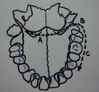
The surgery was performed only by the faculty consultants (AM, VV, HPS and SK). The surgical procedure currently practised at our centre is a combination of two techniques, as previously described by SardanaReference Sardana 5 and Mishra et al.,Reference Mishra, Shukla, Bhatia and Pant 6 which is suitable for the majority of large extensions (Figure 1). The classical transpalatal incision can be extended more anteriorly in the form of a tongue-shaped flap if needed. An endoscope is used to visualise the sphenopalatine foramina area to assist in complete exposure of the tumour, and often to assess the need for additional osteotomy or myotomy. Accordingly, four surgical components are occasionally adopted: (1) resection of the pterygoid muscles and plate, (2) nibbling of the pterygoid plates to open the sphenopalatine foramina for better exposure of the juvenile nasopharyngeal angiofibroma stalk, (3) nibbling of the hard palate for better visualisation of the sphenopalatine foramina, and (4) avoidance of the last two components and instead using an angled sinuscope for better visualisation post-excision. Hence, the prototype incision comprises either a pure transpalatal approach or extension across the retromolar region (circumaxillary component), or even extension further laterally and towards the sublabial region.
Diagrammatic representation of the modified transpalatal approaches. Incision ‘A’ constitutes the transpalatal approach. Extension ‘B’ along with ‘A’ constitutes the transpalatal-circumaxillary (transpterygopalatine) approach. Incision ‘A’ if extended to include ‘B’ and ‘C’ constitutes the transpalatal-circumaxillary-sublabial approach.

It is not difficult to detach the tumour from its surroundings by blunt finger dissection, assisted by a periosteal elevator or dissector through the nose, using palatal or sublabial incisions, while simultaneously maintaining a constant traction to deliver the tumour completely. The mucosal incision is stitched without much effort over the packing. The packing is removed after 36–48 hours through the nose.
Following the excision, the tumour volume was assessed by calculating the amount of water displaced in a calibrated beaker. The intra-operative blood loss was determined by adding the amount of suctioned blood to the amount of diluted blood (obtained using the blood-soaked sponges employed for washing) adjusted to the pre-operative haemoglobin status of the patient. The actual blood loss corresponding to the diluted blood was determined by the formula 1000 xy/z, where x = total diluted blood volume (in litres), y = haemoglobin per cent of the diluted blood (measured using a haemoglobinometer) and z = pre-operative haemoglobin status of the patient (haemoglobin per cent).
The patients were discharged at least 5 days after surgery. The majority of patients could not be radiologically assessed post-operatively owing to the cost factor involved in imaging. They were followed up at three-month intervals for at least two consecutive visits, during which formal endoscopy was performed to rule out residual or recurrent disease and to assess surgical morbidities. Subsequent assessment was conducted after one year. The patients were encouraged to attend our out-patient clinic in the event of a fresh bleeding episode or a nasal obstruction possibly denoting recurrence. A telephonic enquiry in this regard was attempted twice at six-monthly intervals.
Results
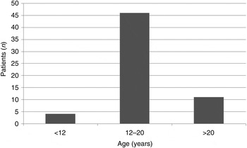
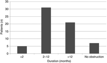
All patients were male; their age distribution is depicted in Figure 2. The presenting symptoms were: nasal blockage, in 58 cases (62 per cent); epistaxis, in 62 cases (98 per cent); headache, in 40 cases (63 per cent); and altered facial profile, in 10 cases (25 per cent). The epistaxis was moderate in amount (50–100 ml) in 31 cases (49 per cent), while severe epistaxis was noted in only 2 cases (3 per cent). The durations of nasal obstruction are shown in Figure 3. The positive clinical findings included: nasal mass on anterior rhinoscopy, in 80 per cent; a mass in the postnasal space or nasopharynx on posterior rhinoscopy, in 97 per cent; cheek swelling, in 18 per cent; a mass in the temporal area, in 3 per cent; and proptosis, in 3 per cent.
Age distribution of the juvenile nasopharyngeal angiofibroma patients.

Duration of nasal obstruction in the juvenile nasopharyngeal angiofibroma patients.

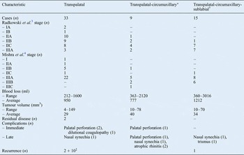
The characteristics of the patients as per the three surgical approaches are depicted in Table I. None of the patients in this series underwent pre-operative selective embolisation or external carotid artery ligation, or even intra-operative Gelfoam embolisation (the latter has in the past been a routine practice at our facility).
Characteristics of cases by surgical approach

*Transpalatal + retromolar extension; †transpalatal + retromolar + sublabial extension. ‡These 10 patients had previously undergone a transpalatal procedure in this hospital or elsewhere.
The transpalatal approach was found to be more appropriate for the tumours located in the nasopharynx and postnasal space, and those with medial involvement of the pterygopalatine fossa. The transpalatal-circumaxillary (transpterygopalatine) approach, on the other hand, was more appropriate for tumours with complete pterygopalatine fossa involvement as evidenced by its widening radiologically, or even tumours with minimal extension to the infratemporal fossa. Here, the palatal incision was extended around the last molar to expose the root of the pterygoids. Visualisation of the sphenopalatine foramina area was facilitated using a 30-degree sinuscope or at times by nibbling the posterior edge of the hard palate. Finally, the transpalatal-circumaxillary-sublabial approach provided better access to the cheek and infratemporal fossa area (Figure 4). Although the tumour could be completely excised, its delivery as a single complete mass was possible in only one-third of cases using this approach.
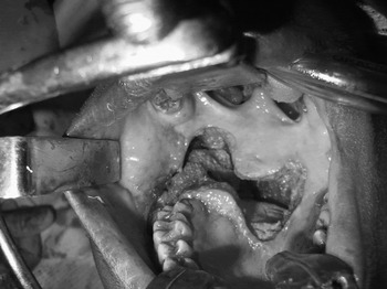
Transpalatal-circumaxillary-sublabial approach, with haemostatic packing in infratemporal fossa and cheek following complete tumour excision.

It is noteworthy that residual disease was encountered only when the transpalatal approach was employed and only in two cases. In one of these cases, there was residual disease in the orbit that was purposely left for the second stage of surgery owing to severe intra-operative haemorrhage during primary excision. The other case concerned a seven-year-old boy in whom the dimensions of the skull were too small for the routine instruments or finger manipulation. Because of severe haemorrhage with partial excision, the lateral extension was left, to be dealt with in future.
The complication rate was minimal. The immediate, acute post-operative complications included palatal perforation, in three patients only (two underwent the transpalatal approach and one underwent the transpalatal-circumaxillary approach). One patient who underwent the transpalatal-circumaxillary approach presented with repeated episodes of haemorrhage with removal of haemostatic packs. The blood investigations revealed abnormal international normalised ratio and a diagnosis of dilutional coagulopathy was made. Subsequent infusion of fresh frozen plasma stabilised his haemostatic status and the patient was discharged in a good condition. Surprisingly, this patient was a 23-year-old with a tumour of small volume (11 mm3) in stage IIB (Radkowski et al.Reference Radkowski, McGill, Healy, Ohlms and Jones 3 ) or IIIA (Mishra et al.Reference Mishra, Shukla, Bhatia and Pant 4 ). The pterygopalatine fossa was completely occupied by the tumour and a simple transpalatal approach was insufficient to deliver the lateral most part of the extension. This resulted in excessive haemorrhage (1383 ml), necessitating infusions of 5 units of Ringer's lactate solution, 1 unit of Isolyte S® (multi-electrolyte solution) and 1 unit of packed red blood cells. As this particular case was a recurrence, the corresponding fibrosis of the tumour bed resulting from previous surgery may have contributed to difficult dissection along the tumour bed and attachment, leading to the prolonged and difficult excision. None of the patients that underwent the transpalatal-circumaxillary-sublabial approach had any acute complication.
Long-term complications, although uncommon, were seen with all the approaches. Nasal synechia occurred as a result of the opposition of the two raw surfaces of nose, possibly induced by intra-operative instrumentation or tight nasal packing. These synechia were easily resolved within a few weeks. Two patients who underwent the transpalatal approach showed gaping in the palatal stitch line. These ‘immediate’ post-operative palatal perforations healed well following secondary suturing. However, another patient who underwent the transpalatal-circumaxillary approach showed incomplete closure of post-operative palatal perforation despite secondary suturing, performed twice.
The partially reversible complications included atrophic rhinitis, in two cases. The patients were advised to use topical lubricants and nasal douches long-term. In addition, there was a single case of post-operative trismus, which improved significantly within a month of jaw-stretching exercises. In this latter case, there was incomplete excision of the pterygoid muscles, inducing inflammation in the post-operative period. A small degree of fibrosis was anticipated and the patient was advised active physiotherapy, to be continued after discharge from hospital.
It is noteworthy that recurrence was seen only in two patients that underwent the transpalatal approach and in one patient that underwent the transpalatal-circumaxillary-sublabial approach. It is also worth mentioning that 10 patients had a previous history of transpalatal surgery for juvenile nasopharyngeal angiofibroma (in addition to the 33 patients that underwent the transpalatal approach in this study). Hence, these 10 patients may represent cases of recurrence. In one patient, the transpalatal approach revealed pea-sized tumour recurrence twice and he underwent transnasal endoscopic resection. One patient who underwent the transpalatal-circumaxillary-sublabial approach had recurrence within six months; this was dealt with via the transpalatal approach as the site of recurrence was different. No recurrence was subsequently noted within one year of follow up.
It is worth summarising the relevant points of the pure endoscopic approach adopted. We have undertaken many successful pure endoscopic resections for tumours restricted to the nasopharynx, nasal cavity, pterygopalatine fossa or the medial part of the infratemporal fossa. These were often in those patients who either had a very limited recurrence in the postnasal space or in affluent patients with large tumours who could afford the cost of selective embolisation. There have been a very few occasions when the embolising material was extruded and excessive haemorrhage in the operative field made further resection extremely difficult. However, the advantages of a pure endoscopic technique include the surety of complete resection, the avoidance of blood transfusion and reduced hospital stay (1–2 days).
Discussion
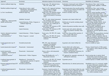
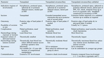
This paper attempts to establish the acceptability of extended transpalatal techniques for the majority of extensive juvenile nasopharyngeal angiofibromas, as per our institutional experience. The literature on these techniques is lacking relative to other open techniques (Table II).Reference Howard, Lloyd and Lund 7 – Reference Mickey, Close, Schaefer and Samson 21 Our facility has been able to manage the majority of juvenile nasopharyngeal angiofibroma cases with these approaches, without selective pre-operative embolisation. A summary of our experience is presented in Table III.
Open surgical approaches for juvenile nasopharyngeal angiofibroma (other than transpalatal)

NP = nasopharynx; PPF = pterygopalatine fossa; ITF = infratemporal fossa; CSF = cerebrospinal fluid
Characteristics of modified transpalatal approaches
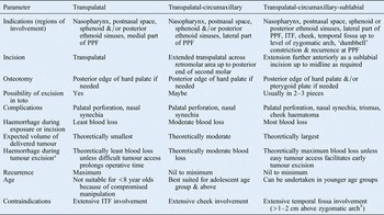
*Depends on tumour characteristics and vascularity. †Open anterior approach preferred. PPF = pterygopalatine fossa; ITF = infratemporal fossa
The presentation of stage I tumours was a rare event in our series, possibly because the associated mild degree of epistaxis and/or nasal obstruction may not significantly concern patients. Such symptoms are often overlooked, particularly given that the dry atmosphere in this part of the world does predispose to repeated epistaxis episodes, especially in summer.
Regarding the 10 patients with a past history of a transpalatal surgery, it may not be appropriate to label these as cases of recurrence, as there is an equal likelihood that 5 are cases of residual disease as they were possibly operated incompletely elsewhere (no records available).
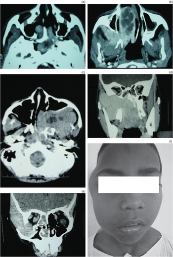
The various staging systems describe the lateral extensions of juvenile nasopharyngeal angiofibroma quite broadly. This broad definition is important for planning the modified transpalatal approaches, to achieve a complete resection. The lateral extensions can accordingly be classified as per six levels (Figure 5a–f): (1) medial pterygopalatine fossa involvement, (2) complete pterygopalatine fossa involvement (Holman Miller sign), (3) partial infratemporal fossa involvement, (4) complete infratemporal fossa involvement, (5) extension to the cheek (widened mandibulomaxillary tunnel or Hondusa sign) and temporal fossa up to the level of the zygomatic arch, and (6) temporal fossa involvement above the zygomatic arch.
Levels of lateral extension. Axial computed tomography (CT) scans of the paranasal sinuses showing: (a) medial pterygopalatine fossa, (b) complete pterygopalatine fossa with partial infratemporal fossa, and (c) complete infratemporal fossa involvement. (d) Coronal CT scan of the paranasal sinuses showing juvenile nasopharyngeal angiofibroma extension to the cheek with a widened mandibulomaxillary tunnel and temporal fossa involvement up to the level of the zygomatic arch. The tumour was successfully excised through a transpalatal-circumaxillary-sublabial approach. (e) Coronal CT scan of the paranasal sinuses, showing temporal fossa involvement above the zygomatic arch, and (f) physical appearance of the same patient; an anterior facial (Weber–Ferguson) approach was utilised for this patient.

Although the transpalatal approach was carried out in the majority of cases (52 per cent), tumours staged as IIC (Radkowski et al.Reference Radkowski, McGill, Healy, Ohlms and Jones 3 ) or IIIB (Mishra et al.Reference Mishra, Shukla, Bhatia and Pant 4 ) were better dealt with using extended approaches. Based on our experience, we recommend the transpalatal approach for the first level of lateral extension, the transpalatal-circumaxillary (transpterygopalatine) approach for the second and third levels, and the transpalatal-circumaxillary-sublabial approach for the fourth and fifth levels. As per the world literature, massive lateral extension, especially that involving the cheek and lower temporal area, is better dealt with using anterior and lateral open approaches (Table II). However, our experience favours the transpalatal-circumaxillary-sublabial approach for lateral extensions up to the zygomatic arch, as it is associated with less morbidity than the other approaches (Table II vs Table III). In cases of a widened pterygopalatine fossa, we discourage the resection of pterygoid muscles and plates, but encourage the manipulation of the cheek component through sublabial incision and, if possible, negotiation of the tumour through a widened sphenopalatine foramina into the nasopharynx. In cases where the tumour is significantly large, it can be excised in two pieces (cheek with infratemporal fossa component, and intranasal component). The excision of pterygoid muscles and plates in order to open the sphenopalatine foramina posteriorly may produce transient trismus. It is best avoided unless: the pterygopalatine fossa is minimally widened, producing an hourglass-shaped constriction of the tumour; predisposition to residual disease is anticipated; or there is recurrent disease.
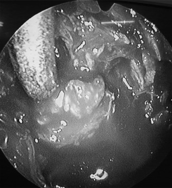
There were only three recurrences in this study. The possible reason for recurrence in two patients that underwent transpalatal approach was residual disease, especially of the most lateral aspect of the extension, which was not amenable to excision. It is quite possible to have a small region of residual disease at the sphenopalatine foramina area, even with the transpalatal-circumaxillary-sublabial approach. An endoscope may sometimes fail to visualise very small residue at the most lateral part of the pterygopalatine fossa through the sphenopalatine foramina, particularly in the presence of oozing. Such residual devascularised tumour may undergo frank necrosis or spontaneous resolution, or even result in recurrence. Figure 6 shows small disease residue at the medial part of the pterygopalatine fossa after total excision following a transpalatal-circumaxillary-sublabial approach, which emphasises the importance of post-excision endoscopic visualisation of the cavity. Herman et al. too have advocated the combination of an open technique with an endoscopic approach for inspection of the nasal cavity, to reduce recurrence.Reference Herman, Bublik, Ruiz and Younis 22 As a policy, we have adopted the latter to remove gross residual disease. Such methods of post-operative visualisation have been used in our department since the late 1970s. Holm's nasopharyngoscope was initially used for visualising the surgical field post-operatively to ensure complete excision, especially when undertaken per via naturalis approach. This instrument is a right-angled prism that facilitates a ‘holistic’ visualisation. The earlier method of pre-operative external carotid artery ligation was later replaced with the selective embolisation technique used today.
Endoscopic view of residual disease following the transpalatal-circumaxillary (transpterygopalatine) approach that was subsequently excised with endoscopic assistance.

It is possible for tumour recurrence to develop at some other site in the nasopharynx (not involving the sphenopalatine foramina area), as evidenced in one of the two cases that recurred after a transpalatal approach (A Mishra, in press).Reference Mishra 23 Hence, recurrence depends not only on residual disease, but also on the approach selected, the aetiopathological origin or site of juvenile nasopharyngeal angiofibroma, and the tumour biology including maturation status.
The range of intra-operative blood loss was high with the extended procedures (blood loss was greatest in the transpalatal-circumaxillary-sublabial approach, followed by the transpalatal-circumaxillary approach and the transpalatal approach). However, although mean blood loss was highest with the transpalatal-circumaxillary-sublabial approach, it was lowest with the transpalatal-circumaxillary procedure (with the transpalatal approach falling in the middle in terms of mean blood loss). A complete tumour excision is suggested by the cessation of bleeding, while a continued haemorrhage is expected if the tumour is incompletely excised. Accordingly, with the relatively limited access in the transpalatal approach, a prolonged manipulation time is likely to result in more bleeding than the transpalatal-circumaxillary approach. Superior extensions of tumours in the anterior ethmoid, orbit, and cribriform or parasellar regions, which upstage the disease, may have contributed to excessive haemorrhage as well. Hence, the selection of the transpalatal approach to manage non-lateral extensions may have further contributed to the enhanced blood loss. The transpalatal-circumaxillary-sublabial approach, on the other hand, facilitated complete excision, in one piece (in the majority of cases), but the extent and size of the delivered tumour probably resulted in more blood loss as compared to the other two procedures.
The use of a raspatory, a Farabeuf periosteal elevator and a tonsillar dissector is routine in our practice. The ‘transpalatal pterygopalatine fossa dissector’ is a novel addition to our armamentarium for the removal of tumour adhesions from the pterygopalatine fossa and sphenopalatine foramina. The principal author intends to file a patent application regarding its design. Use of these instruments lessens the chances of incomplete avulsion of the tumour at this area, which is known to cause recurrent foci. Being a firm (fibrotic) tissue, a moderate traction with subperiosteal elevation enables easy delivery. Manipulation of the tumour is better facilitated by its palpation during dissection, rather than by visualisation; the latter is often not possible amidst the pool of oozing blood.
Owing to its firm consistency, the tumour seldom breaks during the traction process, unless the distal adhesion is inaccessible for manipulation. Not uncommonly, the narrow part of the tumour across the sphenopalatine foramen may break, facilitating the removal of the juvenile nasopharyngeal angiofibroma in two pieces. Occasionally, the tumour is more fragile or its attachment more fibrotic, necessitating its delivery piecemeal. This situation in particular requires immediate endoscopic evaluation of the post-operative cavity, as the continued haemorrhage anticipated from the raw mucosal surfaces may mask residual disease. Unless residual disease is grossly visible, further management is not immediately indicated. Given the low recurrence rate in our series and the significant chances of spontaneous regression of a small residue, we agree with Banhiran and Casiano,Reference Banhiran and Casiano 24 and recommend close monitoring of the disease, treating only when symptoms reappear.
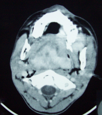
The size (volume) of the tumour may not directly correlate with the extent of surgery in every case. Moreover, the transpalatal-circumaxillary approach is in no way inferior to the transpalatal-circumaxillary-sublabial approach for non-lateral extensions of juvenile nasopharyngeal angiofibroma. For example, in a patient with a large juvenile nasopharyngeal angiofibroma with oropharyngeal extension, who presented with stridor, a transpalatal approach was employed and the tumour was delivered into the oropharynx (Figure 7). Hence, tumour size does not strictly reflect the disease stage or define the surgical approach. Accordingly, the mean size of delivered tumours in our series was greater with the transpalatal-circumaxillary approach than with the transpalatal-circumaxillary-sublabial approach. It is advisable to convert from the transpalatal approach to the transpalatal-circumaxillary approach for large volume tumours, to avoid undue traction and possible avulsion of the lateral palatal pedicle during manipulation. The additional advantage of the transpalatal-circumaxillary-sublabial approach is complete visualisation of the sphenopalatine foramina following excision of the pterygoid muscles and nibbling of the root of the pterygoid plates. The latter opens up the pterygopalatine foramina to facilitate delivery of the narrowed juvenile nasopharyngeal angiofibroma stalk or a recurrent tumour in that area, as stated earlier.
Axial computed tomography scan of an extremely rare case of juvenile nasopharyngeal angiofibroma extending in the oropharynx and presenting with stridor.

The complications with transpalatal techniques (Table III) are minimal compared to other open approaches (Table II) employed to deal with tumours of a similar profile (similar stage and size or volume). The early complications are seldom troublesome, while the late complications, such as atrophic rhinitis, can be comfortably managed. Only a single case of a very small palatal perforation due to excessive palatal fibrosis (associated with multiple surgical procedures) persisted in our series.
Previously reported recurrence rates vary, with one study (of a series of 30 cases) reporting 16 per centReference Iovanescu, Ruja and Cotulbea 25 and another reporting rates of 30–50 per cent.Reference McCombe, Lund and Howard 26 Hence, our recurrence rate with extended transpalatal approaches of less than 5 per cent indicates better chances of complete tumour excision with such approaches. Nevertheless, it is possible that our variable follow-up period (which ranged from three months to three years) may have contributed to the low recurrence rate. Frequent recurrences have been associated with extensions to the sphenoid sinus, the base of the pterygoid and the clivus bone.Reference Lloyd, Howard, Phelps and Cheesman 27 The use of a stout periosteal elevator inside the sphenoid sinus and along the clivus, a curved raspatory, and a specially designed transpalatal pterygopalatine fossa dissector facilitated near-complete subperiosteal and submucosal dissection. Hence, the chance of residual disease is theoretically lower with our techniques.
Endoscopic techniques are undoubtedly the procedures of choice today, but their routine acceptance depends on the wide availability of selective embolisation. It is selective embolisation rather than endoscopy per se that has revolutionised the conservative surgical approaches for vascular tumours with skull base extensions. Hence, in the absence of embolisation, the completeness and speed of excision, and minimal intra-operative haemorrhage, are the main parameters to rank a skilled surgeon in a setup like ours. As the majority of our population cannot afford embolisation, we opt for open approaches that are associated with the least morbidity. This situation is apparent throughout the developing world, including India and Africa where the major burden of juvenile nasopharyngeal angiofibroma resides. Thus, this paper attempts to convey our experience for any centre that frequently utilises open approaches. The common limitations as compared to an endoscopic approach include a moderate degree of immediate post-operative morbidity and a somewhat longer hospital stay (of 4–5 days). This policy works very well, with low care costs available for the majority of patients who present at our facility. Hence, the main reasons for not routinely employing endoscopic techniques are the lack of resources and affordability, rather than lack of expertise or otherwise. However, a pea-sized tumour that is a suspected recurrence is always dealt with by pure endoscopy, even without embolisation.
-
• Endoscopic resection is currently the procedure of choice for juvenile nasopharyngeal angiofibroma
-
• The additional cost-burden of pre-operative selective embolisation makes this technique impractical at many centres
-
• ‘Cost-effective’ open surgery options with minimal morbidity seem justified
-
• Six levels of juvenile nasopharyngeal angiofibroma lateral extensions were identified and treated using modified approaches
-
• The modified approaches were transpalatal, transpalatal-circumaxillary and transpalatal-circumaxillary-sublabial
-
• With endoscopic assistance, and without embolisation, these techniques can be superior to other open procedures regarding morbidity and recurrence
In less well developed countries (where facilities for selective embolisation seldom exist), and in cases of advanced disease or delayed presentation, open techniques are more appropriate for treating juvenile nasopharyngeal angiofibroma. Endoscopic-assisted open techniques may be more practical in such situations. Overall, transpalatal modifications under hypotensive anaesthesia offer a genuine superior option over other open surgical techniques for juvenile nasopharyngeal angiofibroma in terms of the long-term outcomes (especially morbidity and recurrence).
Acknowledgement
The principal author would like to acknowledge Dr Girish Kumar Shukla, professor and past chairman of the department (currently an executive member of the King George Medical University), for his moral support.