Implications
There have been no studies reported on the effects of orally ingested ovine serum immunoglobulin (Ig) on growth performance in pathogen-challenged animal models. The results of this study clearly demonstrate that the feeding of an ovine serum Ig negates the acute impact of orally administered Salmonella enteritidis (a pathogen) to such a degree that the profound loss of body weight observed in the Salmonella-challenged animals was completely avoided when the challenged animals were fed the freeze-dried ovine Ig (FDOI). The weight of some digestive organs, and intestinal mucin content were significantly (P < 0.05) enhanced in the FDOI-challenged rats and the morphology was more intact. The growing rat was used here as a generalized mammalian model. Ovine Ig may offer a novel prophylactic against S. enteritidis.
Introduction
Animal plasma (AP) is a potent source of immunoglobulins (Igs) and, when included in the diets of healthy weaned production animals, has been shown to improve voluntary feed intake, growth rate and intestinal growth (Hansen et al., Reference Hansen, Nelssen, Goodband and Weeden1993; Kats et al., Reference Kats, Nelssen, Tokach, Goodband, Hansen and Laurin1994). This effect may be due to a greater availability of nutrients and energy for growth, consequent upon a lower degree of immune cell activation (Demas et al., Reference Demas, Chefer, Talan and Nelson1997). For animals that have undergone an immune challenge such as infection with Escherichia. coli, the effects of AP on growth were even more pronounced (Bosi et al., Reference Bosi, Casini, Finamore, Cremokolini, Merialdi, Trevisi, Nobili and Mengheri2004). It has also been suggested that Ig present in AP may bind to bacteria in the lumen of the gastrointestinal tract (GIT) and prevent their attachment to the mucosa (Bosi et al., Reference Bosi, Casini, Finamore, Cremokolini, Merialdi, Trevisi, Nobili and Mengheri2004), thereby reducing the degree of adhesion of pathogenic bacteria to the gut wall (Coffey and Cromwell, Reference Coffey and Cromwell1995).
Orally administered Ig appears to be at least partially resistant to digestion in the GIT and intact active Ig has been reported in the stools of low birth weight infants given Ig orally (Blum et al., Reference Blum, Phelps, Ank, Krantman and Stiehm1981). Roos et al. (Reference Roos, Mahé, Benamouzig, Sick, Rautureau and Tomé1995) demonstrated that as much as 19% of ingested IgG and IgM remain immunologically active in the intestine of adult humans. Orally administered human Ig has been shown to reduce the subclinical symptoms of necrotizing enterocolitis (Eibl et al., Reference Eibl, Wolf, Furnkranz and Rosenkranz1988), whereas Ig has also been used therapeutically against Campylobacter jejuni (Hammarström et al., Reference Hammarström, Smith and Hammarström1993) and Clostridium difficile (Tjellstrom et al., Reference Tjellstrom, Stenhammar, Eriksson and Magnusson1993), a major cause of diarrhoea in immunocompromised patients as well as chronic diarrhoea of unknown aetiology in normal infants (Casswall et al., Reference Casswall, Hammarström, Veress, Nord, Bogstedt, Brockstedt and Dahlstrom1996). The successful treatment of Helicobacter pylori, rotavirus and cryptosporidial infection using bovine Ig has also been reported (Bogstedt et al., Reference Bogstedt, Johansen, Hatta, Kim, Casswall, Svensson and Hammarström1996). Moreover, reports have been published demonstrating the positive effects of Ig concentrate in rats challenged with Staphylococcus aureus enterotoxin (Pérez-Bosque et al., Reference Pérez-Bosque, Miro, Polo, Russell, Campbell, Weaver, Crenshaw and Moretó2008 and Reference Pérez-Bosque, Miro, Polo, Russell, Campbell, Weaver, Crenshaw and Moretó2010).
Recently, our own group has reported that feeding freeze-dried ovine serum Ig (Ig extracted from lamb's blood) to normal growing rats improved growth performance, increased the weight of some digestive organs and improved the gut architecture (Balan et al., Reference Balan, Han, Rutherfurd, Singh and Moughan2009). It also supported a higher degree of immunological activity (Balan et al., Reference Balan, Han, Rutherfurd-Markwick, Singh and Moughan2010), selectively altered the composition of the gut microbiota and modulated the transcription and translation levels of mucin in the GIT (Balan, Reference Balan2010).
There are no reports in the literature, however, regarding the effects of oral ovine Ig in pathogen-challenged animal models. The objective here, therefore, was to extend our recent work to animals challenged with Salmonella enteritidis and this study thus examined the effect of dietary ovine Ig on acute growth performance, digestive organ weights, gut morphology and intestinal mucin in rats following challenge with S. enteritidis.
Material and methods
Preparation and quantitation of ovine serum immunoglobulin
FDOI and inactivated ovine Ig (IOI) were prepared as described by Balan et al. (Reference Balan, Han, Rutherfurd, Singh and Moughan2009). Briefly, ovine Ig was precipitated from fresh lamb's blood using ammonium sulphate and the resulting precipitate freeze-dried. A portion of the FDOI was inactivated by autoclaving (121°C, 15 min). The inactivation of Ig was confirmed using circular dichroism (Chirascan, Applied Photophysics, Surrey, UK) and based on the method of Vermeer and Norde (Reference Vermeer and Norde2000).
The IgG content in the ovine Ig fraction was determined in quadruplicate by direct ELISA using a rabbit anti-sheep IgG (AbD Serotec, Oxford, UK) as described by Balan et al. (Reference Balan, Han, Rutherfurd, Singh and Moughan2009). The respective protein and Ig concentrations of the FDOI were 96.5% and 73.6%, whereas in the IOI they were 94.3% and 71.9%, respectively.
Experimental diets
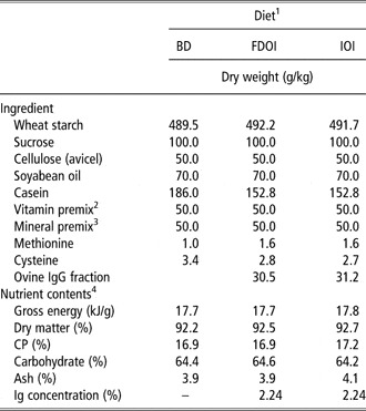
Three semi-synthetic diets were formulated: (1) a basal casein-based diet containing no Ig; (2) the basal casein-based diet supplemented with FDOI; and (3) the basal casein-based diet supplemented with IOI. The ingredient composition and determined nutrient composition of the diets are shown in Table 1. All diets exceeded the recommended nutrient requirement levels for the growing rat for all nutrients (National Research Council, NRC, 1995). The FDOI and IOI fractions were included in the respective diets at a concentration approximately equal to the amount of spray-dried porcine plasma (SDPP) IgG used in previous studies (Pierce et al., Reference Pierce, Cromwell, Lindemann, Russell and Weaver2005; Balan et al., Reference Balan, Han, Rutherfurd, Singh and Moughan2009).
Ingredient composition and determined nutrient and energy contents of the experimental diets

BD = basal diet; FDOI = freeze dried ovine immunoglobulin; IOI = inactivated ovine immunoglobulin; IgG = immunoglobulin G.
1All diets were formulated to meet or exceed the nutrient requirements for the growing rat.
2The mixture supplied (mg/kg diet): retinol acetate 5.0, dl-α-tocopheryl acetate 200, menadione 3.0, thiamin hydrochloride 5.0, riboflavin 7.0, pyridoxine hydrochloride 8.0, d-pantothenic acid 20, folic acid 2.0, nicotinic acid 20, d-biotin 1.0, myo-inositol 200, choline chloride 1500; (μg/kg diet) ergocalciferol 25, cyanocobalamin 50.
3The mixture supplied (g/kg diet): calcium hydrogen phosphate 6.29, magnesium oxide 1.06, potassium chloride 5.24, sodium chloride 1.97; (mg/kg diet) chromic potassium sulphate 1.97, cupric corbonate 10.7, ferric citrate 424, manganese sulphate 78.0, zinc oxide 48.2; (μg/kg diet) cobalt chloride 29.0, potassium iodate 151, ammonium molybdate 152, sodium selenite 151.
4Values for nutrients were from chemical analysis except for carbohydrate that was calculated by difference using the formula (100 − (moisture + ether extract + ash + CP) = carbohydrate).
Animal study
This work was approved by the Massey University Animal Ethics Committee (MUAEC 09/29) and procedures complied with the New Zealand Code of Recommendations and Minimum Standards for the Care and Use of Animals for Scientific purposes (New Zealand Animal Welfare Advisory Committee, 1995).
Sixty Sprague–Dawley male rats (8 weeks of age) were housed individually in stainless steel cages in a room maintained at 22 ± 2°C with a 12 h light/dark cycle. The animals had unrestricted access to water at all times. During the first week, the rats were acclimatized to the cages and received the basal diet. The diet was provided ad libitum as a dry powder. At the end of the acclimatization period, the rats were randomly allocated to the four experimental treatments such that there were 15 rats per treatment. The experimental treatments were as follows: (1) non-gavaged rats fed the basal diet (BD); (2) rats gavaged with S. enteritidis and fed BD; (3) rats gavaged with S. enteritidis and fed the FDOI-containing diet; and (4) rats gavaged with S. enteritidis and fed the IOI-containing diet. The rats received their respective diets ad libitum for 18 days (experimental period). On day 15, the rats on treatments 2, 3 and 4 were gavaged with a single dose of S. enteritidis ATCC 13076 (1 ml of saline solution containing 3% sodium bicarbonate with 1 × 10⁷ viable S. enteritidis). Prior to use, S. enteritidis was subcultured three times in LB (Luria-Bertani) broth (Difco, USA). The exact viable count of S. enteritidis inoculum was determined by plating on Brilliant Green Agar (Oxoid, England). The body weights (BWs) of the rats were measured at days 1, 7, 14, 15, 16, 17 and 18 of the experimental period, and the food intake was recorded daily. Average daily gain (ADG) and average daily feed intake (ADFI) were determined. Gain : feed ratio (GF) was calculated by dividing the daily gain by the daily feed intake. Since many of the gavaged rats showed symptoms of S. enteritidis infection, including nasal discharge, and red crusts around the eyes and nostrils after 3 days of gavage, for ethical reasons the study was culminated after 3 days of challenge. However, the effects of S. enteritidis were expected to be acute and marked and a 3-day challenge period was considered adequate to test the effects of Ig.
On the final day (day 18) of the experimental period, the rats were anaesthetized by an intra-peritoneal injection (0.1 ml/100 g live weight) of a mixture containing acepromazine maleate BP (British Pharmacopoeia) (ACP, 0.2%, w/v) two parts, ketamine (10%, w/v) five parts, xylazine (10%, w/v) one part and sterile water two parts. Rats were then killed by exsanguination.
The stomach of each rat was removed and its contents emptied. The tissues were then dried with absorbent paper and weighed. The entire small intestine from the pylorus to the ileocaecal valve was removed, dissected free of mesenteric attachments and divided approximately into the duodenum (the upper portion 11 cm from the pylorus end), and the rest was divided into two parts, that is, the jejunum (the central portion of the small intestine) and the ileum (the portion anterior to the ileocaecal valve). The contents were flushed out using phosphate buffer saline. The tissues were then blotted dry and weighed as described above. The same procedure was followed for the caecum and colon. The liver and spleen were also removed and weighed. The remaining portion of the animal was weighed and is defined here as being the empty BW. The relative weights of the organs were calculated with respect to empty BW.
Chemical analysis
Dietary dry matter, ash, crude protein (CP), total fat and gross energy were determined according to methods described by the Association of Official Analytical Chemists, AOAC (2003). Carbohydrate was determined by difference using the formula (100 − (moisture + ether extract + ash + CP) = carbohydrate).
Gut morphology
Tissue sections, 2 cm in length, were collected from the middle of the duodenum, jejunum and ileum and immediately placed (and subsequently stored) in 10% buffered formalin for 24 h. After fixation, each tissue was excised, dehydrated, and embedded in paraffin wax. From each tissue, transverse sections were cut, stained with haematoxylin, eosin and alcian blue and examined under a light microscope. The five longest and straightest villi and their associated crypts from each transverse tissue section were selected from 10 rats for each experimental treatment, and the villus height, crypt depth and villus surface area (VSA) for each section measured as described previously (Balan et al., Reference Balan, Han, Rutherfurd, Singh and Moughan2009). Colon tissue samples were prepared as described for the small intestine samples and the crypt depths were measured for five elongated and straight crypts from each transverse tissue section selected from 10 rats for each experimental treatment.
Intact goblet cell count
Goblet cells were measured in intact villi (from the tip of the villus to the villus-crypt junction) and crypts (from the villus-crypt junction to the base of the crypt). Positive staining with Alcian blue indicated goblet cells containing acid mucins. For the duodenum, jejunum and ileum, the five longest and straightest villi and their associated crypts from each transverse tissue section of 10 randomly selected rats in each group were quantified. For the colon tissue, goblet cells in crypts were measured from five elongated and straight crypts from each transverse tissue section of 10 randomly selected rats in each group.
Enzyme-linked lectin assay (ELLA) for mucin
Ileal and colonic digesta of ten randomly selected rats from each group were freeze-dried and assayed for glycosylated mucin by ELLA using wheat germ agglutinin (WGA) as the lectin, as described by Trompette et al. (Reference Trompette, Blanchard, Zoghbi, Bara, Claustre, Jourdan, Chayvialle and Plaisance2004) with some modification. Porcine gastric mucin (Sigma, Auckland, New Zealand) was used as the standard. Briefly, various dilutions of standards and samples were prepared in carbonate buffer (0.5 M, pH 9.6) and coated on 96-well microtiter plates (NUNC microplates, NUNC A/S, Roskilde, Denmark). After incubating overnight at 4°C, the plates were washed three times with PBS (phosphate buffered saline) Tween (PBS-T, pH 7.2). Microplates were then blocked with 300 μl of a PBS-T solution containing bovine serum albumin (BSA, 5 g/l, Sigma, Auckland, New Zealand; PBS-T-BSA) and incubated for 1 h at 37°C. Plates were washed again, and 100 μl of biotinylated WGA (Sigma, Auckland, New Zealand) in PBS-T-BSA was added and the plates were incubated for 1 h at 37°C. The plates were then washed and incubated with 100 μl of avidine-peroxidase conjugate (Sigma, Auckland, New Zealand) for 1 h at room temperature. After washing, 100 μl OPD solution (Sigma) was added to each well and the plates incubated in the dark at room temperature for 10 min. The reaction was stopped by adding 50 μl of sulphuric acid (1 M). Absorbance was read at 492 nm using an ELISA reader (Anthos 2010; Austria). Glycosylated mucin concentration in samples was calculated with reference to a porcine gastric mucin standard curve. Data were expressed as μg or ng mucin per ml digesta.
Statistical analysis
Results are presented as means ± s.e.m. Data were analysed using a one-way ANOVA (GLM procedures of SAS, version 9.1, SAS Institute, Cary, NC, USA) and means were compared using Tukey's method. ADG, ADFI and GF were also analysed before and after gavage with S. enteritidis using a repeated measures analysis (SAS). For the goblet cell numbers, the Mann–Whitney U-test was used. For a comparison of the results obtained from the four groups, the Kruskal–Wallis test was applied; P < 0.05 was considered to be statistically significant.
Results
Growth performance
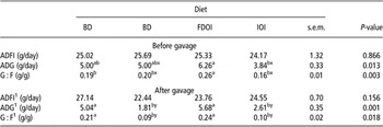
ADFI, ADG and GF were determined for the period prior to gavaging with S. enteritidis (days 1 to 14) and after gavaging with S. enteritidis (days 15 to 18) and are shown in Table 2. Prior to gavaging, there was no significant (P > 0.05) effect of diet on ADFI. For ADG, there was no significant (P < 0.05) difference between the rats fed the BD and FDOI diets; however, ADG was significantly (P = 0.013) lower for the rats receiving the IOI diet compared to the rats fed the FDOI diet. G : F ratio was higher (P = 0.003) for the rats consuming the FDOI diet than for those fed the BD or IOI diet. After gavaging with S. enteritidis, ADFI was not significantly (P > 0.05) different among treatments but ADG and GF (3 days) were significantly (P = 0.001 and P = 0.018, respectively) and markedly lower for the gavaged rats fed the BD diet when compared to the ungavaged rats receiving the BD diet and there was no significant (P > 0.05) difference between the gavaged rats fed either BD or IOI. In contrast, the ADG and GF were significantly (P > 0.05) higher for the gavaged rats fed the FDOI diet in comparison with the gavaged BD- and IOI-diet fed rats. When comparing the pre- and post-gavaging periods, gavaging with S. enteritidis resulted in a significant (P < 0.05) lowering of both ADG and GF for the rats fed either BD or IOI, but this effect was not observed (P > 0.05) in rats fed the FDOI diet.
Feed intake and growth performance of rats before and after gavaging (3 days) with Salmonella enteritidis

BD = basal diet; FDOI = freeze dried ovine immunoglobulin; IOI = inactivated ovine immunoglobulin; ADFI = average daily feed intake; ADG = average daily gain.
1Final 3 days without gavaging for rats fed the BD diet.
Values are means ± s.e.m (n = 15), a,bmeans in a row without a common superscript differ and x,ymean in a column (pre- and post-gavaging) for same variable without a common superscript differ at P < 0.05.
Organ weights
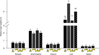
The mean selected organ weights expressed relative to empty BW of the rats on the final day of the study are shown in Figure 1. Gavaging with S. enteritidis resulted in a significantly (P < 0.05) lower stomach and small intestine and higher caecum, liver and spleen weights for the rats fed the BD diet compared to ungavaged rats fed the BD diet. Furthermore, the organ weights in the gavaged rats fed the IOI diet were not significantly (P > 0.05) different from those in the gavaged rats fed the BD diet. In contrast, the weights of the organs in the gavaged FDOI-fed rats were not significantly (P > 0.05) different from those in the ungavaged BD diet fed rats but were significantly (P < 0.05) different from the gavaged BD- and IOI-diet fed rats.
(colour online) Mean (n = 15) relative organ weights (%, empty BW) for the growing rat fed diet BD and the growing rat gavaged with Salmonella enteritidis and fed diet BD, FDOI or IOI. BD = basal diet; FDOI = freeze-dried ovine immunoglobulin; g, rats gavaged with S. enteritidis; IOI = inactivated ovine immunoglobulin and fed with inactivated ovine Ig. a,bMeans in an organ without a common superscript differ at P < 0.05.

Gut morphology
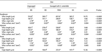
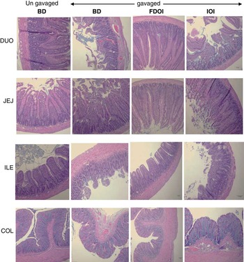
Gut morphology data are shown in Table 3 and stained transverse sections of gut tissue in Figure 2. The villus length, crypt depth, villus : crypt ratio and VSA of the duodenum and jejunum were generally significantly (P ⩽ 0.001) greater in rats gavaged with S. enteritidis and receiving the FDOI diet compared to either the ungavaged rats fed the BD diet (except duodenal and jejunal VSA) or the gavaged rats fed the BD or IOI diet. In the ileum, gavaging with S. enteritidis resulted in significantly (P < 0.001) lower villus length, villus : crypt ratio and VSA for the gavaged rats receiving the BD, FDOI or IOI diet when compared to the ungavaged rats fed the BD diet. In contrast, gavaged rats fed the FDOI diet had greater crypt depth than the rats in the other treatment groups. The colon crypt depth was also significantly greater (P < 0.001) in the ungavaged rats receiving the BD diet and the FDOI diet-fed gavaged rats when compared to the gavaged rats fed the BD or IOI diet. In the stained transverse sections, severe mucosal histological damage (villus shortening and fusion, crypt loss, inflammatory infiltration) was found for the gavaged rats fed the BD diet when compared to the ungavaged rats receiving the BD diet and a similar trend was observed for the gavaged IOI-fed rats. In contrast, these effects were not observed for the gavaged rats fed the FDOI diet.
Intestinal morphology characteristics in the growing rat after gavaging Salmonella enteritidis

BD = basal diet; FDOI = freeze dried ovine immunoglobulin; IOI = inactivated ovine immunoglobulin; VSA = villus surface area.
1VSA (mm2) = (3.14 × mh × h) + 3.14 × (mh/2)2, where mh (μm) = villus width at midvillus height, h (μm) = villus height measured from the tip of the villus to the villus-crypt junction and crypt depth is measured from villus-crypt junction to the base of the crypt.
Values are means ± s.e.m. (n = 10), a,b,cmeans in a row without a common superscript differ at P < 0.05.
(colour online) Transverse sections of the rat duodenum (DUO), jejunum (JEJ), ileum (ILE) and colon (COL). Section was stained with haematoxylin, eosin and alcian blue. BD = basal diet; FDOI = freeze-dried ovine immunoglobulin; IOI = inactivated ovine immunoglobulin.

Goblet cell count
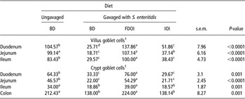
Goblet cell count data are shown in Table 4. The number of goblet cells in the villi and crypts of duodenal, jejunal and ileal segments was significantly (P < 0.05) lower for the gavaged rats receiving diet BD compared to their ungavaged counterparts. This lowering of goblet cell numbers was completely overcome for the gavaged rats fed the FDOI diet. Moreover, the latter rats had a significantly (P < 0.05) higher number of goblet cells compared to ungavaged rats fed the BD diet in villi and crypts for some of the intestinal segments.
Goblet cell counts in the intestine of growing rats after gavaging with Salmonella enteritidis

BD = basal diet; FDOI = freeze dried ovine immunoglobulin; IOI = inactivated ovine immunoglobulin.
1Number of goblet cells per five complete villi and their associated crypts.
Values are means ± s.e.m. (n = 10), a,b,cmeans in a row without a common superscript differ at P < 0.05.
Quantification of luminal N-Acetylglucosamine (GlcNAc) glycosylated mucin protein
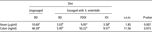
Data for the glycosylated mucin protein contents of intestinal digesta are shown in Table 5. In the ileum and colon, the glycosylated mucin protein content was significantly (P < 0.05) lower for the rats gavaged and fed either the BD or IOI diet than for the ungavaged rats fed the BD diet. In contrast, in both segments, for the gavaged rats fed the FDOI diet, the glycosylated mucin protein content was significantly (P < 0.05) higher than for the gavaged rats fed either the BD or IOI diet, but was not different (P > 0.05) from the ungavaged rats fed the BD diet.
Concentration of GlcNAc glycosylated mucin protein in the ileal and colonic luminal digesta for the growing rat following gavaging with Salmonella enteritidis
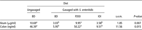
BD = basal diet; FDOI = freeze dried ovine immunoglobulin; IOI = inactivated ovine immunoglobulin.
Values are means ± s.e.m. (n = 10), a,b,cmeans in a row without a common superscript differ at P < 0.05.
Discussion
Prior to S. enteritidis administration, rats fed the FDOI diet had a greater G : F ratio than the rats fed either the BD or IOI diet. This finding is consistent with the earlier findings of Balan et al. (Reference Balan, Han, Rutherfurd, Singh and Moughan2009) who reported that rats fed an FDOI-based diet had a higher G : F ratio than rats fed with a BD containing no Ig or a diet containing IOI.
The experimental period during which time the rats were gavaged with S. enteritidis was restricted to 3 days for ethical reasons. After gavaging with S. enteritidis over 3 days, the decrease in ADG and feed conversion efficiency were drastic and highly statistically significant. In contrast, feed intake and water intake (data not shown) did not vary (P > 0.05) across the diets. Although ADG declined, we have no information on the composition of the BW loss. This relatively short gavaging period was deemed to be sufficient since the effects of S. enteritidis were expected to be acute. The dramatic effects of gavaging on the BD-diet-fed rats compared to the ungavaged counterparts observed for most of the parameters measured in this study clearly support this assumption. It is worth emphasizing, however, that the results discussed here relate to the acute (over 3 days) effects of the pathogen. After gavaging with S. enteritidis, the rats fed the IOI and BD diets grew considerably more slowly than either the gavaged rats fed the FDOI diet or the ungavaged rats fed the BD diet. Furthermore, the gavaged FDOI diet-fed rats and the ungavaged BD diet-fed rats grew at the same rate, suggesting that dietary Ig may be able to prevent colonization of S. enteritidis in the first place or reduce the S. enteritidis load post-challenge to that of a normal ungavaged animal. When comparing the pre- and post-gavage periods, the ADG of the gavaged rats fed either the BD or IOI diet decreased by 64% and 32%, respectively. Similarly, the G : F ratio of the same rats decreased by 55% and 27%, respectively. In contrast, for the ungavaged rats fed the BD diet and the gavaged rats fed the FDOI diet, there was no significant reduction in growth performance between the pre- and post-gavage periods. These results clearly show that the dietary FDOI first enhanced the growth performance of the normal rat and then completely negated any effects of S. enteritidis administration on growth, at least in the short term (3 days following pathogen administration).
Similar results have been reported in other studies using healthy and infected animals and given diets containing FDOI (Balan et al., Reference Balan, Han, Rutherfurd, Singh and Moughan2009), SDPP (Gatnau et al., Reference Gatnau, Cain, Drew and Zimmerman1995; Owen et al., Reference Owen, Nelssen, Goodband, Tokach, Friesen, Ricert, Smith and Russell1995) and spray-dried animal plasma (SDAP) (Corl et al., Reference Corl, Harrell, Moon, Phillips, Weaver, Campbell, Arthington and Odle2007). Owusu-Asiedu et al. (Reference Owusu-Asiedu, Baidoo, Nyachoti and Marquardt2002) reported effects of SDAP and SDPP supplemented with or without egg yolk antibodies in piglets challenged with enterotoxigenic E. coli (ETEC) strain F18. Since the growth performance, visceral organ weights and small intestine histology were not affected (P > 0.05) by dietary treatment, they suggested that SDPP and SDAP contain specific anti-ETEC antibodies, which is one of the factors responsible for its superior growth-enhancing effects in the presence of pathogen challenge. Pierce et al. (Reference Pierce, Cromwell, Lindemann, Russell and Weaver2005) conducted a comprehensive study examining the effects of dietary SDPP, SDBP (spray-dried bovine plasma), porcine Ig and bovine Ig in healthy, early-weaned piglets and showed that all Ig preparations resulted in higher growth rates. They concluded that the improved growth performance was due to a nutrient-sparing effect as a result of the action of the consumed Ig. Johnson (Reference Johnson1997) reported that oral administration of antigens such as either pathogenic or non-pathogenic organisms induced production of the pro-inflammatory cytokines IL-1, IL-6 and TNF-α and acute phase proteins, thereby increasing the demand for amino acids for non-growth functions and reducing overall animal growth. Consequently, a lowered activation of the immune system may lead to a greater amount of dietary nutrients and energy being available for growth (Coffey and Cromwell, Reference Coffey and Cromwell1995).
As for BW gain, administration (3 days) of the pathogen had an immediate and marked effect upon the weights of certain digestive organs and the spleen. The small intestine and colon of the rats gavaged with S. enteritidis and receiving the FDOI-containing diet were heavier (possibly due to the presence of more, and larger, villi) when compared to similarly gavaged rats fed the control diet but were the same weight as those for the ungavaged rats fed diet BD. In addition, in this study the weight of the caecum, liver and spleen were higher in the gavaged rats fed either the BD or IOI diet compared to gavaged rats fed the FDOI diet or ungavaged rats fed the BD diet. Some of our results are similar to the recent findings of Lu et al. (Reference Lu, Wu, Liu, Xia, Zhang and Sun2010) who found that mice gavaged with S. tyhpimurium had a significant increase in both liver and spleen weights. Feeding the FDOI diet prophylactically may have reduced the S. enteritidis attachment to the gut wall, which may have avoided infection and thus led to the normal weights observed for the liver and spleen in the FDOI pathogen-challenged rats.
In this study, gavaged rats fed the FDOI diet had longer villi, greater crypt depths and greater villi surface area in most of the small intestinal sections, which may have resulted in a greater absorption of available nutrients from the gut leading to the higher growth rate observed for the FDOI-fed S. enteritidis – gavaged rats. In contrast, gavaging with S. enteritidis appeared to cause acute villus atrophy for those rats fed either BD or IOI. This was similar to the findings of Gatnau et al. (Reference Gatnau, Cain, Drew and Zimmerman1995) who reported that feeding either SDPP or its corresponding Ig fraction to healthy weaned pigs resulted in an increased villus surface area. Balan et al. (Reference Balan, Han, Rutherfurd, Singh and Moughan2009) showed that in normal rats fed the FDOI-based diet, there was an increase in the villus length, crypt depth, villus : crypt ratio and VSA compared to similar rats fed diet BD (containing no Ig) or a diet containing inactivated ovine Ig.
In this study, gavaging with S. enteritidis dramatically and immediately reduced the amount of GlcNAc glycosylated mucin in the gut lumen and this was likely caused, at least in part, by the observed reduction in the number of villi and crypt goblet cells in the small and large intestine. These results are consistent with the findings of other workers (Lambert et al., Reference Lambert, Schofield, Ironside and Mandal1979). In contrast, dietary freeze-dried ovine Ig, but not inactivated ovine Ig, appeared to completely negate the negative effects of S. enteritidis on mucin content and goblet cell numbers. The potential effects on the rate of mucin breakdown and reabsorption may also underline these observed differences. It should be noted that in the presently described work, only GlcNAc glycosylated mucins were determined, and changes in other forms of mucins cannot be excluded.
Mack et al. (Reference Mack, Michail, Wei, McDougall and Hollingsworth1999) in in vitro work have reported that dietary supplementation with probiotics inhibits enteropathogenic E. coli adherence to intestinal epithelial cells by increasing intestinal mucin gene (Muc2 and Muc3) expression. This is similar to our previous finding (Balan, Reference Balan2010) where we showed that feeding ovine Ig to healthy rats resulted in an increase in beneficial bacteria such as Lactobacillus sp. (in both healthy and S. enteritidis-gavaged rat models) and Muc2, Muc3 and Muc4 gene expression in the ileum. FDOI-based diets have also been shown to increase villi and crypt goblet cell numbers throughout the gut in healthy rats (Balan, Reference Balan2010). A decline in goblet cell numbers after infection has also been reported in a murine model infected with Citrobacter rodentium (King et al., Reference King, Morel, Pluske and Hendriks2008; Lindén et al., Reference Lindén, Florin and McGuckin2008).
In conclusion, it appears that feeding freeze-dried ovine Ig to rats negated the negative impact of orally administered S. enteritidis to such a degree that the growth performance of the S. enteritidis gavaged rats fed FDOI, at least in the short term, was the same or superior to that in normal ungavaged rats.