The herpesviridae family is subclassified into three subfamilies (the alpha-, beta- and gamma-herpesvirinae), based originally on biological properties and, more recently, on genome sequence and structure (Ref. Reference McGeoch, Rixon and Davison1). Herpesviruses have been found in most animal species examined and, to date, eight have been identified in humans. Although the biology of herpesviruses is quite varied, one common property is their ability to persist for the lifetime of the host as a latent infection.
Human cytomegalovirus (HCMV) is the prototypic member of the beta-herpesvirinae subfamily and has one of the largest genomes of all herpesviruses at 235 kb. HCMV causes a widespread persistent infection in humans: seroprevalence for this virus is tightly linked to socioeconomic status and can be up to 90% for some populations. In common with all herpesviruses, primary infection with HCMV results in an initial productive infection resulting from extensive transcription of the viral genome, viral DNA replication and release of infectious virus. This rarely results in clinical disease if the host is immunocompetent (Ref. Reference Zanghellini2). The lytic transcription programme comprises a temporal cascade of viral gene expression termed immediate early (IE), early (E) and late (L), with IE genes playing a pivotal role in the induction of E and L viral gene expression and optimising the cellular environment for viral DNA replication and virus production (Ref. Reference Stenberg3). However, subsequent to primary infection, HCMV can persist as a latent infection resulting from transcription of only a few viral genes and carriage of the viral genome in the absence of infectious virus production (Ref. Reference Sinclair and Sissons4). The identity and function of these latency-associated transcripts is far from complete and is the subject of intense investigation by a number of laboratories (Ref. Reference Sinclair5). Importantly, latent virus can routinely reactivate, which can result in sporadic production of infectious virus (Fig. 1).
Human cytomegalovirus latent and lytic infection. (a) Lytic infection results from extensive viral gene expression (green arrows) from all regions of the viral genome, shown as long unique (UL) and short unique (US) regions. This program of viral gene expression results in viral DNA replication and virus production. By contrast, latent infection results from a minimal transcription program in which viral immediate early (IE) expression is repressed and only specific latency-associated genes are transcribed. Latent viral genomes can reactivate (dotted arrow) by induction of the lytic transcription program resulting in full induction of the lytic cycle. (b) Model of HCMV latency in vivo. Following primary infection, HCMV establishes a latent infection in CD34+ bone marrow progenitor cells. The viral genome persists in the CD34+ cells in the absence of viral lytic gene expression. However, upon differentiation of CD34+ cells to a mature DC phenotype, reactivation of lytic gene expression occurs from the previously quiescent genome. This ultimately results in productive infection and the release of infectious virus progeny.

Primary infection or reactivation of HCMV in an immunocompetent individual rarely results in clinically obvious disease. However, high morbidity and mortality can occur in patients with a suboptimal immune response, and clinical disease is frequently observed when infection occurs in solid organ or bone marrow transplant recipients (Refs Reference Rubin6, Reference Drew7, Reference Falagas and Snydman8, Reference Griffiths, Clark and Emery9, Reference Yoshikawa10). Reactivation of virus from a seropositive solid organ graft can lead to disease, particularly in seronegative recipients, but serious disease can also result from reactivation of the recipient's own latent virus after a seronegative bone marrow graft (Refs Reference Hughes11, Reference Stratta12).
Recurrent HCMV infection or HCMV reactivation produces a range of complications resulting in significant morbidity and mortality in individuals with immunosuppression due to human immunodeficiency virus (HIV)/AIDS (Refs Reference Stewart13, Reference Griffiths14). Indeed, HCMV represents one of the most frequent opportunistic infections in HIV/AIDS patients, although the use of highly active antiretroviral therapy (HAART) has led to a substantial reduction in the severity of HCMV disease (Refs Reference Deayton15, Reference Varani16, Reference Steininger, Puchhammer-Stockl and Popow-Kraupp17).
As well as immmunosuppressed individuals, HCMV infection represents a particular problem for the immunologically naive fetus. Infection of the fetus during pregnancy occurs transplacentally, either from a primary infection of the mother or recurrent reactivation of latent infection, and can lead to congenital deafness and neurological disorders in the newborn. Infections in early pregnancy result in the worst outcome (Refs Reference Fowler18, Reference Gaytant19, Reference Adler20, Reference Griffiths21) and HCMV is the most common infective cause of mental handicap (Ref. Reference Hassan and Connell22). Infection with HCMV can also be acquired perinatally from cervical secretions during birth or postnatally from breast milk, although this is routinely less severe because of the presence of maternal antibodies to the virus (Refs Reference Stagno23, Reference Numazaki24).
The importance of HCMV as a human pathogen is underscored by the fact that a study by the US Institute of Medicine, which gave particular weighting to quality life years (QUALYS) lost in consequence of an infection, concluded that HCMV infection was one of the highest priorities for vaccine development (Ref. Reference Arvin25).
Targets of human cytomegalovirus infection in vivo and in vitro
Upon infection in immunocompromised individuals, HCMV antigens can be detected in several cell types, including fibroblasts, epithelial cells, endothelial cells and vascular smooth muscle cells, suggesting that there is little tissue specificity for virus infection (Ref. Reference Sinzger26). However, some cell types, such as B cells and T cells (Ref. Reference Sinzger26), do not show any evidence of HCMV infection, whereas others, including polymorphonuclear cells, phagocytose viral particles or take up virus during coculture with other infected cells, but do not themselves support productive infection (Refs Reference Grefte27, Reference Skarman28).
Many of the in vivo observations of virus infection and cell specificity are reflected in vitro. Cultured primary fibroblasts, endothelial cells, smooth muscle cells and retinal pigment epithelial cells are all fully permissive for HCMV infection – although the level of infection varies and is very dependent on the virus isolate used. For example, laboratory-adapted strains of HCMV, routinely grown for extended periods in primary human fibroblasts, do not grow in myeloid and endothelial cells because of mutation or loss of the region encoding UL128–UL131A from the viral genome. By contrast, clinical isolates of virus grown in endothelial cells, or for only a limited time in fibroblasts, efficiently infect myeloid and endothelial cells (Refs Reference Sinzger29, Reference Hahn30, Reference Gerna31, Reference Wang and Shenk32, Reference Cha33).
Finally, some cell types, particularly cells of the early myeloid lineage such as bone marrow progenitor cells and monocytes, take up virus but do not support viral replication because of differentiation-specific repression of the viral IE gene (Refs Reference Rice, Schrier and Oldstone34, Reference Ibanez35, Reference Lathey and Spector36, Reference Sinclair37, Reference Reeves38, Reference Maciejewski and St Jeor39, Reference Kondo, Kaneshima and Mocarski40). Such repression of viral lytic transcription is believed to underpin the mechanism by which these cell types act as sites of latent infection in vivo (Ref. Reference Sinclair5). However, terminally differentiated cells of the myeloid lineage, including some dendritic cell (DC) subsets, are sites of productive infection of HCMV (Refs Reference Ibanez35, Reference Lathey and Spector36, Reference Reeves38, Reference Maciejewski and St Jeor39, Reference Kondo, Kaneshima and Mocarski40, Reference Riegler41, Reference Hertel42). Consistent with this permissiveness of DCs for experimental infection, DCs are also sites of reactivation of HCMV from latency: myeloid progenitor cells carry latent HCMV in vivo (Refs Reference Sinclair and Sissons4, Reference Sinclair5, Reference Bego and St Jeor43) and differentiation of these cells into DCs reactivates the latent virus (Ref. Reference Reeves38).
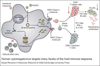
This strategy of persistence of HCMV as a result of long periods of latent infection and intermittent reactivation helps to prevent clearance of virus from the infected individual and results in the coexistence of virus and host. It is also underpinned by the ability of HCMV to be carried in vivo, despite potent antiviral responses. This ability of HCMV to avoid host immune surveillance relies on the expression of several viral genes that specifically target critical host immune functions (Fig. 2). Virus gene products have been identified that can disrupt antigen presentation to T cells, inhibit natural killer cell activity and blunt other facets of the innate antiviral immune response (Refs Reference Wilkinson44, Reference Mocarski45, Reference Mocarski46). These effects arguably have the greatest effect in DCs – the professional antigen-presenting cells of the immune system. This review will discuss the effects of HCMV infection on DCs and in particular how DC function is modified to prevent initiation of their normal potent antiviral immune response in the human host.
Human cytomegalovirus targets many facets of the host immune response. HCMV enters the cell where viral structural proteins are processed by the endolysosome to generate viral polypeptides that are presented to T cells via MHC Class II. Alternatively, virus can be transported to the nucleus where extensive transcription occurs resulting in de novo expression of viral proteins. These can be processed through the cellular proteasome resulting in presentation of viral polypeptides via MHC Class I. HCMV infection of target cells inhibits (red crosses) the interferon response and natural killer cell recognition. Inhibition of presentation of viral peptide antigen mediated by MHC Class I and Class II also ultimately prevents (red arrows) stimulation of CD4+ and CD8+ T cells, inhibiting the Th1 and Th2 immune response (Refs Reference Riegler41, Reference Hertel42, Reference Bego and St Jeor43). Abbreviations: HCMV, human cytomegalovirus; IFN-γ, interferon-gamma; IL-2, interleukin-2; MHC, major histocompatibility complex.

Dendritic cells – sentinels of the immune system
Although all DCs are believed to arise from leukocytes derived from the haematopoietic compartment, they are not represented by any single uniform population of cells: location, phenotype and specific immune function identify particular DC subsets (Fig. 3). The ability to study DCs directly in vivo has long been hampered by the relative paucity of these cells in different tissues and hence, difficulty of isolation. However, our understanding of DC phenotype and function has been substantially advanced by the ability to culture DC progenitors ex vivo and differentiate these cells into DC subsets, which closely resemble the phenotype of DC subtypes found in vivo (Refs Reference Gatti47, Reference Strobl48, Reference Romani49, Reference Sallusto and Lanzavecchia50, Reference Strobl51). Other specific DC subsets can be isolated directly from peripheral blood (Refs Reference Kvale52, Reference O'Doherty53).
Location, phenotype and immune function identify specific dendritic cell subsets. CD34+ stem cells give rise to myeloid or lymphoid progenitors, which produce two major dendritic cell (DC) subtypes, CD11c+ (DC1) myeloid-derived DCs and CD11c– lymphoid-derived DCs (DC2). Myeloid DCs include interstitial DCs, found in the dermis and submucosa and Langerhans DCs, which reside in the dermis and mucosal epithelia. Langerhans-like DCs can be generated ex vivo from culture of CD34+ progenitors and interstitial-like DCs can be generated ex vivo from peripheral blood monocytes. Lymphoid progenitors give rise to plasmacytoid DCs, which can be isolated directly from peripheral blood. One other subset, the CD11c+ blood myeloid DCs, can also be isolated directly from peripheral blood. Although phenotypically most similar to interstitial DCs, they are not identical (Refs Reference Pepperl-Klindworth106, Reference Moutaftsi107).

In humans, two major DC subsets are found (Refs Reference Cella, Sallusto and Lanzavecchia54, Reference Ueno55), CD11c+ myeloid DCs (DC1) and CD11c– lymphoid DCs (DC2). The lymphoid DC subset includes the plasmacytoid DCs (pDCs) which are CD123+, are found in the circulation and characteristically secrete large amounts of type I interferons in response to viral DNA or RNA. The myeloid DC subset contains two types of DCs: Langerhans DCs, which reside in the epidermis and stratified mucosal epithelia and interstitial DCs, which reside in the dermis and other deep mucosal tissues (Refs Reference Romani56, Reference Banchereau57, Reference Caux58). All myeloid DCs secrete IL-12 upon activation (Refs Reference Gatti47, Reference Langenkamp59, Reference Steinman and Hemmi60).
In most tissues, DCs are present as immature cells that are unable to efficiently present antigen to T cells. This inability to stimulate antigen-specific T cell responses stems from the fact that these immature DCs do not express, or have low levels of key cell surface molecules such as CD80, CD86 and CD40, which are responsible for binding and stimulating T cells; they also have low levels of surface MHC molecules, which present peptide antigen to T cells. This low-level cell surface expression of T cell costimulatory molecules may result from both transcriptional and post-transcriptional control (Ref. Reference Granucci61). MHC Class II is present at high levels in immature DCs, but in intracellular compartments rather than on the cell surface (Ref. Reference Stern, Potolicchio and Santambrogio62).
Immature DCs respond to inflammatory chemokines and cytokines that are often secreted at sites of pathogen invasion, and traffic to these sites where they capture antigen and sense the invading micro-organisms through several pattern recognition receptors (PRRs) (Refs Reference Janeway and Medzhitov63, Reference Iwasaki and Medzhitov64). For example, pDCs routinely respond to viral pathogens through PRRs such as Toll-like receptor 7 (TLR7) and TLR9 (Refs Reference Ito, Liu and Kadowaki65, Reference Asselin-Paturel66), which sense viral DNA and RNA. This drives high levels of type I interferon secretion, which can initiate a potent antiviral cascade in many cell types (Ref. Reference Randall and Goodbourn67), activate natural killer cells (Refs Reference Katze, He and Gale68, Reference Colonna, Trinchieri and Liu69) and DCs. Langerhans DCs or interstitial myeloid DCs, which do not express TLR9, respond to pathogens by other PRRs (Ref. Reference Ueno55), activating IL-12 secretion and driving DC maturation.
DC activation substantially increases cell surface expression of MHC Class I and II, several T cell costimulatory molecules such as CD80 and CD86 (Refs Reference Granucci70, Reference Lanzavecchia and Sallusto71), as well as specific lymph-node-homing chemokine receptors such as CCR7 (Refs Reference Sallusto and Lanzavecchia72, Reference Sozzani73, Reference Caux74). As these DCs migrate to secondary lymphoid tissues they mature, whereupon they are able to efficiently present antigen to T cells, which stimulates antigen-specific T cell proliferation and the initiation of a potent B cell- and T cell-specific immune response to the invading pathogen (Fig. 4) (Refs Reference Cella, Sallusto and Lanzavecchia54, Reference Lanzavecchia and Sallusto71, Reference Tan and O'Neill75).
Immature dendritic cells generate dendritic cells that can present antigen to T cells in lymph nodes. Tissue-resident or blood-resident immature dendritic cells expressing high levels of receptors such as CCR1 and CCR5 migrate (black arrow) to sites of pathogen invasion by responding to inflammatory cytokines (e.g. CCL1–CCL4) where they sense infection through pathogen recognition receptors (PRRs) such as Toll-like receptors (TLRs). This results in induction (green arrows) of cell surface expression of many T cell costimulatory molecules (e.g. CD80, CD86, CD83) and MHC Class I and Class II; increased expression of maturation cytokines, such as IL-12 and IL-6; chemokine receptor switching with inhibition (red arrow) of expression of inflammatory chemokine homing receptors, such as CCR1 and CCR5 and induction of secondary lymphoid tissue homing receptors such as CCR7. Cells then migrate (dotted arrow) to areas with high levels of CCL19 and CCL21 in lymph nodes and efficiently present antigen in the context of MHC Class I and Class II to T cells resulting in stimulation of CD8+ and CD4+ T cells and induction of potent Th1 and Th2 responses to presented antigen. Abbreviations: DC, dendritic cell; IL, interleukin; MHC, major histocompatibility complex.

The presence of immature DCs in the skin, blood and mucosal surfaces make them likely to be the first cells to contact viruses upon infection. Consequently, there has been considerable interest in the effects of HCMV infection on DCs and, in particular, the ability of the virus to prevent these sentinels from carrying out their normal immunological functions. Langerhans-like DCs and interstitial-like DCs from ex vivo differentiation of CD34+ haematopoietic progenitors or peripheral blood monocytes, respectively (Refs Reference Gatti47, Reference Strobl48, Reference Romani49, Reference Sallusto and Lanzavecchia50, Reference Strobl51), as well as pDCs directly from peripheral blood, have all been used to analyse the effects of HCMV infection on DC phenotype and function.
Effect of HCMV infection on interstitial-like dendritic cells
Infection of immature dendritic cells
Interstitial DCs naturally reside in the dermis and deep tissues, and substantial numbers can be generated from adherent peripheral blood mononuclear cells or CD14+ cells by ex vivo culture with IL-4 and GM-CSF (Refs Reference Sallusto and Lanzavecchia50, Reference Herbst76, Reference Gieseler77). This model has been used by a number of groups to analyse the effects of HCMV infection on this DC subset, which was generated in vitro but is relevant in vivo.
Although it was originally reported that monocyte-derived immature DCs were nonpermissive for viral IE expression upon infection (Ref. Reference Soderberg78), it is now generally accepted that these cells fully support productive HCMV infection (Ref. Reference Riegler41). The ability of DCs to bind virus appears, at least in part, to be mediated by interaction between viral glycoprotein B (gB) with the DC-specific lectin DC-SIGN (Ref. Reference Halary79), although viral entry also requires the UL128–UL131A genes (Ref. Reference Gerna31). This binding of infectious virions to DCs has also been suggested to mediate trafficking of infectious virions to other permissive cells (Ref. Reference Halary79).
HCMV infection of immature monocyte-derived DCs clearly has profound effects on their phenotype (Fig. 5). Instead of inducing maturation of these DCs, as might be expected from our knowledge of the effects of infection of other viruses (Refs Reference Janeway and Medzhitov63, Reference Macagno80), work from a number of laboratories has led to a consensus that HCMV infection of these cells results in downregulation of cell surface expression of key T cell costimulatory molecules such as CD86, CD80 and CD40, as well as MHC Class I at the very earliest times of infection and MHC Class II later in infection (Refs Reference Grigoleit81, Reference Beck82, Reference Senechal83, Reference Raftery84, Reference Moutaftsi85). US11 is one of the HCMV gene products that inhibits MHC Class I cell surface expression in a number of cell types (Refs Reference van der Wal, Kikkert and Wiertz86, Reference Tortorella87) including immature monocyte-derived DCs (Ref. Reference Rehm88). The mechanism by which virus downregulates MHC Class II in these cells has not yet been attributed to any specific viral gene product but probably targets multiple stages of MHC Class II maturation (Ref. Reference Kessler, Reich and Jahn89).
Human cytomegalovirus has pronounced effects on dendritic cells. (a) HCMV infection of immature myeloid dendritic cells results in: inhibition of CCR1 and CCR5 expression; inhibition of expression of T cell costimulatory molecules such as CD80, CD86, CD83 as well as MHC Class I and II, and reduction of CCR7 expression. All these HCMV-mediated effects (red crosses), generally, blunt the ability of dendritic cells (DCs) to migrate to lymph nodes and present antigen to T cells resulting in a lack of antigen-specific stimulation of T cells and inhibition of the Th1 and Th2 response. Many of these effects appear to depend on viral immediate-early or early gene expression-mediated induction of soluble factors, which include soluble cmvIL-10 and/or soluble CD83. (b) HCMV infection of mature plasmacytoid DCs (pDCs) results in changes in phenotype that resemble partial differentiation. This includes HCMV-mediated increases (red arrow) in expression of MHC Class II as well as increases in expression of T cell costimulatory molecules such as CD83 and increases in interleukin-6 (IL-6) and interferon-α (IFN-α). These HCMV-mediated effects blunt the ability of pDCs to present antigen to CD8+ T cells (red cross) resulting in a lack of antigen-specific stimulation of the Th1 response but induce (red arrow) T-cell-dependent B cell activation and stimulation.

Consistent with these effects, HCMV-infected immature monocyte-derived DCs are unable to stimulate T cell proliferation in a mixed lymphocyte reaction (Refs Reference Grigoleit81, Reference Beck82, Reference Raftery84, Reference Moutaftsi85) and infection also prevents proliferation and cytotoxicity of antigen-specific T cells to recall antigen (Ref. Reference Moutaftsi85). One report, however, suggests that at the very earliest times of infection (2–24 h), immature monocyte-derived DCs can efficiently stimulate both CD4+ and CD8+ cells (Ref. Reference Gerna31), suggesting that the effects of virus on DC function may vary profoundly at different times of the infection cycle. Perhaps consistent with this are reports that HCMV-infected fibroblasts secrete factors early in infection, which induce DC maturation, whereas at later times of infection, HCMV-induced soluble factors inhibit DC maturation (Refs Reference Arrode90, Reference Mandron91). Others, however, have reported that soluble factors released throughout the course of infection induce maturation of immature monocyte-derived DCs (Ref. Reference Kis92).
Some investigators have also noted that although infected immature monocyte-derived DCs (i.e. viral-antigen-positive cells) clearly show HCMV-mediated downregulation of T cell costimulatory molecules, uninfected bystander cells in the same infected cultures appear to increase cell surface expression of the same molecules (Ref. Reference Senechal83). However, other investigators have not observed such bystander effects (Ref. Reference Grigoleit81).
HCMV infection of immature monocyte-derived DCs has other major effects on the phenotype of these cells. For example, infection inhibits expression of CCR1 and CCR5, which are normally found at high levels on the surface of these cells, presumably preventing their further migration to sites of inflammation (Ref. Reference Varani93); and it reduces cellular IL-10 expression, even in the presence of a HCMV-mediated increase in TNF-α secretion, which normally might be expected to increase cellular IL-10 (Ref. Reference Grigoleit81). Many of these effects appear to require viral gene expression, because purified UV-inactivated virus does not induce any of these HCMV-induced changes (Refs Reference Grigoleit81, Reference Beck82, Reference Senechal83, Reference Raftery84, Reference Moutaftsi85, Reference Varani93).
Recent work has also shown that HCMV infection of immature monocyte-derived DCs has profound effects on the expression of cellular CD1a, CD1b and CD1c (Ref. Reference Raftery94), which are non-classical MHC Class I molecules believed to be involved in presenting hydrophobic antigens such as lipids (Ref. Reference Major, Joyce and Van Kaer95). The CD1 family also has major modulatory effects on both the adaptive and innate immune response by mediating rapid cytokine release (Ref. Reference Yue96). This HCMV-mediated downregulation of CD1, at least in part, results from expression of the HCMV UL111A open reading frame (Ref. Reference Raftery94), which is believed to function as an IL-10 homologue (see below).
Immature monocyte-derived DCs are also known to be capable of acquiring viral antigens, not by direct interaction with infectious virus, but by their ability to take up virally infected cell debris, which is then presented to T cells in the context of MHC Class I. This, so-called cross-presentation of viral antigens to T cells can induce certain characteristics of DC maturation and can also result in T cell stimulation (Refs Reference Cresswell97, Reference Heath and Carbone98, Reference Larsson, Fonteneau and Bhardwaj99, Reference Arrode and Davrinche100). Interestingly, it has been suggested that the release of soluble factors at different times of infection, which differentially inhibit or stimulate DC maturation (soluble factors expressed at IE/E times of infection are stimulatory, whereas soluble factors induced later in infection are inhibitory) may have profound effects on which species of HCMV proteins generate T cell responses by cross-presentation (Refs Reference Arrode90, Reference Mandron91). Such cross-presentation of HCMV antigens by host DCs might enable them to bypass some of the evasion mechanisms HCMV has developed to avoid T cell recognition – T cells are activated by the cross-presenting DCs, but no viral gene expression occurs in these DCs. Consequently, maturation is not perturbed and the DCs are still able to stimulate T cells (Refs Reference Arrode90, Reference Tabi, Moutaftsi and Borysiewicz101).
Infection and dendritic cell maturation
HCMV clearly encodes viral functions that downregulate the normal endogenous levels of T cell costimulatory molecules, MHC Class I and Class II on the surface of immature monocyte-derived DCs, as well as expression of cellular IL-12 and other chemokines, although normally these would all be increased upon DC activation by pathogen sensing.
On the basis that these cell surface molecules should also be induced upon normal maturation of immature DCs, a number of analyses have determined whether HCMV infection compromises the ability of immature monocyte-derived DCs to respond to maturation signals (Fig. 5).
Most, if not all, studies show that infection with HCMV prevents lipopolysaccharide (LPS)- or CD40-induced maturation of these immature DCs in vitro (Refs Reference Beck82, Reference Senechal83, Reference Raftery84, Reference Moutaftsi85). Consistent with this, these cells also fail to stimulate T cell proliferation in mixed lymphocyte reactions (Ref. Reference Raftery84). The exact mechanisms by which HCMV infection of immature DCs downregulates the resident levels of T cell costimulatory molecules, MHC Class I and Class II, and also prevents the induction of expression of these molecules by maturation signals is, as yet, unclear; however, as mentioned earlier, expression of viral genes does appear to be required (Refs Reference Grigoleit81, Reference Beck82, Reference Senechal83, Reference Raftery84, Reference Moutaftsi85, Reference Varani93). Supernatants from HCMV-infected immature monocyte-derived DCs have also been shown to prevent some aspects of DC maturation, suggesting that viral gene expression may be required to induce soluble factors, which then mediate at least some of these inhibitory effects (Refs Reference Beck82, Reference Senechal83, Reference Moutaftsi85).
One candidate for the identity of virus soluble factors that might mediate this type of inhibition of DC maturation is virally encoded IL-10, which has potent immunosuppressive activity (Refs Reference Enk102, Reference De Smedt103). The UL111A open reading frame of HCMV encodes an IL-10 homologue (cmvIL-10) and experiments have shown that the cmvIL-10 present in supernatants of infected fibroblasts or the purified protein can prevent LPS-induced maturation of immature monocyte-derived DCs at the phenotypic and functional level (Refs Reference Chang104, Reference Raftery105). However, other experiments have failed to confirm a role for cmvIL-10 in the ability of HCMV infection to prevent cells from presenting antigen, at least in the context of MHC Class I (Ref. Reference Pepperl-Klindworth106).
The profound effect of HCMV on the normal induction of DC maturation is also observed at the level of expression of chemokines and cytokines that are normally associated with maturation of immature monocyte-derived DCs. For example, induction of IL-6, TNF-α and IL-12, which are all associated with DC maturation, is inhibited if immature DCs are infected with HCMV before induction of maturation (Refs Reference Beck82, Reference Moutaftsi85) and this does not require viral gene expression, because UV-inactivated virions appear to have similar effects (Ref. Reference Moutaftsi85).
Receptor homing switch is also associated with maturation of DCs: LPS-stimulated maturation normally results in the downregulation of inflammatory receptors such as CCR5 and concomitant induction of lymphoid homing CCR7 receptors (Refs Reference Sallusto and Lanzavecchia72, Reference Sozzani73). This results in mature DCs responding to chemokines such as CCL21 and CCL19, which are found at high levels in secondary lymphoid tissue. HCMV infection of immature DCs inhibits CCR7 expression (Ref. Reference Varani93), even in the presence of LPS (Ref. Reference Moutaftsi107), suggesting that HCMV prevents migration of infected immature monocyte-derived DCs to lymph nodes after maturation signals. It is believed that IE or E viral gene products are required for this, because uninfected bystander cells show no such downregulation of CCR7 (Ref. Reference Moutaftsi107).
Infection of mature dendritic cells
In the same way that researchers have examined the effect of HCMV infection on levels of expression of T cell costimulatory molecules on immature DCs and the effect of HCMV infection on LPS-mediated maturation of immature DCs, they have also analysed the effect of HCMV on mature monocyte-derived DCs. Results are somewhat less consistent, and this may reflect laboratory to laboratory variation in the levels of HCMV infection different groups observe with mature DCs. Some reports suggest that mature monocyte-derived DCs are virtually nonpermissive for infection (Refs Reference Moutaftsi85, Reference Varani93), whereas others suggest that maturation results in only a minor decrease in the number of cells permissive for HCMV infection (Refs Reference Beck82, Reference Senechal83). The reason for this is unclear, but may reflect differences in multiplicities of infection, virus strains or the derivation of the DCs used in the different analyses.
Consistent with the known effects of HCMV on immature monocyte-derived DCs, infection of mature monocyte-derived DCs (when infections are efficient) results in the reduction in cell surface expression of CD80, CD86, MHC Class I and Class II (Refs Reference Beck82, Reference Senechal83, Reference Raftery84) and also impairs T cell proliferation in mixed lymphocyte reactions (Refs Reference Beck82, Reference Senechal83, Reference Raftery84). However, it does not appear to affect CCR7 expression (Refs Reference Beck82, Reference Varani93). This impairment of T cell proliferation is not mediated via T cell anergy and is due to downregulation of MHC Class I and Class II and T cell costimulatory molecules. It probably also involves other mechanisms, including the ability of HCMV-infected mature DCs to render activated T cells susceptible to apoptosis (Ref. Reference Raftery84). In most analyses, virus-induced soluble factors released from the infected mature monocyte-derived DCs appear to mediate these effects (Refs Reference Drew7, Reference Iwasaki and Medzhitov64). However, in one case (Ref. Reference Senechal83), this inhibition of T cell proliferation appeared not to be due to downregulation of T cell costimulatory molecules, but rather to the release of soluble CD83. In yet another analysis, soluble viral UL18 altered the DC phenotype and inhibited T cell proliferation (Ref. Reference Wagner108) – whether UL18 acts similarly in the context of virus infection awaits further analysis.
In general, therefore, immature monocyte-derived DCs support a permissive and lytic infection with HCMV and this infection has profound effects on the phenotype and function of this important cell subset (Fig. 5). Changes induced directly by viral gene expression as well as by virus-induced release of soluble factors, inhibits DC maturation, blocks cytokine induction, prevents migration to sites of inflammation, inhibits migration to secondary lymphoid sites and prevents DC signaling to stimulate T cell proliferation, as well as preventing cytotoxicity of antigen-specific T cells to recall antigen. In all, HCMV infection appears to blunt many of the functions necessary for these cells to fulfil their primary role of orchestrating an antiviral immune response.
Most of the observed effects of HCMV on monocyte-derived DCs have been defined using in vitro infection models of these cells after culture or maturation ex vivo. However, analyses of DC subsets from HCMV-infected patients have supported some of these in vitro experimental observations. For example, in patients suffering from HCMV mononucleosis, immature monocyte-derived DCs with and without LPS-mediated maturation appeared to express lower MHC Class II levels than control subjects (Ref. Reference Frascaroli109). In addition, mature DCs from mononucleosis patients, as well as heart transplant patients with active HCMV infection, were less efficient at stimulating T cell proliferation in a mixed lymphocyte reaction (Refs Reference Frascaroli109, Reference Varani110).
Effects of HCMV on CD34+ Langerhans-like dendritic cells
Langerhans DCs reside in the epidermis and stratified mucosal epithelia and, because of this, are a particularly important cell type for pathogens such as HCMV, which can be mucosally acquired naturally. The analysis of the roles of Langerhans DCs in the biology and pathogenesis of HCMV has also been advanced because of the ability to culture large number of Langerhans-like DCs (LCs) by culture of CD34+ haematopoietic progenitors under defined conditions. CD34+ cells from umbilical cord blood, G-CSF-mobilised stem cell donors and bone marrow can all be used as a source of CD34+ cells from which to derive LCs in culture (Refs Reference Gatti47, Reference Strobl48, Reference Caux58): these immature LCs can also be matured by LPS or CD40 ligand (CD40-L). Initial analysis of the permissiveness of immature and mature LCs showed that immature LCs are nonpermissive for HCMV infection, as determined by viral IE gene expression, whereas maturation of these cells by CD40-L resulted in cells fully permissive for viral IE expression and productive infection (Ref. Reference Hertel42).
Because of the lack of permissiveness of immature LCs for HCMV infection, work has tended to concentrate on the effects of virus on mature LCs. As for monoctyte-derived immature and mature DCs, HCMV infection of mature LCs has profound effects on their phenotype and function (Fig. 5a). Cell surface CD86, CD1a, CD80, ICAM-1, as well as MHC Class I and Class II are all downmodulated after HCMV infection and, in general, this appears to be dependent on viral gene expression (Ref. Reference Hertel42). However, intriguingly, exposure of cells to virus particles appears to be sufficient to cause MHC Class II cell surface downregulation (Ref. Reference Kessler, Reich and Jahn89) – this appears to involve a virus-mediated effect on DC morphology, probably through perturbation of the microtubule structure, which results in incorrect trafficking of MHC Class II to the cell surface (Ref. Reference Lee111). Consistent with this, bystander uninfected cells in the population not expressing IE antigens also show a decrease in cell surface MHC Class II expression (Ref. Reference Kessler, Reich and Jahn89). Viral genes already known to be responsible for downregulation of MHC Class I and Class II in other cell types (US2-11) appear not to be responsible for this HCMV-mediated downregulation of MHC Class II by HCMV on mature LCs (Ref. Reference Lee111).
Consistent with HCMV mediating downregulation of cell surface expression of known T cell costimulatory markers on infected cells, infected mature LCs are also attenuated for their ability to stimulate T cell proliferation in mixed lymphocyte reactions (Ref. Reference Lee111).
Although no decrease in CCR7 appears to accompany infection of mature LCs with HCMV, these mature cells are no longer able to migrate to CCL19 and CCL21 chemokines (Ref. Reference Lee111). How HCMV perturbs CCL19/CCL21 homing is not clear, but may be due to HCMV-induced changes in LC morphology.
Effects of HCMV on peripheral blood myeloid dendritic cells
The analyses of the effects of HCMV on DCs described above have, for technical reasons, used populations of DCs derived ex vivo after differentiation of monocytes or CD34+ cells. However, one other defined DC1 subtype can also be isolated directly from peripheral blood and has also been used to analyse the effects of HCMV infection (Ref. Reference Kvale52). This subset of DCs are characteristically CD1c+ and, consistent with their CD11c+ and CD123– phenotype, have some characteristics of monocyte-derived immature DCs (Ref. Reference Jarrossay112). However, they are clearly not identical, underscored by their different responses to maturation signals. For example, maturation of immature monocyte-derived DCs routinely results in induction of IL-6 and IL-12, which is not observed upon maturation of peripheral blood isolated CD1c+ DCs (Ref. Reference Krug113).
Surprisingly, in complete contrast to the ability of HCMV to prevent maturation of monocyte-derived immature DCs, HCMV infection appears to induce changes in peripheral blood-derived CD1c+ cells, consistent with maturation of these cells. This includes induction of interferon-α, an increase in IL-12 and IL-6, and increases in cell surface expression of CD83, MHC Class I and Class II (Ref. Reference Kvale52).
Again, in complete contrast to HCMV infection of immature or mature monocyte-derived DCs or mature LCs, infection of peripheral blood-derived CD1c+ DCs has no effect on their ability to stimulate T cell proliferation (Ref. Reference Kvale52). Exactly what role these cells play during HCMV infection and how the virus overcomes their antiviral activity, if at all, awaits further analysis.
Effects of HCMV on plasmacytoid dendritic cells
Release of type I interferons upon activation typifies pDC function (Refs Reference Colonna, Trinchieri and Liu69, Reference Kadowaki and Liu114, Reference Varani115, Reference Jego116, Reference Bekeredjian-Ding117) and this release promotes an antiviral cascade in many cell types, as well as activating natural killer cells (Refs Reference Katze, He and Gale68, Reference Colonna, Trinchieri and Liu69). Maturation of pDCs and presentation of antigen results in efficient CD4+ and CD8+ T cell proliferation (Refs Reference Jarrossay112, Reference Krug113, Reference McKenna, Beignon and Bhardwaj118) and recent evidence also suggests an important role for pDCs in B cell differentiation and antibody production (Refs Reference Varani115, Reference Jego116, Reference Bekeredjian-Ding117). Consequently, the importance of these cells in the antiviral immune response has led to a number of studies analysing the effects of HCMV on this important DC subset (Refs Reference Kvale52, Reference Varani115).
In contrast to immature monocyte-derived DCs and mature LCs, pDCs are relatively resistant to infection with HCMV and little viral IE gene expression is observed in infected cells (Refs Reference Kvale52, Reference Varani115). This appears to be independent of the autocrine induction of interferon-α upon virus infection (Ref. Reference Kvale52). However, HCMV infection still has profound effects on pDC phenotype and function. In contrast to infection of myeloid-derived DCs or LCs (Fig. 5b), HCMV infection of pDCs results in some aspects of pDC maturation: the virus induces large amounts of interferon-α, TNF-α and IL-6 (Ref. Reference Varani115). This apparent induction of changes associated with DC maturation markers by HCMV appears to require only the sensing of virus particles rather than viral gene expression (Ref. Reference Varani115), which is consistent with these cells responding to HCMV infection, even though they are relatively resistant to initiation of viral gene expression.
Soluble factors released from these infected pDCs are likely to mediate at least some of these effects, because infected pDC supernatants are also capable of inducing this ‘semi-matured’ phenotype (Ref. Reference Varani115). Intriguingly, although these HCMV-induced maturation signals appear to prevent stimulation of T cell proliferation in mixed lymphocyte reactions, they have a totally different effect on B cells. The soluble factors released from HCMV-infected pDCs are able to activate B cells and induce their proliferation in the context of B-cell-receptor stimulation (Ref. Reference Varani115). With CD4+ help, these HCMV-induced soluble factors are also able to induce antibody secretion in B-cell-receptor-stimulated B cells (Ref. Reference Varani115). Consequently, it appears that HCMV infection of pDCs results in a differential ability of these DCs to regulate B cell and T cell responses. Whether such B cell hyperactivation is simply an epiphenomenon of infection or whether it has any significance for viral pathogenesis or any clinical relevance is unclear.
It is also worth noting that all pDCs might not be equal. Recent work has shown that the ability of HCMV to induce type I interferons upon infection of pDCs isolated from adult peripheral blood is not observed upon infection of pDCs isolated from umbilical cord blood (Ref. Reference Danis119). This might be relevant to the known increased susceptibility of newborns to viral infection (Ref. Reference Danis119) and acts as a warning on the interpretation of results obtained using different analysis systems.
Dendritic cell function during reactivation from latency – an unknown quantity
Clearly, interactions between infectious HCMV and DCs during primary infection of the cell have profound effects on host cell phenotype and function. Many of these effects are mediated by virus binding to the cell, the direct effects of ensuing viral gene expression or a result of viral genes inducing potent regulators of DC function. However, DCs have another crucial role as sites of HCMV latency and reactivation.
Undifferentiated cells of the myeloid lineage are one confirmed site of latent carriage of HCMV in vivo (Refs Reference Sinclair and Sissons4, Reference Sinclair5, Reference Bego and St Jeor43). CD34+ progenitor cells and their monocyte derivatives carry HCMV in the absence of any appreciable lytic gene expression and without the production of infectious virus (Refs Reference Sinclair and Sissons4, Reference Sinclair5, Reference Bego and St Jeor43). Differentiation of CD34+ cells to LCs or peripheral blood monocytes to monocyte-derived DCs results in reactivation of naturally latent HCMV as a result of differentiation-dependent changes in chromatin structure around the viral major IE promoter (Ref. Reference Reeves120). Therefore, knowledge of the effects of HCMV on DCs during reactivation from latency is of crucial importance for a complete understanding of the pathogenesis of this persistent human herpesvirus. Thus far, there are no published data that detail the effects of virus reactivation on the phenotype and function of any of the DC subsets we have discussed above. In part, this lack of information results from the low frequency of myeloid cells that carry latent HCMV in vivo: estimated to be as low as 1 in 10–4 to 1 in 10–5 (Ref. Reference Slobedman and Mocarski121). However, a number of experimental latency systems have been developed that rely on the in vitro infection of various types of myeloid progenitor cells, which, when cultured in their undifferentiated state, carry viral DNA for 2–3 weeks in the absence of production of infectious virus. Differentiation of these cells can, however, reactivate viral lytic gene expression and result in the production of infectious virions (Refs Reference Reeves38, Reference Maciejewski and St Jeor39, Reference Kondo, Kaneshima and Mocarski40). These types of experimental latency models are now tractable systems that can be used to examine the role of HCMV reactivation in modification of the DC phenotype and any effect this has on DC function. Unpublished observations from our laboratory using one such system of experimentally latent CD34+ cells (Ref. Reference Reeves38) suggests that reactivating LCs secrete a number of factors, which, somewhat counterintuitively, would be predicted to recruit myeloid cells. Why reactivating cells might recruit these types of immune cells to sites of reactivation is under investigation.
Research in progress and outstanding research questions
DCs are crucial for the initiation and maintenance of the innate and adaptive antiviral immune response and represent an important portal of entry for HCMV. Infection of these key immune regulatory cells clearly subverts many of their immune functions, often preventing DC maturation and inhibiting their ability to stimulate T cells. Not only is this a powerful strategy for the virus to evade the host immune response, but it may well blunt the immune response of the host to any concomitant pathogen infection (Ref. Reference Ljungman122). Although outside the scope of this review, analyses of murine cytomegalovirus (MCMV) infection of DCs also appears to show, in general, that murine DCs are a target for MCMV and this also results in profound impairment of murine DC function (Refs Reference Andrews123, Reference Benedict124).
It is, however, worth remembering that even though HCMV uses numerous mechanisms to evade immune surveillance, more often than not in the immunocompetent host, primary infection rarely results in overt disease. Consequently, how the immunocompetent host is still able to mount a robust immune response to HCMV infection in the face of a virus that has evolved such potent immune avoidance mechanisms remains unclear (Ref. Reference Manley125). This is likely, at least in part, to reflect the ability of uninfected DCs to crosspresent antigen, allowing antigen-specific stimulation of T cells by DCs that are not infected and hence not immune-subverted by HCMV infection (Ref. Reference Arrode and Davrinche100) – a characteristic that is being exploited for the expansion of CD4+ and CD8+ T cells for adoptive immunotherapy (Ref. Reference Kleihauer126). However, we also do not know whether the ability of HCMV to infect DCs and prevent the general expansion of T cells in experimental mixed lymphocyte reactions is also reflected in the quality and quantity of HCMV antigen-specific T cell responses. For instance, although HCMV infection of DCs clearly targets conventional T cell costimulatory receptors (e.g. CD80, CD86) and antigen presentation by downregulation of MHC Class I and Class II, we have no information on the effect of HCMV on the expression of other alternative costimulatory ligands, such as 4-1BB ligand, for activation of specific T cell populations (Ref. Reference Waller127). It is also possible that the timing of virus infection may be important: some aspects of HCMV subversion of DC function clearly require viral gene expression. Consequently, one way of circumventing virus immunomodulatory effects is for the DC to mount a response before these viral genes are expressed, or to have a DC repertoire that includes some cell types in which HCMV cannot modify their T cell costimulatory functions; clearly there is some evidence that T cell proliferation in response to HCMV infection of DCs is not hugely affected at the very earliest times of infection (Ref. Reference Gerna31) and HCMV infection of peripheral blood myeloid DCs has little effect on their ability to stimulate T cell proliferation (Ref. Reference Kvale52). Whichever is the case, virus-encoded immune evasion functions are clearly circumvented by the host, at least enough to prevent serious disease. This fine balance of host antiviral and viral antihost functions probably reflects millennia of co-evolution of this virus with humans and could be looked upon as the epitome of a viral persistence strategy: success of the virus ultimately relies on a balance between the disease it causes and its long-term ability to reinfect new hosts.
The immune-subversion functions of HCMV will be of great importance in reactivation of the virus, especially from DCs. The mechanism by which latent genomes initiate lytic gene expression is, necessarily, quite different from the initiation of lytic gene expression from incoming virions. For instance, incoming virion proteins are absent from the scene in reactivation. Consequently in de novo infection, processing and presentation of dominant viral epitopes, such as those from incoming structural proteins, including pp65 (Refs Reference Kern128, Reference Wills129), which are presented in the context of MHC Class I (Ref. Reference Besold130), are unlikely to be perturbed by expression of any immunomodulatory functions of HCMV that will only occur later in infection. By contrast, during reactivation, expression of viral immune evasion genes will probably precede or coincide with sensing of viral reactivation by DCs. This would result in a short, but crucial, window of opportunity for the reactivating virus to initiate localised virus production. Such continual localised reactivation of virus from DCs perhaps also allows the continual reseeding of virus to latent sites and probably underpins the mechanisms by which HCMV persists so effectively in the host. It might also explain the staggering quantity and quality of the host T cell immune response to this persistent virus (Ref. Reference Borysiewicz131).
T cells play a fundamental role in limiting HCMV infection in vivo (Ref. Reference Reusser132) and DCs play a crucial role in the initiation and maintenance of this adaptive arm of the anti-HCMV immune response. Our understanding of HCMV–DC interactions has increased considerably over the past decade, not least because of advances in the ability to culture DC progenitors ex vivo and to analyse the effects of HCMV infection directly on DC phenotype, homing and function. These types of analyses have started to give new insights into HCMV biology, which will be important for our complete understanding of the immunosuppressive effects and pathology of HCMV and should also help the development of cellular immunotherapy and vaccines for prevention of HCMV disease.
Acknowledgements and funding
The author's work is funded by the British Medical Research Council and the Wellcome Trust. I thank Drs Mark Wills and Mark Bain for discussions and critical reading of the manuscript. I would also like to thank the reviewers for their helpful comments.