Introduction
The trans-sphenoidal route is the standard approach for removal of benign pituitary tumours.Reference Joshi and Cudlip1 Regardless of the trans-sphenoidal surgical technique adopted (sublabial, trans-septal or endoscopic), elimination of sinonasal infection or polyps is necessary to improve access and reduce the risk of post-operative meningitis.Reference Lubbe and Semple2
Pituitary apoplexy is an acute clinical syndrome characterised by sudden headache, nausea, vomiting and visual impairment, due to infarction or haemorrhage in a pre-existing pituitary adenoma. The syndrome is associated with pathological conditions, or occurs spontaneously. Pituitary apoplexy has also been reported after surgery, mainly heart operations.
We describe a patient with a known pituitary adenoma and chronic rhinosinusitis with polyps, who developed pituitary apoplexy following functional endoscopic sinus surgery.
To our knowledge, this is the first report of pituitary apoplexy after a nasal operation. Rhinologists involved in pituitary surgery should be aware of this rare, potentially life-threatening syndrome which jeopardises vision and necessitates urgent neurosurgical intervention.
Clinical report
A 67-year-old man with a known, non-secreting pituitary macroadenoma presented for management of chronic rhinosinusitis with nasal polyps, prior to final resection of his tumour (Figure 1). He complained of nasal stuffiness and purulent nasal discharge for the past two years. He had already undergone two previous nasal polypectomies. His medical history revealed hypertension, diabetes mellitus, coronary artery disease and anti-platelet drug therapy.
Pre-operative, axial computed tomography scan of the paranasal sinuses, showing inflammatory and polypoid changes in the ethmoidal cells bilaterally. A space-occupying lesion can be seen arising from the pituitary fossa and protruding into the sphenoid sinuses.

After discontinuation of his anti-platelet therapy, the patient underwent bilateral endoscopic middle meatal antrostomy, ethmoidectomy and polypectomy. The sphenoid sinus was left intact, and nasal packing was inserted on both sides.
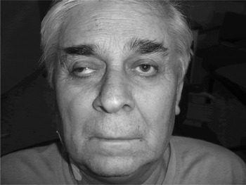
Post-operatively, the patient complained of headache, and the nasal packing was removed and analgesics administered. No improvement was noted, and on the following day the patient reported reduced vision and diplopia. Paresis of the right oculomotor and trochlear nerves was evident on clinical examination (Figure 2). An urgent ophthalmological assessment revealed diminished visual acuity bilaterally, worse on the right side (2/10).
The patient's clinical appearance after functional endoscopic sinus surgery; note right eyelid ptosis and lateral deviation, and mydriasis of the right pupil.

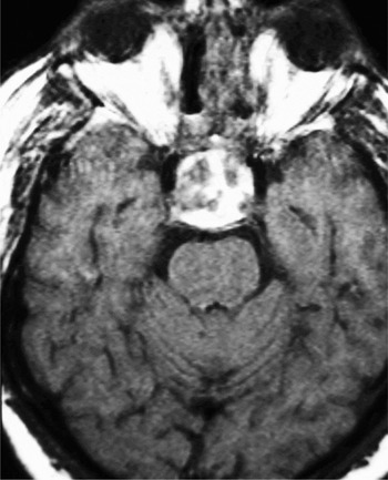
Computed tomography (CT) and magnetic resonance imaging indicated haemorrhage within the pituitary tumour (Figure 3). A diagnosis of pituitary apoplexy was made. In view of the patient's deteriorating vision, he was returned to the operating theatre two days after his initial endoscopic sinus surgery.
Axial, T1-weighted, gadolinium-enhanced magnetic resonance imaging scan taken in the early post-operative period. High signal areas in the sellar mass represent recent haemorrhage within the pituitary adenoma, and suggest pituitary apoplexy.

The patient's haemorrhagic pituitary tumour was resected completely through a combined microscopic endoscopic trans-sphenoidal approach, performed in cooperation with the neurosurgeons.
Corticosteroid replacement therapy was instituted.
In the first post-operative week, the patient regained adequate vision (7/10 bilaterally) and oculomotor nerve function.
Although ptosis of the right upper eyelid persisted and diabetes insipidus developed, the patient returned to his normal daily activities a few weeks after the episode.
Discussion
Symptomatic pituitary apoplexy has an incidence of 0.6–9.1 per cent in cases of surgically treated pituitary adenoma.Reference Randeva, Schoebel, Byrne, Esiri, Adams and Wass3 The true incidence is higher because haemorrhage or infarction within a pituitary tumour, as seen in surgical specimens, can occur without acute symptoms (i.e. subclinical pituitary apoplexy). Rarely, pituitary apoplexy may occur in pituitary glands without tumours.
The commonest presenting symptom of pituitary apoplexy is sudden, retro-orbital headache. Other symptoms, in decreasing order of frequency, are: nausea, reduction of visual fields, ocular paresis, reduction of visual acuity, vomiting, photophobia, fever and altered level of consciousness. The full clinical picture develops within hours to days.
The pathophysiology of pituitary apoplexy is still under speculation. Four mechanisms, leading to pituitary infarction and haemorrhage, have been proposed.Reference Biousse, Newman and Oyesiku4
The first involves hypoperfusion of a pituitary adenoma. Possible triggers include fluctuations in blood pressure (e.g. during cardiac surgery), raised intracranial pressure (due to coughing or lumbar puncture) and vascular changes after pituitary gland irradiation.
The second proposed mechanism is an acute increase in pituitary blood flow. Malignant hypertension and diabetic ketosis have been implicated, but there is no convincing evidence that uncomplicated hypertension and diabetes mellitus definitely lead to pituitary apoplexy.
The third mechanism involves stimulation of the pituitary gland. Relative ischaemia of the pituitary gland may result when blood flow demand exceeds supply, for example during exogenous administration of oestrogens, dynamic testing of pituitary function with gonadotrophin-releasing hormone, or surgical stress.
The fourth proposed mechanism of pituitary infarction and haemorrhage is anticoagulation.
During a surgical procedure, the confluence of several mechanisms acting simultaneously can lead to pituitary apoplexy. Fluctuations in blood pressure, hypotension, blood dilution due to crystalloid fluid infusion, anticoagulation, excessive steroid secretion and transient increases in intracranial pressure may all play a role in the pathogenesis of the syndrome. Functional endoscopic sinus surgery requires a bloodless field, particularly when polyps are present. Our patient had a number of factors that possibly rendered him prone to an acute vascular pituitary event. He was hypertensive, diabetic and receiving anticoagulation therapy, and he underwent an operation which required prolonged intra-operative hypotension.
We believe that a trans-sphenoidal approach to the sella turcica through the nose was the least invasive and hazardous strategy for this patient. In addition, functional endoscopic sinus surgery was justified in this patient, because elimination of nasal infection and polyps reduces the risk of post-operative meningitis and improves access to the pituitary fossa.Reference Lubbe and Semple2 Infection of muscle grafts and nasal packs in the sphenoid sinus in the presence of a cerebrospinal fluid leak has been reported as the main cause of meningitis after trans-sphenoidal pituitary surgery.Reference Gransden, Wickstead and Eykyn5
External approaches to the sellar region, namely frontal craniotomy, are required only for extensive sellar tumours and so are infrequently used.Reference Liu, Weiss and Couldwell6 The majority of pituitary adenomas can be resected via a trans-sphenoidal approach. The sublabial and trans-septal approaches are time-consuming and technically demanding in revision cases, result in a longer hospital stay, and cause functional nasal problems.Reference Nasseri, Kasperbauer, Strome, McCaffrey, Atkinson and Meyer7 The trans-sphenoidal endoscopic approach interferes little with nasal structures and is better tolerated by patients.Reference Dusick, Esposito, Mattozo, Chaloner, McArthur and Kelly8 Potential disadvantages are the lack of stereoscopic vision, the need for meticulous control of bleeding, and the surgical ‘learning curve’ involved. Severe complications comprise cerebrospinal fluid leakage (8.42 per cent) and meningitis (2.11 per cent).Reference Santos Rde, Zymberg, Abucham Filho, Gregorio and Weckx9
Diagnosis of pituitary apoplexy requires a high index of clinical suspicion. A patient with a known pituitary adenoma who develops headache and visual impairment should be investigated for pituitary apoplexy.Reference Semple, Jane and Laws10 Although CT is readily available in most departments, haemorrhage in a pituitary adenoma may be missed in 50 per cent of patients.Reference Onesti, Wisniewski and Post11 Magnetic resonance imaging is the radiological modality of choice, with a sensitivity of 88 per cent.Reference Randeva, Schoebel, Byrne, Esiri, Adams and Wass3
• Pituitary apoplexy is a rare, potentially life-threatening event which may occur spontaneously or following various types of surgical intervention
• Safe, minimally invasive resection of pituitary adenomas via an intranasal approach requires the elimination of polyps and other sources of sinonasal infection
• This case report shows that preparative endoscopic sinus surgery can be complicated by pituitary apoplexy, necessitating immediate neurosurgical consultation
• The intranasal trans-sphenoidal approach to the pituitary fossa is effective in minimising the consequences of an expanding, haemorrhagic pituitary adenoma and preventing a fatal outcome
The management of pituitary apoplexy is controversial. A conservative medical approach is advocated when vision is unaffected, or when the visual deficit is stable or improving at presentation, because spontaneous recovery may ensue.Reference Ayuk, McGregor, Mitchell and Gittoes12 Patients managed conservatively did not differ from those treated surgically in terms of the endocrine outcome.Reference Ayuk, McGregor, Mitchell and Gittoes12 Other authors have proposed early surgical intervention (within eight days), on the grounds that pituitary apoplexy has an unpredictable course, the trans-sphenoidal approach has a low morbidity rate, visual and oculomotor impairments improve post-operatively, and conservative treatment rarely reverses hypopituitarism.Reference Randeva, Schoebel, Byrne, Esiri, Adams and Wass3, Reference Zhang, Fei, Zhang, Cao, Liu and Zhang13 Surgical decompression may also prevent a second apoplectic event or residual tumour growth.Reference Randeva, Schoebel, Byrne, Esiri, Adams and Wass3 Regardless of the chosen approach, long term follow up is required to monitor for tumour recurrence. Gamma knife radiotherapy may be necessary in select cases.
Rhinologists are substantially involved in pituitary surgery, by providing (1) minimally invasive access with preservation of nasal function, and (2) eradication of sinonasal infection. It is important to be aware that pituitary apoplexy can follow nasal procedures performed to facilitate the final trans-sphenoidal approach to the pituitary fossa in high risk patients. In the case of an apoplectic event, prompt neurosurgical consultation is required. Early combined surgical intervention by rhinologists and neurosurgeons will eliminate headache, improve visual impairment and prevent a fatal outcome in deteriorating patients. The endoscopic trans-sphenoidal approach is a safe and effective technique in the acute management of pituitary apoplexy.