Introduction
Cyanobacteria are prokaryotes that can perform oxygenic photosynthesis and fix nitrogen. Throughout the history of Earth, cyanobacteria have been significant components of benthic marine ecosystems, played important ecological roles (Schopf, Reference Schopf and Whitton2012; Liu et al., Reference Liu, Wu, Jiang and Riding2016a; Schirrmeister et al., Reference Schirrmeister, Patricia and David2016), and influenced patterns of carbonate sedimentation (Riding, Reference Riding2006). Body fossils of Archean and Proterozoic cyanobacteria are mostly preserved as condensed carbon in kerogen, embedded in silica, or phosphatized, and are rarely calcified. In contrast, Phanerozoic fossil cyanobacteria are usually calcified (Golubic and Seong-Joo, Reference Golubic and Seong-Joo1999; Riding, Reference Riding2006; Schopf, Reference Schopf and Whitton2012; Min et al., Reference Min, Hua, Liu, Sun, Cui and Dai2020), a process that is often microbially induced and is dependent on water chemistry and physiological and morphological factors (Riding, Reference Riding2006; Konhauser and Riding, Reference Konhauser, Riding, Knoll, Canfield and Konhauser2012). Calcified cyanobacteria are relatively common on Paleozoic and Mesozoic marine carbonate platforms (Riding, Reference Riding1992) and played an important role in the formation of lime mud in ancient shallow-marine carbonates, especially in the early Paleozoic (Pratt, Reference Pratt2001).
Great radiations of marine invertebrates occurred during the Ordovician, known as the Great Ordovician Biodiversification Event (GOBE) (Webby et al., Reference Webby, Paris, Droser and Percival2004). However, the evolutionary and ecological history of Ordovician cyanobacteria and superficially similar associated microfossils has been hindered by difficulties in their taxonomy (Nitecki et al., Reference Nitecki, Webby, Spjeldnaes, Zhang, Webby, Droser, Paris and Percival2004). Riding (Reference Riding1992) concluded that the abundance and diversity of cyanobacteria and other microfossils declined in the Middle and Late Ordovician, whereas Webby (Reference Webby, Kiessling, Flügel and Golonka2002) stated that new types of calcified cyanobacteria and associated microfossils appeared from the Darriwilian onwards, resulting in more complex and diverse associations, especially with metazoans. Assemblages of calcified cyanobacteria and associated microfossils are mainly known from Europe, North America, South China, North China (Ordos Basin), and Northwest China (Tarim Basin) (Liu et al., Reference Liu, Wu, Yang and Riding2016b), which were all adjacent to the equator during the Ordovician (Fig. 1.1). None of these floras exceeds eight genera (see review of Liu et al., Reference Liu, Wu, Yang and Riding2016b). In the last few years, an assemblage of calcified cyanobacteria and associated microfossils with 20 genera was discovered in the Ordovician carbonate platform of the Tarim Basin (Liu et al., Reference Liu, Wu, Yang and Riding2016b). Research on this flora suggests that cyanobacteria and associated microfossils played important ecological roles (Liu et al., Reference Liu, Wu, Jiang and Riding2016a, Reference Liu, Wu, Jiang, Wu and Jia2017). Additional detailed investigations of calcified cyanobacteria worldwide are needed to reveal their global diversity and ecological roles during the Middle and Late Ordovician.
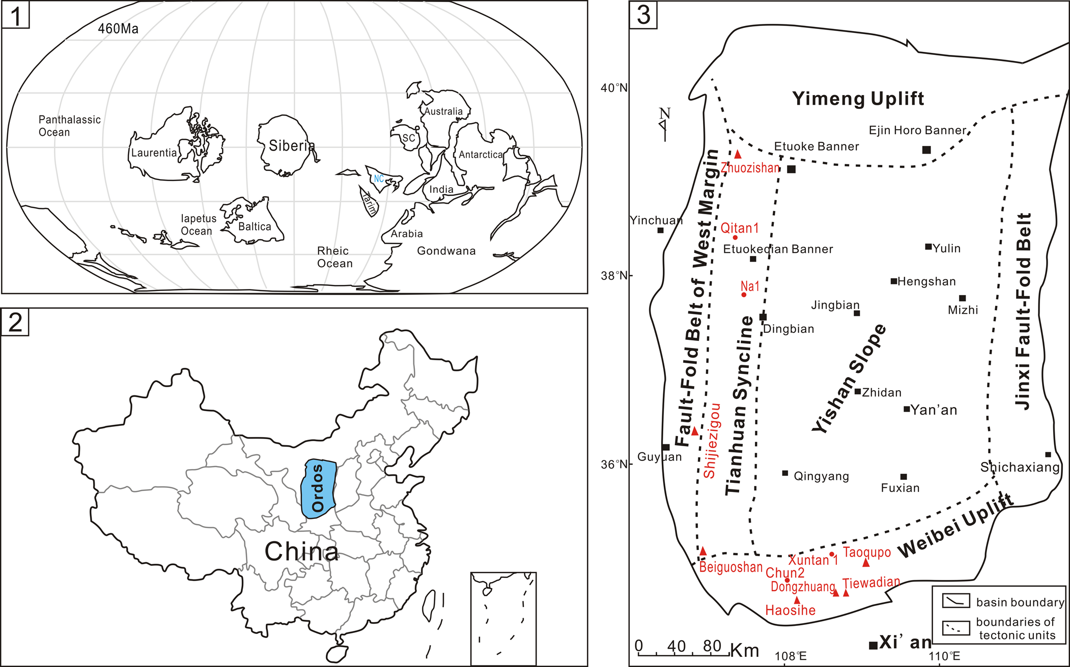
(1) Late Ordovician (460 Ma) paleogeographic positions of paleocontinents (after Huang et al., Reference Huang, Yan, Piper, Zhang, Yi, Yu and Zhou2018). Laurentia includes North America, Greenland, and slivers of crust in Norway and the British Isles (Servais et al., Reference Servais, Owen, Harper, Kröger and Munnecke2010). (2) Location of the Ordos Basin in China. (3) Current regional structure of Ordos Basin (according to Guo et al., Reference Guo, Zhao, Shi and Gao2014 and Zheng et al., Reference Zheng, Bao, Wu, Sun, Jiang, Ren, Huang and Liu2018) showing tectonics and locations of outcrops and drill cores involved in this study.

In this study, we carried out a systematic study on calcified cyanobacteria and associated microfossils from the Middle and Late Ordovician of the Ordos Basin, North China (Fig. 1.2), based on 1330 thin sections from seven outcrop profiles and four drill cores (Fig. 1.3). The purpose of this paper is to investigate the composition, diversity, and facies occurrences of calcified cyanobacteria and associated microfossil flora in North China and to compare them with other contemporaneous floras globally. The discovery of new materials in this area provides key evidence for global diversity and ecological roles of cyanobacteria in benthic communities on carbonate platforms during the Middle and Late Ordovician.
Geological setting
The North China Block was largely confined to the tropical zone near the equator during the Middle and Late Ordovician (Zhen et al., Reference Zhen, Zhang, Wang and Percival2015), and is interpreted as a peri-Gondwanan terrane located on the northeast Gondwana margin (Metcalfe, Reference Metcalfe2006) (Fig. 1.1). The Ordos Basin is located in the western part of the North China Block (Fig. 1.2; Hou et al., Reference Hou, Fang, Dong, Li, Lu, Wu and Chen2003), and is an intercratonic basin with stable subsidence, depression migration and multicyclic tectonic evolution (Wang et al., Reference Wang, Zhou, Wang, Jing, Zhang, Yuan and Shen2016). The Ordos Basin consists of six tectonic units: the Yimeng Uplift, the Jinxi Fault-Fold Belt, the Weibei Uplift, the Tianhuan Syncline, the Fault-Fold Belt, and the Yishan Slope (Xie et al., Reference Xie, Zhang and Guo2003; Liu et al., Reference Liu, Zhao, Wang and Chen2005; Zheng et al., Reference Zheng, Bao, Wu, Sun, Jiang, Ren, Huang and Liu2018) (Fig. 1.3).
Ordovician rocks on the North China Block can be subdivided into two major depositional settings: the broad North China platform and the narrow western slope to the southwest marginal platform (Yang et al., Reference Yang, Li and Ma2005; Cao et al., Reference Cao, Feng, Zhao, Zhou, Wang, Liu, Lu and Li2011; Feng et al., Reference Feng, Zheng, Bao, Jin, Wu, He, Peng, Yang, Zhang and Zhang2014; Zhen et al., Reference Zhen, Zhang, Wang and Percival2015; Jing et al., Reference Jing, Zhou and Wang2016). The broad North China platform was very shallow-marine tidal flats, dominated by carbonate deposition of 500–1000 m in thickness (Jing et al., Reference Jing, Zhou and Wang2016), where calcified cyanobacteria and associated microfossils are absent. Along the western to southwestern margin of the Ordos Basin, there are even thicker accumulations (up to 2600 m) of mixed carbonates and siliciclastics. Carbonates were deposited in slope, marginal, open platform, and restricted platform environments during the Middle–Late Ordovician (Jing et al., Reference Jing, Zhou and Wang2016; Zheng et al., Reference Zheng, Bao, Wu, Sun, Jiang, Ren, Huang and Liu2018). Carbonate strata of the Middle and Upper Ordovician in the western to southwestern margin of the Ordos Basin contain various reefs built by corals, stromatoporids, calcareous red and green algae, and calcified microbes (Ye et al., Reference Ye, Yang, Xu, Zheng, Zuo, Zhou, Li, Li, Song, Yong, Zhang and Zhang1995; Wang et al., Reference Wang, Deng, Wang and Li2012; Jiang et al., Reference Jiang, Bao, Sun, Wu and Diao2013; Lee et al., Reference Lee, Sun, Choh and Lee2014, Reference Lee, Hong, Lee and Choh2016).
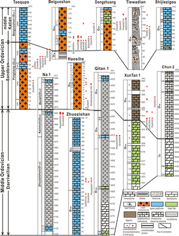
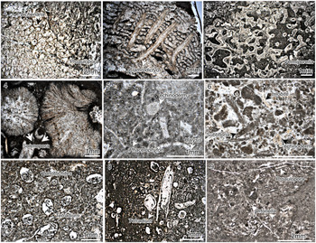
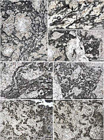
Carbonate strata of the Middle and Upper Ordovician in seven outcrop profiles and 4 drill wells in the southern and western edges of the Ordos Basin were examined in this study (Figs. 1.3, 2). The profiles of Shijiezigou (36°12'53.6"N, 106°36'36.1"E) and Zhuozishan (39°22'49"N, 106°52'5"E) and wells of Na 1 and Qitan 1 are located on the western edge of the Ordos Basin. The profiles of Taoqupo (34°58'58"N, 108°54'14"E), and the profiles of Beiguoshan (35°1'20"N, 106°39'42"E), Dongzhuang, Haosihe, and Tiewadian (34°41'38.3"N, 108°37'57.4"E), and wells of Xuntan 1 and Chun 2 are located on the south edge of the Ordos Basin (Fig. 1.3). On the western edge of the Ordos Basin, four formations were investigated, listed in ascending order: Zhuozishan (O2zh), Kelimoli (O2k), Wulali (O3w), and Beiguoshan (O3b) (Fig. 2). On the south edge of the Ordos Basin, three formations were investigated, in ascending order: Majiagou (O2m), Pingliang (O3p), and Beiguoshan (O3b). The Zhuozishan, Kelimoli, and Majiagou formations are attributed to the Darriwilian Stage of the Middle Ordovician, according to conodont biostratigraphy (Fig. 2; Jing et al., Reference Jing, Zhou and Wang2016). The Wulali and Pingliang formations are Sandbian (Late Ordovician) in age according to sequence stratigraphy, conodont zones, and U-Pb geochronology (Fig. 2; Guo et al., Reference Guo, Zhao, Fu, Xu, Shi, Sun, Gao, Zhang, Zhang and Liu2012, Reference Guo, Zhao, Shi and Gao2014; Wang et al., Reference Wang, Bergström, Zhen and Zhang2013, Reference Wang, Zhou, Wang, Jing and Zhang2015). The conodont zone of the Beiguoshan Formation demonstrates a middle Katian age (Fig. 2; Jin et al., Reference Jin, Rong and Zhan2011; Zhang et al., Reference Zhang, Zhan, Zhen, Wang, Yuan, Fang, Ma and Zhang2019). Marginal reef (Fig. 3.1–3.4), open platform (Fig. 3.5, 3.6), and restricted platform, including lagoon (Fig. 3.7, 3.8) and tidal flat (Fig. 3.9), facies are developed in these outcrops and wells (Figs. 2, 3). Many calcified cyanobacteria and associated microfossils are included in these facies (Ye et al., Reference Ye, Yang, Xu, Zheng, Zuo, Zhou, Li, Li, Song, Yong, Zhang and Zhang1995; Lee et al., Reference Lee, Sun, Choh and Lee2014, Reference Lee, Hong, Lee and Choh2016; Lee and Riding, Reference Lee and Riding2016; this study) (Figs. 2, 3).
Stratigraphic column of the Ordovician successions of outcrops and drill cores involved in this study and horizons and facies occurrences of calcified cyanobacteria and associated microfossils.

Sedimentary facies types of strata from outcrops and wells involved. (1–4) Reef facies; (1) reef limestone containing the coral Lichenaria and calcified cyanobacteria Proaulopora, O3p, sample No. HSHB-2-2; (2) reef limestone containing the stromatoporid Clathrodictyon, O3b, sample No. LJPA-1; (3) reef limestone containing the calcareous green alga Vermiporella, O3b, sample No. LJP6 (2)-12; (4) reef limestone formed by the red alga Solenopora, O3b, sample No. LJPBGS-1 (d). (5, 6) Open platform; (5) packstone, containing many bioclastics, such as echinoderms, possible alga Halysis, and many calcified cyanobacteria Phacelophyton, O2K, Na1-3918.03m; (6) packstone, containing many bioclastics, such as trilobite, echinoderms, and calcified cyanobacteria Subtifloria, O2K, ZZS-K-3, scale bar = 1 mm. (7, 8) Lagoonal facies; micritic limestone, containing some coral Tetradium and calcified cyanobacteria Zonotrichites, O3p, sample No. XT1-7-33-25. (9) Tidal flat facies; micritic limestone, containing calcified cyanobacteria Proaulopora and Ortonella, O3b, samle No. DZ-2.

Previous investigation
The most important report of calcified cyanobacteria and associated microfossils from the Ordos Basin to date was published in Chinese more than 20 years ago (Ye et al., Reference Ye, Yang, Xu, Zheng, Zuo, Zhou, Li, Li, Song, Yong, Zhang and Zhang1995). This work reported seven genera—Girvanella, Ortonella, Hedstroemia, Proaulopora, Phacelophyton, Renalcis, and Epiphyton—from Upper Ordovician outcrop profiles located in Taoqupo, Tiewadian, Dongzhuang, Haosihe, and Beiguoshan (Ye et al., Reference Ye, Yang, Xu, Zheng, Zuo, Zhou, Li, Li, Song, Yong, Zhang and Zhang1995). These fossils were identified at the genus level and were simply attributed to algae, without detailed analysis of their systematic positions (Ye et al., Reference Ye, Yang, Xu, Zheng, Zuo, Zhou, Li, Li, Song, Yong, Zhang and Zhang1995). Moreover, few photographs were included in this report, and all show only part of the fossils, without illustration of the overall morphology of the genera (Ye et al., Reference Ye, Yang, Xu, Zheng, Zuo, Zhou, Li, Li, Song, Yong, Zhang and Zhang1995), hindering further comparison and analysis. Those genera with pictures were obtained from the Middle Ordovician Yaoxian Formation in the Taoqupo section, the Middle Ordovician Jinghe Formation in the Tiewadian section, and the Upper Ordovician Beiguoshan Formation in the Dongzhang section (Ye et al., Reference Ye, Yang, Xu, Zheng, Zuo, Zhou, Li, Li, Song, Yong, Zhang and Zhang1995). The Yaoxian Formation has since been re-attributed to the Pingliang Formation, which belongs to the Sandbian stage (Guo et al., Reference Guo, Zhao, Fu, Xu, Shi, Sun, Gao, Zhang, Zhang and Liu2012, Reference Guo, Zhao, Shi and Gao2014). The Tiewadian breccias have subsequently been ascribed to the Beiguoshan Formation (Jiang et al., Reference Jiang, Bao, Sun, Wu and Diao2013; Lee and Riding, Reference Lee and Riding2016), which is early Katian in age. Girvanella-like and Ortonella fossils were reported from the reef of the Haosihe profile from the Pingliang Formation (Lee et al., Reference Lee, Sun, Choh and Lee2014). Girvanella, Epiphyton-like, and Renalcis-like fossils have been reported from bivalve, siliceous sponge-microbe reefs in the Pingliang Formation (Lee et al., Reference Lee, Hong, Lee and Choh2016). Subsequently, a new genus and species of calcified cyanobacterium, Xianella hongii, was reported by Lee and Riding (Reference Lee and Riding2016) from the Beiguoshan Formation in the Tiewadian profile of the Ordos Basin. In total, there have been eight genera reported from the Ordos Basin.
Materials and methods
Samples were systematically collected from the seven outcrop profiles and four drill cores (Figs. 1.3, 2). More than 1330 large (size 5 × 7 cm) thin sections were prepared. The thin sections were investigated using transmitted light microscopy and microphotography, revealing numerous calcified cyanobacteria and associated calcified microfossils. These fossils occur in ~100 thin sections from six profiles and three drill cores.
Fossils in the thin sections were measured, and more than 1500 microphotographs were taken. The calcified cyanobacteria and associated microfossils were identified based on morphological features, such as the pattern of arrangement of filaments or chambers, branching pattern of filaments, and sizes of filaments and chambers (Lee and Riding, Reference Lee and Riding2016; Liu et al., Reference Liu, Wu, Yang and Riding2016b). The systematic positions of most genera are based on Liu et al. (Reference Liu, Wu, Yang and Riding2016b and references therein) and on further comparison with modern analogues. Sedimentary facies types of cyanbobacteria and associated microfossils in these outcrops and drill cores were determined based on lithological features, sedimentary structures, and community components (Flügel, Reference Flügel2004; Liu et al., Reference Liu, Wu, Jiang, Wu and Jia2017).
Eighteen species within 16 genera, including a new species, were identified. Girvanella, Subtifloria, Xianella, Acuasiphonoria, and Oscillatoriaceae gen. indet. Are attributed to Osillatoriales of cyanobacteria; Ortonella, Hedstroemia, Cayeuxia, Zonotrichites, Proaulopora, and Phacelophyton are attributed to Nostocales of cyanobacteria, and Renalcis, Izhella, Rothpletzella, Garwoodia, and Wetheredella are attributed to calcified Microproblematica. Most of the fossils are from the Sandbian and middle Katian of the Late Ordovician. Girvanella and Phacelophyton also occur in the Darriwilian Stage, Middle Ordovician. Subtifloria is the only genus that was only discovered in the Darriwilian Stage. The location, horizons, and sedimentary facies of calcified cyanobacteria and microfossils are illustrated in Figure 2.
Repositories and institutional abbreviations
Specimens illustrated here are housed in the collection of the Institute of Geology and Geophysics (IGG), Chinese Academy of Sciences (CAS), Beijing, China. The specimens are identified according to the slide numbers cited in the figure captions.
Systematic paleontology
Cyanobacteria Stanier, Reference Stanier, Buchanan and Gibbons1974
Order Osillatoriales Elenkin, Reference Elenkin1949
Genus Girvanella Nicholson and Etheridge, Reference Nicholson and Etheridge1878
Type species
Girvanella problematica Nicholson and Etheridge, Reference Nicholson and Etheridge1878 emend. Wood, Reference Wood1957; Upper Ordovician, Scotland.
Remarks
Nicholsonia Korde, Reference Korde1973 is regarded as a junior synonym of Girvanella (Danielli, Reference Danielli1981). Girvanella was very common in the Paleozoic and Mesozoic (Riding, Reference Riding and Riding1991). Riding (Reference Riding1977) compared it with the present-day calcified sheaths of Plectonema due to its sinuous to irregularly tangled filaments, and this identification has been followed by many researchers (Feng et al., Reference Feng, Gong and Riding2010; Liu et al., Reference Liu, Wu, Yang and Riding2016b, fig. 3).
Girvanella problematica Nicholson and Etheridge, Reference Nicholson and Etheridge1878 emend. Wood, Reference Wood1957
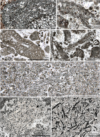
Figure 4.1
- Reference Nicholson and Etheridge1878
Girvanella problematica Nicholson and Etheridge, p. 23, pl. 9, fig. 24.
- Reference Wethered1890
Girvanella ducii Wethered, p. 280, pl. 11, fig. 2a–c.
- Reference Høeg1932
Girvanella problematica; Høeg, p. 64, pl. 1, figs. 4–6.
- Reference Bourque, Mamet and Roux1981
Girvanella problematica; Bourque et al., p. 95, p1. 1, figs. 2–4.
- Reference Ye, Yang, Xu, Zheng, Zuo, Zhou, Li, Li, Song, Yong, Zhang and Zhang1995
Girvanella; Ye et al., p. 15, pl. 8, fig. 4.
- Reference Riding and Fan2001
Girvanella problematica; Riding and Fan, p. 789, text-figure 3B, C.
- Reference Liu, Yang and Wu2011
Girvanella problematica; Liu et al., p. 495, pl. I, fig. 4.
- Reference Liu, Wu, Yang and Riding2016b
Girvanella problematica; Liu et al., p. 188, fig. 4B.
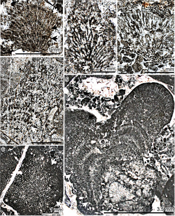
(1) Girvanella problematica, O3b, sample No. JJSBGS-2-C, oblique longitudinal sections. (2, 3) Girvanella kasakiensis, O2k; (2) sample No. ZZS-Z-2, oblique longitudinal sections; (3) sample No. Na1-3917.51m, oblique longitudinal sections. (4, 5) Subtifloria delicata, O2k; (4) sample No. ZZS-K-3, longitudinal sections, (5) sample No. ZZS-Z-5, longitudinal sections. (6, 7) Acuasiphonoria ordovica, from O3p, sample No TQPQ-4-1, longitudinal and cross sections.

Holotype
Specimen number V. 34566, British Museum collection; illustrated by Nicholson and Etheridge (Reference Nicholson and Etheridge1878, pl. 9, fig. 24), Stinchar Limestone (Upper Ordovician), Ayrshire, Scotland.
Description
Calcareous tubular filaments with uniform external diameter; filaments long and packed, sinuous to irregularly tangled in mesh-like masses; tube wall thin, micritic; external diameter 14~17 μm; wall thickness 2~4 μm.
Material
LJPBGS-2, LJPBGS-3, O3b, packstone, middle Katian, Beiguoshan; TQPBD-3, O3p, packstone, Sandbian, Taoqupo.
Remarks
Researchers have defined numerous species of Girvanella, mainly based on tube diameter. However, many of these species appear to have overlapping sizes, as summarized by Fournie (Reference Fournie1967). Mamet and Roux (Reference Mamet and Roux1975) proposed just four species—G. kasakiensis Maslov, Reference Maslov1949; G. problematica, G. wetheredeii Chapman, Reference Chapman1908, and G. staminea Garwood, Reference Garwood1931—according to their internal diameter and wall thickness. However, Wood (Reference Wood1957) showed that detailed measurements of Girvanella from the type area did not reveal clear subgroups. Nevertheless, Liu et al. (Reference Liu, Wu, Yang and Riding2016b) found that Ordovician species of Tarim Basin on the whole can be confirmed as belonging to four species according to diameter size range of filaments, as in Mamet and Roux's approach (Reference Mamet and Roux1975). We now also identify Girvanella species according to Mamet and Roux's approach (Reference Mamet and Roux1975).
Girvanella sp. reported by Ye et al. (Reference Ye, Yang, Xu, Zheng, Zuo, Zhou, Li, Li, Song, Yong, Zhang and Zhang1995) can likely be further identified as Girvanella problematica, which occurred in the Pingliang Formation of the Taoqupo section. The Girvanella-like fossil reported by Lee et al. (Reference Lee, Sun, Choh and Lee2014) from the Pingliang Formation of the Haosihe Profile is different from Girvanella in its non-tangled and coarse filaments.
Girvanella kasakiensis Maslov, Reference Maslov1949 emend. Mamet and Roux, Reference Mamet and Roux1975
Figure 4.2, 4.3
- Reference Maslov1949
Girvanella ducii var. kasakiensis Maslov, S. 6 (ohne Abb).
- Reference Maslov1956
Girvanella ducii var. kasakiensis; Maslov, p. 47, pl. 5, figs. 2, 4, pl. 4, fig. 2.
- Reference Chuvashov1965
Girvanella aff. ducii; Chuvashov, p. 74, pl. 17, fig. 3.
- Reference Wray1967
Girvanella ducii; Wray, p. 34, pl. 7, fig. 5.
- Reference Mamet and Roux1975
Girvanella kasakiensis; Mamet and Roux, p. 142, pl. 4, fig. 10, pl. 5, figs. 1, 2, 9, 10.
- Reference Liu, Yang and Wu2011
Girvanella kasakiensis; Liu et al., p. 495, pl. 1, fig. 3.
- Reference Liu, Wu, Yang and Riding2016b
Girvanella kasakiensis; Liu et al., p. 188, fig. 4A.
Holotype
Specimen illustrated by Maslov (Reference Maslov1956, pl. 5, figs. 2, 4, pl. 4, fig. 2); Silurian, Kazakhstan.
Description
Calcareous tubular filaments with uniform external diameter; filaments long and loose, sinuous to irregular and tangled, prostrate; tube wall thin, micritic; external diameter of filaments 22~24 μm; wall thickness 4~6 μm.
Material
LSD-K-3, LSD-K-5, LSD-K-7, O2k, LSD-Z-2, O2z, Darriwilian, packstone, Zhuozishan; 9-95-33, O2k, Qitan1, Darriwilian, wackstone; 3917.26, 3917.39, 39717.51, 3917.65, 3917.92, 3918.03, 3918.22, O2k, Darriwilian, Na1.
Remarks
Girvanella kasakiensis has the thickest filaments (20–28 μm) among all the species according to Mamet and Roux (Reference Mamet and Roux1975).
Genus Subtifloria Maslov, Reference Maslov1956
Type species
Subtifloria delicata Maslov, Reference Maslov1956; lower Cambrian; Siberian Platform, Russia.
Remarks
Botominella Reitlinger, Reference Reitlinger1959 is regarded as a junior synonym of Subtifloria (Luchinina, Reference Luchinina1975). For a long time, the only confirmed reports of Subtifloria were from the early Cambrian (Maslov, Reference Maslov1956). The other confirmed records were in the Darriwilian and early Katian (Middle and Upper Ordovician) of the Tarim Basin (Riding and Fan, Reference Riding and Fan2001; Liu et al., Reference Liu, Wu, Yang and Riding2016b). The report in the Ordos Basin (this study) is also from the Darriwilian stage. In younger strata, Subtifloria is similar to some Girvanella in tube size, but differs in its overall cable-like, bundled arrangement of aligned filaments. Subtifloria closely resembles modern oscillatoriaceans, such as Microcoleus (Chuvashov et al., Reference Chuvashov, Luchinina and Shuysky1987). This comparison is corroborated by Liu et al. (Reference Liu, Wu, Jiang and Riding2016a, fig. 3).
Subtifloria delicata Maslov, Reference Maslov1956
Figure 4.4, 4.5
- Reference Maslov1956
Subtifloria delicata Maslov, p. 85, text-fig. 24, pl. 27, fig. 4.
- Reference Racki and Sobon-Podgorska1992
?Botominella Racki and Sobon-Podgorska, fig. 3f.
- Reference Riding and Fan2001
?Subtifloria sp. Riding and Fan, p. 790, text-fig. 3, fig. D.
- Reference Wang, Li, Zhang, Yang and Huang2009
Girvanella sp. Wang et al., fig. 2a.
- Reference Liu, Yang and Wu2011
Subtifloria sp. Liu et al., p. 496, pl. 1, fig. 5.
- Reference Rong, Jiao, Wang, Wu and Wang2014
Girvanella; Rong et al., figs. 5a, b.
- Reference Liu, Wu, Jiang and Riding2016b
Subtifloria delicata; Liu et al., p. 191, fig. 4F.
Holotype
Specimen illustrated by Maslov (Reference Maslov1956, text-fig. 24, pl. 27, fig. 4.), lower Cambrian, Siberian Platform, Russia.
Description
Calcareous tubular filaments with uniform diameter; filaments subparallel (Fig. 3.4, 3.5), often slightly sinuous, aggregated into bundles (Fig. 3.4, 3.5); filaments typically closely spaced, sometimes interlaced; tube wall thin, micritic; filaments up to 2 mm long; external diameter 17–23 μm; wall thickness ~3 μm.
Material
LSD-K-3, LSD-K-5, LSD-K-7, O2k, packstone, LSD-Z-4, LSD-Z-5, O2k, Darriwilian, Zhuozishan section.
Remarks
Subtifloria delicata is the only species of the genus.
Genus Acuasiphonoria Liu et al., Reference Liu, Wu, Jiang and Riding2016b
Type species
Acuasiphonoria ordovica Liu et al., Reference Liu, Wu, Yang and Riding2016b; Upper Ordovician, Katian, Lianglitag Formation; Tarim Basin, Xinjiang, Northwest China.
Remarks
The genus Acuasiphonoria was recently erected based on Tarim Basin material from the early Katian of the Upper Ordovician (Liu et al., Reference Liu, Wu, Yang and Riding2016b). This report in the Ordos Basin is its first record in the Sandbian stage of the Upper Ordovician. Acuasiphonoria resembles Girvanella and Subtifloria in tubular filaments, but is distinguished from them by its long, straight, or only slightly curved, filaments that appear to taper, resembling needles. Acuasiphonoria is regarded as an oscillatoriacean sheath (Liu et al., Reference Liu, Wu, Yang and Riding2016b).
Acuasiphonoria ordovica Liu et al., Reference Liu, Wu, Yang and Riding2016b
Figure 4.6, 4.7
- Reference Liu, Wu, Yang and Riding2016b
Acuasiphonoria ordovica Liu et al., p. 191, fig. 5A–C
Holotype
Specimen No. TZ42-4-53-49, collection of Institute of Geology and Geophysics, Chinese Academy of Sciences; illustrated by Liu et al. (Reference Liu, Wu, Yang and Riding2016b, fig. 5A–C), Lianglitag Formation (lower Katian, Upper Ordovician), Tarim Basin, Xinjiang, Northwest China.
Description
Calcareous long tubular filaments; filaments possibly branched (Fig. 4.7), separated (Fig. 4.6), straight, or gently curved, ending in a sharp point (Fig. 4.7); tube wall thin, micritic; filaments extending straight for up to 0.5 mm or more; external diameter 19~29 μm; wall thickness up to 2 μm.
Material
TQBPXX, TQPQ-4-1, O3P, Sandbian, microbial reef limestone, Taoqupo.
Remarks
Acuasiphonoria ordovica is the only species of the genus.
Genus Xianella Lee and Riding, Reference Lee and Riding2016
Type species
Xianella hongii Lee and Riding, Reference Lee and Riding2016; Ordovician, Katian; North China.
Remarks
The organization of Xianella into relatively thick, anastomosing, cable-like strands most closely resembles Cladogirvanella Ott, Reference Ott1966 from the Ladinian (Middle Triassic) of the Italian Dolomites (Lee and Riding, Reference Lee and Riding2016). Xianella is distinguished from Cladogirvanella by its larger tube size and the overall prostrate alignment of its cable-like strands (Lee and Riding, Reference Lee and Riding2016). Razumovskia also resembles Xianella in the size of tube, but it is distinguished from Xianella with prostrate, thin, felted layers with upturned ends, and short vertical tangled to curved tufts and outgrowths that arise from its upper surface (Liu et al., Reference Liu, Wu, Yang and Riding2016b). ?Kordephyton Li et al., Reference Li, Li and Kiessling2015 resembles Xianella in its individual filament, but is distinguished from it in overall organization of filaments and the presence of coarser filaments. Xianella is considered to be calcified sheath of a relatively large, filamentous, mat-forming cyanobacterium (Lee and Riding, Reference Lee and Riding2016).
Xianella hongii Lee and Riding, Reference Lee and Riding2016
Figure 5.1–5.6
- Reference Lee and Riding2016
Xianella hongii Lee and Riding, p. 4, figs. 4, 5.
(1–6) Xianella hongii, O3b. (1, 2) Sample No. TWDA-1-2-9; (1) overall morphology, longitudinal sections; (2) detailed morphology of Xianella filaments, longitudinal sections; black arrows pointing to tubular filaments; (3–6) sample No. TWDA-1-1-a; (3, 4) overall morphology; (5) enlargement of part of (3), longitudinal sections; (6) enlargement of (4), cross sections; (7, 8) Oscillatoriaceae gen. indet., from O3p, sample No HSH-5-3; (7) overall morphology; (8) enlargement of part of (7), oblique and cross sections.

Holotype
Specimen NIGPAS159413, illustrated by Lee and Riding (Reference Lee, Hong, Lee and Choh2016, figs. 4, 5), Beiguoshan Formation (middle Katian, Late Ordovician), Ordos Basin, North China.
Description
Calcareous tubular filaments; filaments unbranched, straight to sinuous, interlaced into cable-like strands forming an open anastomosing network in which layers of prostrate strands, joined by more erect branched strands, define relatively large spar-filled areas (Fig. 5.1); tube wall relatively thick, micritic (Fig. 5.2–5.6); filaments commonly extend 1–2 mm in length (Fig. 5.2, 5.5); tube external diameter 24–29 μm; wall thickness 4–5 μm.
Material
TWDA-1-2-9, TWDA-1-1-a, TWDA-1-1-b, O3b, middle Katian, reef block in breccia, Tiewadian section.
Remarks
Xianella hongii is the only species of the genus. They are generally (24–29μm) smaller than the specimen (20–60μm) reported by Lee and Riding (Reference Lee and Riding2016).
Oscillatoriaceae gen. indet.
Figure 5.7, 5.8
Description
Calcareous tubular filaments with uniform external diameter; filaments unbranched, straight to sinuous (Fig. 5.8); filaments relatively short and loose (Fig. 5.7); tube wall thin, micritic; filaments extending straight for up to 1 mm or more; external diameter ~90 μm; wall thickness up to 8 μm.
Material
HSH-5-3, O3p, Sandbian, coral reef limestone.
Remarks
The microfossil is similar to Girvanella in some curly filaments, but it is different from Girvanella in non-tangled, relatively short and coarse filaments. It resembles ‘Girvanella-like’ specimens reported in the same coral reefs by Lee et al. (Reference Lee, Sun, Choh and Lee2014, p. 36, fig. 5G, H) in filament morphology and large-filament diameter. However, many relatively long and parallel filaments in the ‘Girvanella-like’ form have not been observed in our specimens, perhaps due to poor preservation (Fig. 5.7, 5.8). Perhaps this specimen can be regarded as a new genus in the future. Based on similarities with sheaths of oscillatoriaceans, we regard it as an Oscillatoriaceae gen. indet.
Order Nostocales (Borzi) Geitler, Reference Geitler1925
Genus Zonotrichites Bornemann, Reference Bornemann1887
Type species
Zonotrichites lissaviensis Bornemann, Reference Bornemann1887; Upper Triassic; Poland.
Remarks
Ortonellina Bian and Zhou, Reference Bian and Zhou1990 is regarded as a junior synonym of Zonotrichites (Liu et al., Reference Liu, Wu, Jiang and Riding2016a). Zonotrichites was previously reported from the Upper Triassic (Bornemann, Reference Bornemann1887; Elliott, Reference Elliott1964). Recently, it has been reported from the early Katian Stage of the Upper Ordovician in the Tarim Basin (Liu et al., Reference Liu, Wu, Jiang and Riding2016a). Occurrences in the Ordos Basin (this study) are the first records from the Sandbian and middle Katian (Upper Ordovician). Zonotrichites also occurs in Ordovician strata in other areas of the world, but it has often been misidentified as Ortonella (Liu et al., Reference Liu, Wu, Yang and Riding2016b).
Its organization of small juxtaposed radial filaments links Zonotrichites to a wide variety of common Palaeozoic and Mesozoic fossils, including Hedstroemia, Ortonella, Bija, Cayeuxia, Botomaella, and Apophoretella (Liu et al., Reference Liu, Wu, Jiang and Riding2016a). Zonotrichites is characterized by its closely packed radial filaments, which are still subparallel after branching (Fig. 5.1; see Liu et al., Reference Liu, Wu, Yang and Riding2016b, fig. 6G, H). Zonotrichites closely resembles Apophoretella (see Liu et al., Reference Liu, Wu, Yang and Riding2016b, fig. 6), but has a larger thallus and coarser filaments (Liu et al., Reference Liu, Wu, Yang and Riding2016b, fig. 3). Bornemann (Reference Bornemann1887) first correctly compared Zonotrichites with the extant cyanobacterium Rivularia. Monty (Reference Monty1967) compared Ortonella with the extant cyanobacterium Scytonema, an identification that was widely followed (Dragastan, Reference Dragastan, Toomey and Nitecki1985; Riding, Reference Riding and Riding1991). Dragastan (Reference Dragastan, Toomey and Nitecki1985, Reference Dragastan1993) insisted on the affinities among Cayeuxia, Apophoretella, Zonotrichites, some Ortonella, and extant Rivularia. Riding and Voronova (Reference Riding, Voronova, Toomey and Nitecki1985) and Riding (Reference Riding and Riding1991) also noted the similarities among Botomaella, Cayeuxia, Hedstroemia, and extant calcified Rivularia, but suggested that fossil names should be retained for these taxa, especially because sheath calcification can preserve distinctive morphology (Riding, Reference Riding and Riding1991). All these fossils are compared with calcified sheaths of extant Rivularia (see Liu et al., Reference Liu, Wu, Yang and Riding2016b, fig. 3). Moreover, associations of Hedstroemia, Ortonella, Zonotrichites, and Cayeuxia often occurred abundantly in restricted lagoons and similar environments in the Late Ordovician (Pratt and Haidl, Reference Pratt, Haidl, Pratt and Holmden2008; Liu et al., Reference Liu, Wu, Jiang and Riding2016a) and at other times (Liu et al., Reference Liu, Wu, Jiang and Riding2016a and herein references). This association is interpreted to be related to their abilities to fix nitrogen and utilize organic phosphate, which are common in modern rivulariaceans (Liu et al., Reference Liu, Wu, Jiang and Riding2016a). This similar ecological characteristic provides new evidence for their Rivularia affinity.
Zonotrichites lissaviensis Bornemann, Reference Bornemann1887
Figure 6.1
- Reference Bornemann1887
Zonotrichites lissaviensis Bornemann, p. 126, pl. 5, figs. 1, 2, pl. 6, figs. 1, 2.
- Reference Elliott1964
Zonotrichites lissaviensis; Elliott, pl. 1, figs. 1, 2, pl. 2, figs. 1–3.
- Reference Walker1972
Hedstroemia; Walker, fig. 14F, G.
- Reference Guilbault and Mamet1976
Ortonella aequalis Høeg, Reference Høeg1932; Guilbault and Mamet, pl. 1, figs. 1–4, 6, 7.
- Reference Bian and Zhou1990
Ortonellina zhuzhaiensis Bian and Zhou, p. 6, pl. 2, fig. 6.
- Reference Mamet and Shalaby1995
Ortonella aequalis; Mamet and Shalaby, pl. 4, figs. 1–3.
- Reference Riding and Fan2001
Botomaella aequalis; Riding and Fan, p. 787, text-fig. 3A.
- Reference Kwon, Park, Choh, Lee and Lee2012
Ortonella; Kwon et al., fig. 5c.
- Reference Lee, Sun, Choh and Lee2014
Ortonella; Lee et al., fig. 5c.
- Reference Liu, Wu, Yang and Riding2016b
Zonotrichites lissaviensis; Liu et al., p. 196, fig. 6F–H.
(1) Zonotrichites lissaviensis, O3p, sample No. XT1-7-33-7, longitudinal sections; the arrows indicate concentric appearance formed by bifurcating uniformly. (2, 3) Hedstroemia halimedoidea, O3p, sample No. XT-7-33-25, longitudinal sections; arrows indicate the positions of bifurcations. (4, 5) Ortonella furcata, O3b, longitudinal sections; (4) sample No. TWDA-1-3, longitudinal sections; the white arrow points to the position of bifurcation; (5) sample No. JJSBGS-1, longitudinal sections; arrows indicate concentric appearance formed by bifurcating uniformly. (6) Cayeuxia piae, O3b, sample No. JJSBGS-1, oblique cross sections; arrow indicates the position of bifurcation.

Holotype
Specimen illustrated by Bornemann (Reference Bornemann1887, pl. 6, figs. 1, 2.), Upper Triassic, Poland.
Description
Calcareous, subrounded thallus composed of tubular filaments; filaments slightly curved, closely packed; elongate filaments of uniform diameter, radiating from a small base; filaments branched dichotomously at angles <20°, subparallel after branching (Fig. 6.1); filaments bifurcate uniformly, producing a concentric appearance in longitudinal section (Fig. 6.1); filaments bifurcate uniformly; thallus 1~2 mm across; filament diameter 20~50 μm.
Material
7-33-31, O3p, Sandbian, lagoonal facies of wackstone and packstone similar to the Lianglitag Formation of Tarim Basin (Liu et al., Reference Liu, Wu, Jiang, Wu and Jia2017), Xuntan 1.
Remarks
Zonotrichites lissaviensis is the only species of the genus.
Genus Hedstroemia Rothpletz, Reference Rothpletz1913
Type species
Hedstroemia halimedoidea Rothpletz, Reference Rothpletz1913; Silurian, Wenlock; Gotland, Sweden.
Remarks
Hedstroemia (Fig. 6.2, 6.3; see Liu et al., Reference Liu, Wu, Yang and Riding2016b, fig. 5D–F) closely resembles Bija (see Liu et al., Reference Liu, Wu, Yang and Riding2016b, fig. 5I, J) in its polygonal cross section, but is distinguished from Bija by its short filaments that expand distally (see Liu et al., Reference Liu, Wu, Yang and Riding2016b, fig. 3). Hedstroemia also resembles Cayeuxia (Fig. 6.6; see Liu et al., Reference Liu, Wu, Yang and Riding2016b, fig. 5G, H) in its branching pattern, but can be distinguished from the latter by its coarser filaments, which are polygon-like in cross section (see Liu et al., Reference Liu, Wu, Yang and Riding2016b, fig. 3).
Hedstroemia halimedoidea Rothpletz, Reference Rothpletz1913
Figure 6.2, 6.3
- Reference Rothpletz1913
Hedstroemia halimedoidea Rothpletz, p. 17, pl. 3, figs. 1–6.
- Reference Guilbault and Mamet1976
Hedstroemia halimedoidea; Guilbault and Mamet, p. 644, pl. 2, figs. 5–9.
- Reference Ye, Yang, Xu, Zheng, Zuo, Zhou, Li, Li, Song, Yong, Zhang and Zhang1995
Hedstroemia; Ye et al., p. 17, pl. 9, fig. 6.
- Reference Mamet and Shalaby1995
Hedstroemia halimedoidea; Mamet and Shalaby, p. 239, pl. 4, figs. 6–10.
- Reference Pratt, Haidl, Pratt and Holmden2008
Hedstroemia; Pratt and Haidl, fig.15E, F.
- Reference Liu, Yang and Wu2011
Hedstroemia halimedoidea; Liu et al., p. 498, pl. 2, figs. 5–9.
- Reference Liu, Wu, Yang and Riding2016b
Hedstroemia halimedoidea; Liu et al., p. 193, fig. 5D.
Holotype
Specimen collection in Geological Survey of Sweden; illustrated by Rothpletz (Reference Rothpletz1913, pl. 3, figs. 1–6.); Wenlock, Silurian, Järnvägsklinten near Visby, Gotland, Sweden.
Description
Calcareous elongate to subrounded thallus composed of tubular filaments; filaments closely packed, more or less radially arranged, expanding distally (Fig. 6.2, 6.3); filaments dichotomously branched from base, with multiple branching into clusters at acute angles near 10° (Fig. 6.2, 6.3); thallus 2~3 mm across; filament diameter typically 50~90 μm, average 70~80 μm.
Material
5-29-25, 6-43-11, 7-33-7, 7-33-25, 7-33-31, O3p, wackstone and packstone, Sandbian, Xuntan 1; LJP-7-B, O3b, stromatoporid reef, middle Katian, Beiguoshan; LQDZ-2, LQDZ-3, O3b, packstone, middle Katian, Dongzhuang; TQPBB-9 (2), O3b, packstone, middle Katian, Taoqupo.
Remarks
Hedstroemia halimedoidea is distinguished from other species by frequent branching. Hedstroemia sp. reported by Ye et al. (Reference Ye, Yang, Xu, Zheng, Zuo, Zhou, Li, Li, Song, Yong, Zhang and Zhang1995) can likely be identified as Hedstroemia halimedoidea, which also occurred in the Beiguoshan Formation of the Dongzhuang section.
Genus Ortonella Garwood, Reference Garwood1914
Type species
Ortonella furcata Garwood, Reference Garwood1914; lower Carboniferous; Westmorland, England.
Remarks
Springerella and Deisterella Dragastan and Richter, Reference Dragastan and Richter2011 are regarded as junior synonyms (Liu et al., Reference Liu, Wu, Yang and Riding2016b). Ortonella (Fig. 6.4, 6.5; see Liu et al., Reference Liu, Wu, Yang and Riding2016b, fig. 6D, E) resembles Zonotrichites (Fig. 6.1; see Liu et al., Reference Liu, Wu, Yang and Riding2016b, fig. 6G, H) a bit in its frequent dichotomous branching, but is distinguished from the latter with higher branching angles and a larger thallus (see Liu et al., Reference Liu, Wu, Yang and Riding2016b, fig. 3).
Ortonella furcata Garwood, Reference Garwood1914
Figure 6.4, 6.5
- Reference Garwood1914
Ortonella furcata Garwood, p. 265, pl. 20, figs. 1–4.
- Reference Bian and Zhou1990
Ortonella furcata; Bian and Zhou, pl. 1, fig. 6.
- Reference Ye, Yang, Xu, Zheng, Zuo, Zhou, Li, Li, Song, Yong, Zhang and Zhang1995
Ortonella; Ye et al., p. 16, pl. 8, fig. 8.
- Reference Pratt, Haidl, Pratt and Holmden2008
Ortonella; Pratt and Haidl, fig. 14H, fig. 15A–C.
- Reference Wang, Li, Zhang, Yang and Huang2009
Ortonella; Wang et al., fig. 2f.
- Reference Liu, Yang and Wu2011
Ortonella sp. 2, Liu et al., p. 498, pl. 2, fig. 1.
- Reference Liu, Wu, Yang and Riding2016b
Ortonella furcata; Liu et al., p. 194, fig. 6D.
Holotype
Specimen in British Museum (Garwood collection), illustrated by Garwood (Reference Garwood1914, pl. 20, figs.1–4), lower Carboniferous, Westmorland, England.
Description
Calcareous, elongate to nodular thallus composed of tubular filaments; filaments radiating from the base, more or less loosely arranged, slightly undulating; filaments symmetrically dichotomously branched at angles between 25° and 40° (Fig. 6.4, 6.5); filaments bifurcating uniformly, producing a concentric appearance in longitudinal section (Fig. 6.4, 6.5); tube wall thin, micritic; thallus 4~8 mm across, filament diameter 38~55 μm.
Material
6-43-1, 6-43-12, O3p, wackstone and packstone, Sandbian, Xuntan 1; LJP-6-L-1a; LJP-6-L-b, LJPBGS-1(C), LJPBGS-1(e), LJPBGS-1(s), O3b, packstone, middle Katian, Beiguoshan; LQDZ-1A, O3b, middle Katian, grainstone, Dongzhuang.
Remarks
Ortonella furcate is distinguished from other species by its coarser filaments and frequent branching. Ortonella reported by Ye et al. (Reference Ye, Yang, Xu, Zheng, Zuo, Zhou, Li, Li, Song, Yong, Zhang and Zhang1995) likely can be identified as Ortonella furcata, which also occurred in the Pingliang Formation of the Taoqupo section.
Genus Cayeuxia Frollo, Reference Frollo1938
Type species
Cayeuxia moldavica Frollo, Reference Frollo1938; Upper Jurassic; Bicaz Gorges, East Carpathians.
Remarks
Cayeuxia (Fig. 6.6; see Liu et al., Reference Liu, Wu, Yang and Riding2016b, fig. 5G, H) resembles Hedstroemia and Bija (see Liu et al., Reference Liu, Wu, Yang and Riding2016b, figs. 3, 5) in irregular branching intervals, but is distinguished from the latter two by higher branching angles and thinner filaments (see Liu et al., Reference Liu, Wu, Yang and Riding2016b, fig. 3). Cayeuxia was first reported from the Mesozoic (see review in Liu et al., Reference Liu, Wu, Jiang and Riding2016a) and has more recently been reported from the early Katian of the Upper Ordovician in the Tarim Basin (Liu et al., Reference Liu, Wu, Yang and Riding2016b). The reports in the Ordos Basin (this study) are its first records in the Sandbian and middle Katian stages of the Upper Ordovician.
Cayeuxia piae Frollo, Reference Frollo1938
Figure 6.6
- Reference Frollo1938
Cayeuxia piae Frollo, p. 269, pl. 18, fig. 2.
- Reference Poncét1986
Hedstroemia halimedoidea; Poncét, p. 268, pl. 4, fig. 3.
- Reference Kuss1990
Rivularia piae; Kuss, p. 67, pl. 19, figs. 1, 2.
- Reference Liu, Wu, Yang and Riding2016b
Rivularia piae; Liu et al., p, 193, fig. 5G.
Holotype
Specimens illustrated by Frollo (Reference Frollo1938, pl. 18, fig.2), Upper Jurassic; Bicaz Gorges, East Carpathians.
Description
Calcareous, rounded or nodular thallus composed of tubular filaments; filaments radially arranged from a base, more or less loosely packed, expanding distally, round in cross section (Fig. 6.6); filaments asymmetrically dichotomous at irregular intervals (Fig. 6.6); new filaments extend at an angle of nearly 45° for a short distance (Fig. 6.6), becoming approximately parallel to the parent filament; thallus 2~4 mm; filament length 0.3~0.4mm, diameter 30~40 μm.
Material
7-33-7, 7-33-25, O3p, wackstone and packstone, Sandbian, Xuntan 1; LJPBGS-1(e), LJPBGS-1(s), O3b, packstone, middle Katian, Beiguoshan; LQDZ-1A, O3b, packstone, middle Katian, Dongzhuang.
Remarks
Cayeuxia piae is very similar to C. moldavica and distinguished from C. moldavica, which is about 0.6–0.7 mm long, by its relative shorter filaments.
Genus Proaulopora Vologdin, Reference Vologdin1937
Type species
Proaulopora rarissima Vologdin, Reference Vologdin1937; lower Cambrian; Siberia Platform.
Remarks
Vologdinella Korde, Reference Korde1957 and Amganella Reitlinger, Reference Reitlinger1959 are junior synonyms (Voronova, Reference Voronova, Voronova and Radionova1976). Proaulopora was first reported from the early Cambrian (Vologdin, Reference Vologdin1937; Korde, Reference Korde1961, Reference Korde1973). Recently, Proaulopora was reported in the Floian, Darriwilian, and early Katian stages of the Ordovician of Tarim Basin (Liu et al., Reference Liu, Wu, Yang and Riding2016b). The reports in the Ordos Basin (this study) are its first records in Sandbian and middle Katian stage.
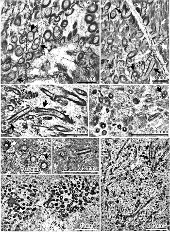
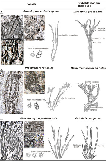
Proaulopora was originally regarded as a red alga (Vologdin, Reference Vologdin1937; Korde, Reference Korde1961, Reference Korde1973). Luchinina (in Chuvashov et al., Reference Chuvashov, Luchinina and Shuysky1987) first compared Proaulopora with the extant cyanobacterium Calothrix, a rivulariacean with collar-like projections of the sheath. However, it is hard to explain the large size of calcareous tubes of Proaulopora, the diameter of which is twice that of Calothrix, as noted by Luchinia (in Chuvashov et al., Reference Chuvashov, Luchinina and Shuysky1987). Proaulopora was recently deduced as sheath fossil of Dichothrix, which is a genus of extant rivulariacean having a thick, laminated sheath (containing one to four trichomes) with collar-like projections (Liu et al., Reference Liu, Wu, Jiang and Riding2016a, fig. 3). In this study, one calcareous tube containing two small tubes can be occasionally seen in the cross section of the tubes of Proaulopora (Figs. 7.1, 8.1, 8.2), which is similar to Dichothrix with two trichomes in one sheath (Fig. 8.1, 8.2). Therefore, the big tube size of Proaulopora can be explained by the characteristics with two or up to four trichomes in one sheath (see Fig. 7.1, 7.2). This structure of Proaulopora indicates its affinity with Dichothrix (Figs. 7.2, 8.1).
(1–3) Proaulopora ordosia n. sp., O3p. (1) Sample No. HSHB-2-2, cross sections; arrows indicate the cross sections where two tubes are included in one tubular sheath; (2, 3) sample No. HSH-5-C, cross section and longitudinal sections; the arrows in (2) indicate the external collars extending from the outer surface; the arrows in (3) point to the non-disjunctionally dichotomous branching. (4–8) Phacelophyton yushanensis; (4) O2k, sample No. Na1-3917.51, cross section and longitudinal sections; arrow indicates the position of dichotomous branching; (5) O2k, sample No. Na1-3918.03, cross sections; arrow points to the swirl-like appearance of the sheath in the cross section; (6) O2k, sample No. Na1-3918.22, cross section and longitudinal sections; (7, 8) O3p, sample No. TQPBD-7, (7) longitudinal section, (8) most are cross sections, the arrow points to a longitudinal section.

Fossil genera of Proaulopora and Phacelophyton and their probable present-day analogues, Dichothrix and Calothrix (rivulariaceans cyanobacteria), based on morphology. (1) Pictures of Proaulopora ordosia n. sp. from Ordos Basin (this study); (2) pictures of Proaulopora rarissina from Tarim Basin (Liu et al., Reference Liu, Wu, Yang and Riding2016b); (3) synthesized pictures of Phacelophyton yushanensis from both Tarim Basin (longitudinal sections, Liu et al., Reference Liu, Wu, Yang and Riding2016b) and Ordos Basin (cross sections, this paper); arrows indicate the swirl-like appearance of sheath and the position of dichotomous branching in the cross section.

Proaulopora ordosia new species
Figure 7.1–7.3
Holotype
Specimen sample no. HSHB-2-2, O3p, Sandbian, Haosihe; and sample No. HSH-5-C; O3p, reef, Sandbian, Haosihe; illustrated in Figure 7.1–7.3.
Diagnosis
Calcified tubes, occasionally and non-disjunctionally branched; wall micritic, apparently layered. Short whorl-like external collars extend from the outer surface at an acute angle and are concentric in cross section.
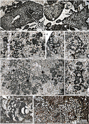
Description
Calcareous tubes; tubes occasionally and non-disjunctionally dichotomously branching (Figs. 7.3, 8.1); tube wall thick, micritic, apparently layered; short whorl-like external collars extend from the outer surface at an acute angle (Fig. 7.3); wall concentric in cross section (Fig. 7.1, 7.2), and occasionally two small tubes included in one large sheath in cross section (Fig. 7.1, 7.2); tubes 1 mm long, external diameter 130–400 μm (based on 50 specimens); wall thickness 50–70 μm.
Etymology
After “ordos”, referring to the Ordos Basin.
Material
HSHB-2-2, HSHB-2-1, HSH-8-1, HSH-8-2, HSH-8-4, O3p, Sandbian, coral limestone, Haosihe; TQPBD-7, O3p, Sandbian, Taoqupo. LQDZ-2, LQDZ-3, O3b, packstone, middle Katian, Dongzhuang.
Remarks
Many species of Proaulopora were defined by Korde (Vologdin, Reference Vologdin1937; Korde, Reference Korde1973). But most of these species, such as P. flexuosa Korde, Reference Korde1973, P. longa Korde, Reference Korde1973, P. extincta Korde, Reference Korde1973, P. composite Korde, Reference Korde1973, P. microspora Korde, Reference Korde1973, P. crassa Korde, Reference Korde1973, and P. recta Korde, Reference Korde1973, have relatively straight tubes without bifurcation and are hard to distinguish from each other due to very little differences. Proaulopora flexuosa is regarded as a synonym of Proaulopora rarissima (Fig. 8.2; Liu et al., Reference Liu, Wu, Jiang and Riding2016a), which has long and flexuous tubes of frequent dichotomous branch. Proaulopora pachydermatica Liu et al., Reference Liu, Wu, Yang and Riding2016b differs from other species in its thick wall and relatively short tubes (Liu et al., Reference Liu, Wu, Yang and Riding2016b).
Proaulopora ordosia n. sp. differs from all other Proaulopora species in its thin wall, relatively larger and shorter tubes, sparse collar-like projections, and non-disjunctional dichotomous branch (Fig. 8.1). Proaulopora sp. reported by Ye et al. (Reference Ye, Yang, Xu, Zheng, Zuo, Zhou, Li, Li, Song, Yong, Zhang and Zhang1995) is different from Proaulopora ordosia n. sp. in irregular collar-like projections, which occurred in the Pingliang Formation of the Tiewadian section.
Genus Phacelophyton Bian and Zhou, Reference Bian and Zhou1990
Type species
Phacelophyton yushanensis Bian and Zhou, Reference Bian and Zhou1990; Upper Ordovician; South China.
Remarks
Phacelophyton has been reported from the Upper Ordovician in South China (Bian and Zhou, Reference Bian and Zhou1990), the Tarim Basin (Liu et al., Reference Liu, Yang and Wu2011, Reference Liu, Wu, Yang and Riding2016b, Reference Liu, Wu, Jiang, Wu and Jia2017), and the Ordos Basin (Ye et al., Reference Ye, Yang, Xu, Zheng, Zuo, Zhou, Li, Li, Song, Yong, Zhang and Zhang1995). In this study, Phacelophyton occurs not only in the Upper Ordovician but also in the Darriwillian Stage of the Middle Ordovician.
Phacelophyton is similar to Proaulopora in having a laminated wall (Fig. 8), but differs from Proaulopora in the absence of whorl-like external collars and presence of pronounced sheath-within-sheath wall structure (Fig. 8). Bian and Zhou (Reference Bian and Zhou1990) regarded Phacelophyton as a cyanobacterium based on the similarities of its laminated wall to the multilayered sheaths of some extant cyanobacteria. Phacelophyton was further compared with the present-day rivulariacean cyanobacteria Calothrix based on relatively thick laminate sheath, and attributed to probable cyanobacteria (see Liu et al., Reference Liu, Wu, Yang and Riding2016b, fig. 3). In this study, we see in cross section a structure in which two packed paratactic tubular filaments are contained in one sheath (Fig. 7.4). It probably represents the position where filament bifurcation occurs, as shown in Calothrix (Fig. 8.3).
Phacelophyton yushanensis Bian and Zhou, Reference Bian and Zhou1990
Figure 7.4–7.8
- Reference Bian and Zhou1990
Phacelophyton yushanensis Bian and Zhou, p. 6, pl. 3, figs. 4, 5, 7.
- Reference Ye, Yang, Xu, Zheng, Zuo, Zhou, Li, Li, Song, Yong, Zhang and Zhang1995
Phacelophyton; Ye et al., p. 16, pl. 9, figs. 1, 2.
- Reference Liu, Yang and Wu2011
Phacelophyton sp. Liu et al., p. 501, pl. 4, fig. 5.
- Reference Liu, Wu, Yang and Riding2016b
Phacelophyton yushanensis; Liu et al., p. 197, fig. 7E–H.
Holotype
Specimen J-05-10, Nanjing University collection; illustrated by Bian and Zhou (Reference Bian and Zhou1990, pl. 3, figs. 5, 7), Sanqushan Formation (upper Katian, Upper Ordovician), Zhejiang and Guizhou provinces, South China.
Description
Calcareous thallus composed of tubes; tubes elongate, straight to gently curved; tubes branched at acute angles 25°~40° (Figs. 7.7, 8.3); tube wall micritic, relatively thick laminate created by a ‘sheath-within-sheath’ interlayered structure, with a smooth interior surface and irregularly patterned external surface, forming a diffuse swirl-like appearance in cross section (Fig. 7.4–7.6), and occasionally two packed paratactic tubes can be seen in cross section (Figs. 7.4, 8.3); overall thallus up to 5 mm wide and 15 mm high; individual tube 4 mm long; external diameter 70~78 μm; internal diameter ~43 μm.
Material
LSD-K-3, LSD-K-5, LSD-K-7, O2k, Darriwilian, packstone, Zhuozishan. 3917.26, 3917.39, 3917.51, 3917.65, 3917.92, 3918.03, 3918.22, packstone, Na1; 9-95-33, 9-95-79, O2z, Darriwilian, packstone, Qitan1; TQPBD-7, O3p, Sandbian, microbial reef, Taoqupo; HSHDSQ-2, O3p, Sandbian, coral limestone.
Remarks
Phacelophyton yushanensis is the only species of the genus. Phacelophyton sp. reported by Ye et al. (Reference Ye, Yang, Xu, Zheng, Zuo, Zhou, Li, Li, Song, Yong, Zhang and Zhang1995) likely can be identified as Phacelophyton yushanensis (Liu et al., Reference Liu, Wu, Jiang and Riding2016a; this study), which occurred in the Beiguoshan Formation of the Tiewadian section.
Calcified Microproblematica
Genus Garwoodia Wood, Reference Wood1941
Type species
Garwoodia gregaria Wood, Reference Wood1941; Carboniferous; Scotland.
Remarks
Due to the similarity of its organization of small juxtaposed radial filaments with Bija, Ortonella, Hedstroemia, and Cayeuxia, Garwoodia was also regarded as a cyanobacterium (Luchinina in Chuvashov et al., Reference Chuvashov, Luchinina and Shuysky1987). However, owing to its large tubes, which distinguish this specimen from other similar cyanobacteria, it is regarded as Microproblematicum by Riding (Reference Riding and Riding1991). Dragastan (Reference Dragastan1993) attributed Garwoodia to green algae, based on its relatively large tube diameter. For these reasons, Liu et al. (Reference Liu, Wu, Jiang and Riding2016a) attributed Garwoodia to calcified Microproblematica. However, relatively small species of Garwoodia were found in the Ordovician of the Tarim Basin (Liu et al., Reference Liu, Wu, Jiang and Riding2016a). Nevertheless, we now still tentatively place it in the cyanobacteria or green algal calcified Microproblematica.
Garwoodia gregaria Nicholson, Reference Nicholson1888, emend. Wood, Reference Wood1941
Figure 9.1, 9.2
- Reference Wood1941
Garwoodia gregaria; Wood, p. 222, pl. 14, figs. 1, 2.
- Reference Mamet and Roux1975
Garwoodia gregaria; Mamet and Roux, p. 151, pl. 8, figs. 6, 8, 9.
- Reference Guilbault and Mamet1976
Garwoodia gregaria; Guilbault and Mamet, p. 642, pl. 2, figs. 2, 3.
- Reference Pratt, Haidl, Pratt and Holmden2008
Garwoodia; Pratt and Haidl, fig. 15G, H.
- Reference Liu, Wu, Yang and Riding2016b
Garwoodia gregaria; Liu et al., p. 204, fig. 9I, J.
(1, 2) Garwoodia gregaria, from O3b, sample No. LJP-7-B, longitudinal sections; the arrows in (2) indicate the position of branching. (3, 4) Renalcis seriata, O3p, sample No. TQPBD-7, oblique longitudinal sections. (5) Renalcis granosus, O3p, TQPBD-7, oblique longitudinal sections. (6) Renalcis seriata and Renalcis granosus growing together, O3p, sample No. TQPBD-7, oblique cross and longitudinal sections. (7) Izhella sp. indet., O3p, sample No. TQPBD-7, longitudinal section. (8) Wetheredella silurica, O3b, sample No. LJPBGS-1(e), cross sections. (9) Rothpletzella sp., O3b, sample No. LJP-5-c-a, cross sections.

Holotype
Slide 70741 in the Geological Survey collection, Carboniferous, Kershopefoot, Roxburghshire, Scotland.
Description
Calcareous, sub-rounded or elongate thallus composed of tubular filaments; filaments packed, radiating from a base, rounded in cross section; filaments locally branching at angles up to 90° and then turning parallel to the parent filament (Fig. 9.1–9.3); thallus 2–4 mm across, diameter 70 μm.
Material
LJP-7-B, LJPBGS-2, O3b, packstone, middle Katian, Beiguoshan.
Remarks
The other species of Garwoodia, such as G. americana Johnson, Reference Johnson1961 and G. media Johnson, Reference Johnson1945, are usually with filament diameters ~30 μm. Therefore, G. gregaria is distinguished from other species by its coarser filaments and larger thallus.
Genus Renalcis Vologdin, Reference Vologdin1932
Types species
Renalcis granosus Vologdin, Reference Vologdin1932; middle Cambrian; Altai Mountains, Russia.
Remarks
Nephelostroma Dangéard and Doré, Reference Dangéard and Doré1957 is regarded as a junior synonym of Renalcis (Reitlinger, Reference Reitlinger1960). The classification of Renalcis has been controversial for many years. Renalcis was considered to be a chroococcalean cyanobacterium for a long time (Hofmann, Reference Hofmann1975; Pratt, Reference Pratt1984; Luchinina in Chuvashov et al., Reference Chuvashov, Luchinina and Shuysky1987; Riding, Reference Riding and Riding1991; Turner et al., Reference Turner, James and Narbonne2000). However, the Renalcis form is also considered to be produced by precipitation induced by communities of phototrophic and heterotrophic bacteria (Chafetz and Guidry, Reference Chafetz and Guidry1999) or heterotrophic bacterial activity only (Stephens and Sumner, Reference Stephens and Sumner2002). Due to the lack of present-day analogues, Renalcis and similar genera, such as Izhella, are attributed to possible bacterial-calcified Microproblematica (Feng et al., Reference Feng, Gong and Riding2010; Liu et al., Reference Liu, Wu, Yang and Riding2016b).
Renalcis seriata Korde, Reference Korde1961
Figure 9.3, 9.4, 9.6
- Reference Korde1961
Renalcis seriata Korde, pl. 2, fig. 3.
- Reference Wayne1971
Renalcis sp. Wayne, fig. 4c.
- Reference Wang, Zhang, Yang, Wang and Li2011
Renalcis; Wang et al., pl. 1, fig. 7.
- Reference Liu, Yang and Wu2011
Renalcis sp. Liu et al., p. 501, pl. 3, fig. 6.
- Reference Liu, Wu, Yang and Riding2016b
Renalcis seriata; Liu et al., p. 199, fig. 9B.
Holotype
Specimen illustrated by Korde (Reference Korde1961, pl. 2, fig. 3), Cambrian, Siberian Platform, Russia.
Description
Calcareous microfossils composed of chamber clusters; chambers hollow and reniform; branches of superposed chambers arising from a common base (Fig. 9.3, 9.4); chambers ~200 μm in external diameter; wall thickness ~50 μm, variable.
Remarks
Renalcis seriata is characterized by its superposed chambers with no clefts on the inner surface.
Renalcis granosus Vologdin, Reference Vologdin1932
Figure 9.5, 9.6
- Reference Vologdin1932
Renalcis granosus Vologdin, p. 15, pl. 9.
- Reference Riding and Toomey1972
Renalcis sp. Riding and Toomey, p. 514, pl. 2, figs. 1–3.
- Reference Bian and Zhou1990
Renalcis devonicus Bian and Zhou, pl. 1, figs. 2, 5.
- Reference Ye, Yang, Xu, Zheng, Zuo, Zhou, Li, Li, Song, Yong, Zhang and Zhang1995
Renalcis; Ye et al., p. 15, pl. 8, fig. 6.
- Reference Wang, Li, Zhang, Yang and Huang2009
Renalcis; Wang et al., fig. 2d.
- Reference Wang, Li, Zhang, Yang and Huang2009
Izhella; Wang et al., fig. 2e.
- Reference Liu, Wu, Yang and Riding2016b
Renalcis granosus; Liu et al., p. 199, fig. 9A.
Holotype
Specimen illustrated by Vologdin (Reference Vologdin1932, p. 15, pl. 9.), Altai Limestone (Cambrian) Siberia.
Description
Calcareous microfossils composed of chambers; chambers hollow, rounded, and reniform; botryoidal clusters with short branches; chambers wider than high, mutually overlapping (Fig. 9.5); wall micritic, moderately thick, may contain clefts on the inner surface (Fig. 9.5); smooth well-defined outer and inner surfaces; chambers relatively large, with external diameter of 200~500 μm, wall ~50 μm thick.
Material
TQPBD-1, TQPBD-7, O3b, microbial reef limestone, middle Katian, Taoqupo.
Remarks
Renalcis reported by Ye et al. (Reference Ye, Yang, Xu, Zheng, Zuo, Zhou, Li, Li, Song, Yong, Zhang and Zhang1995) was identified as Renalcis granosus by Liu et al. (Reference Liu, Wu, Yang and Riding2016b). Renalcis granosus also occurred in the Pingliang Formation of the Taoqupo section. Renalcis granosus and R. seriata are often intergrown together (Fig. 9.5), making them difficult to distinguish.
Genus Izhella Antropov, Reference Antropov1955
Type species
Izhella nubiforms Antropov, Reference Antropov1955; Upper Devonian; Russia.
Remarks
Izhella was originally only reported from the Devonian (Antropov, Reference Antropov1955; Wray, Reference Wray1967; Feng et al., Reference Feng, Gong and Riding2010). Recently, it was reported from the early Katian stage of the upper Ordovician from the Tarim Basin (Liu et al., Reference Liu, Wu, Yang and Riding2016b). Its report in the Ordos Basin (this study) is its first record in the Sandbian stage of the Upper Ordovician. Izhella closely resembles Renalcis, but is distinguished from Renalcis by its much thicker chamber walls and clefts on its inner surface.
Izhella sp. indet.
Figure 9.7
Description
Chambered microfossil composed of chambers; chambers hollow, rounded, and flower-like, compact botryoidal clusters from which short branches arise, small ovoid to reniform chambers in short radial stems; wall lunate, relatively thick, micritic, with deep clefts extending through approximately two-thirds of the wall thickness and opening toward the inner surface (Fig. 9.7); basal chambers up to 0.5 mm in external diameter, with compound lobate cavity ~0.3 mm wide; walls ~0.1 mm thick with clefts up to 30 μm deep.
Material
TQPBD-1, TQPBD-7, O3b, microbial reef limestone, middle Katian, Taoqupo.
Remarks
Due to the lack of observable cross sections (Fig. 9.7), it is difficult to assign this microfossil to a specific species.
Genus Wetheredella Wood, Reference Wood1948
Type species
Wetheredella silurica Wood, Reference Wood1948; Lower Silurian; England.
Remarks
Allonema Ulrich and Bassler, Reference Ulrich and Bassler1904 is a possible synonym of Wetheredella (Jarochowska and Munnecke, Reference Jarochowska and Munnecke2014). Wetheredella is common in many periods of the Paleozoic (Liu et al., Reference Liu, Wu, Jiang, Wu and Jia2017). Wetheredella has variously been suggested to be a foraminifer (Wood, Reference Wood1948), cyanobacterium (Copper, Reference Copper1976; Kazmierczak and Kempe, Reference Kazmierczak and Kempe1992, Reference Kazmierczak and Kempe2004), and green alga (Mamet and Roux, Reference Mamet and Roux1975; Ishchenko and Radionova, Reference Ishchenko and Radionova1981; Chuvashov et al., Reference Chuvashov, Luchinina and Shuysky1987). However, due to lack of modern analogues, it has been attributed to Microproblematicum (Riding, Reference Riding and Riding1991), and this attribution was followed by Liu et al. (Reference Liu, Wu, Yang and Riding2016b) and herein.
Wetheredella silurica Wood, Reference Wood1948
Figure 9.8
- Reference Wood1948
Wetheredella silurica Wood, p. 20, pl. 3, fig. B, pl. 5, fig. B.
- Reference Copper1976
Wetheredella tumulus Copper, p. 277, pl. 1, figs. B, C.
- Reference Liu, Wu, Jiang and Riding2011
Wetheredella silurica; Liu et al., p. 502, pl. 3, fig. 1.
- Reference Liu, Wu, Yang and Riding2016b
Wetheredella silurica; Liu et al., p. 203, fig. 9E, F
Holotype
Specimen illustrated by Wood (pl. 5, fig. B), Wenlock Limestone (Lower Silurian), May Hill, Gloucestershire.
Description
Calcareous microfossils composed of aggregations of encrusting short tubes; tubes generally hemispherical in transverse section (Fig. 9.8), closely packed and irregularly stratiformly arranged; tubes 100–250 μm long and 50–100 μm wide.
Materials
LJBGS-1(e), O3b, packstone, lower Katian, Beiguoshan.
Remarks
Wetheredella silurica is the only species of the genus. Wetheredella tumulus is regarded as a synonym of Wetheredella silurica because of their similarities (Liu et al., Reference Liu, Wu, Yang and Riding2016b).
Genus Rothpletzella Wood, Reference Wood1948
Type species
Rothpletzella gotlandica (Rothpletz) Wood, Reference Wood1948; Silurian, Wenlock and Woolhope Limestones; England and Sweden.
Remarks
Rothpletzella was common in the Paleozoic (see review by Liu et al., Reference Liu, Wu, Jiang, Wu and Jia2017). Rothpletzella has generally, but not for any very compelling reasons, been regarded as an alga or cyanobacterium, and is attributed to Microproblematicum (Riding, Reference Riding and Riding1991). This classification has been followed by many researchers (Riding and Fan, Reference Riding and Fan2001; Feng et al., Reference Feng, Gong and Riding2010; Liu et al., Reference Liu, Wu, Yang and Riding2016b). Therefore, we also attribute Rothpletzella to calcified Microproblematicum.
Rothpletzella sp. indet.
Figure 9.9
Description
Calcareous prostrate and encrusting thallus consists of tubular filaments; filaments bifurcating to form sheet-like flat to undulose layers, packed, and paralleled in longitudinal section, resembling strings of beads in transverse section (Fig. 9.9); wall very thin, micritic; filaments diameter 50–80 μm.
Materials
TQPBD-10, O3b, packstone, Sandbian, Taoqupo; LJP-5-B-a, LJP-5-C-a, LJP-5, LJPBGS-1(d), LJPBGS-1(e), LJPBGS-3(2), packstone, middle Katian, Beiguoshan.
Remarks
The lack of thin sections in which the length and branching of filaments can be measured (Fig. 9.9) makes it is difficult to assign this organism to a species.
Discussion
Comparison with other contemporary flora
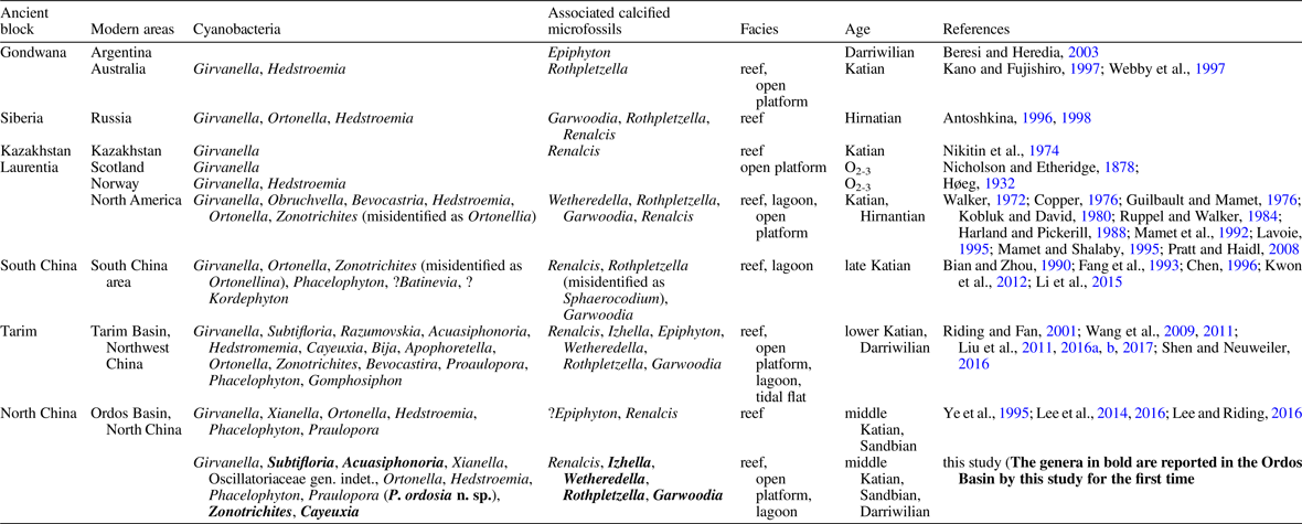
Based on the systematic paleontological analysis above, all genera previously reported in this area (Ye et al., Reference Ye, Yang, Xu, Zheng, Zuo, Zhou, Li, Li, Song, Yong, Zhang and Zhang1995; Lee et al., Reference Lee, Sun, Choh and Lee2014, Reference Lee and Riding2016; Lee and Riding, Reference Lee and Riding2016), except for Epiphyton, were found in this study. Eight genera (Subtifloria, Acuasiphonoria, Oscillatoriaceae gen. indet., Zonotrichites, Cayeuxia, Garwoodia, Izhella, Wetheredella, and Rothpletzella) are reported in this area for the first time (Table 1), increasing the diversity of the Ordos Basin flora to 17 genera (Table 1). We discovered that these calcified cyanobacteria and associated microfossils were widely distributed in the various facies of carbonate platform. This study also provides further evidence for the affinities of Phacelophyton and Proaulopora with modern cyanobacteria.
The main calcified cyanobacteria and associated microfossil floras around the world during the Middle and Late Ordovician.

Table 1 lists the main calcified cyanobacteria and microfossil floras and their facies occurrences from the Middle and Upper Ordovician worldwide (Fig. 1.1). Our findings reveal that the diversity of calcified cyanobacteria and associated microfossils of the Ordos Basin is the second highest in the world, only slightly lower than that of the Tarim Basin (Table 1). The taxonomic composition of the Ordos flora is similar to that of Tarim Basin; except for Xianella and Oscillatoriaceae gen. indet., all 14 of the other Ordos Basin genera are recorded in the Tarim Basin (Table 1). Based on new discoveries in the Ordos Basin, nine genera of calcified cyanobacteria and associated microfossils (Girvanella, Zonotrichites, Hedstroemia, Ortonella, Phacelophyton, Garwoodia, Wetheredella, Rothpletzella, and Renalcis) were distributed in at least three independent blocks during the Middle and Late Ordovician (Table 1), and five genera (Subtifloria, Acuasiphonoria, Cayeuxia, Proaulopora, and Izhella) have been found in two independent blocks (Table 1). Therefore, the high diversity of calcified cyanobacteria and associated microfossils during the Middle and Late Ordovician observed in the Tarim and Ordos basins is possibly a global phenomenon in the facies reported here (reef, open platform, lagoonal, and tidal flat environments) during the Middle and Late Ordovician.
Ecological significance
Modern cyanobacterial floras include taxa such as Oscillatoria, Lyngbya, Microcoleus, Phormidium, Rivularia, Calothrix, Dichothrix, Schizothrix, and Scytonema (Hoffman, Reference Hoffman1999; Whitton, Reference Whitton2012; Liu et al., Reference Liu, Wu, Jiang and Riding2016a) and occur in tropical marine coastal ecosystems. The highest biodiversity of modern cyanobacteria is observed in littoral zones, where they form intertidal and infralittoral mats, live as endoliths in carbonate substrates, or form symbiotic associations (Hoffmann, Reference Hoffman1999). Cyanobacterial communities also inhabit mangrove forests and coastal lagoons, where they reside on leaf litter, root litter, and live roots and often form extensive mats on the surrounding sediments. Especially diverse cyanobacterial communities are found near coral reefs, playing an essential role in the ecology of modern reef ecosystems by forming a major component of epiphytic, epilithic, and endolithic communities (Charpy et al., Reference Charpy, Casareto, Langlade and Suzuki2012). As photosynthetic organisms, cyanobacteria are important contributors to benthic and open ocean primary production, but their main role in tropical marine ecosystems appears to be as nitrogen fixers (Hoffmann, Reference Hoffman1999).
Based on this study, benthic cyanobacteria floras occurred in the tropical littoral zones during the late Middle and Late Ordovician, and were possibly globally composed of Girvanella, Subtifloria, Acuasiphonoria, Hedstroemia, Ortonella, Zonotrichites, Cayeuxia, Proaulopora, Phacelophyton, and others (Table 1). The taxonomic similarity between modern cyanobacteria floras and the Ordovician ones studied here suggests that Middle and Late Ordovician cyanobacteria floras played similar ecological roles in tropical marine coastal ecosystems to modern-day floras, including N2-fixation and hydrolysis of organic phosphorus (Liu et al., Reference Liu, Wu, Jiang and Riding2016a, Reference Liu, Wu, Jiang, Wu and Jia2017). This study strongly suggests that cyanobacteria and associated microfossils were integral to complex benthic ecosystems on carbonate platforms and played greater ecological roles during the late Middle and Late Ordovician than was previously thought.
Acknowledgments
This work was supported by the National Natural Science Foundation of China (Grant Nos. 42072127, 41502004, 41972143, 41502148, 41720104002, 41890844), the Strategic Priority Research Program of the Chinese Academy of Sciences (Grant No. XDB26000000), Natural Science Foundation of Shaanxi Province (Grant No. 2018JM4037), and National Science and Technology Major Project (Grant No. 2016ZX05004006). We thank four anonymous reviewers for helpful comments and suggestions that helped us improve the final manuscript, and E. Currano for expert editorial guidance and encouragement.