There are over 150,000 children in the United States foster care system below five years of age (U.S. Department of Health and Human Services, 2018). Most children in foster care have experienced at least one form of maltreatment, with neglect being the most common (U.S. Department of Health and Human Services, 2018). Internationally, the use of foster care as a form of alternative care varies based on a combination of historical, cultural, structural, and other contextual factors (Petrowski, Cappa, & Gross, Reference Petrowski, Cappa and Gross2017), and the experiences of children in foster care may differ across regions as well. Nevertheless, one common experience among all children in foster care, with the exception of those placed as newborns, is disruptions in caregiving.
Extensive research with children who have histories of foster care placement, and more generally with maltreated children, indicates that a lack of consistent and responsive caregiving in early childhood is often associated with adverse behavioral and neurobiological outcomes (Shonkoff, Boyce, & McEwen, Reference Shonkoff, Boyce and McEwen2009). Such outcomes can include – but are not limited to – increased behavioral difficulties, mental health problems, attachment problems, executive functioning deficits, abnormal diurnal cortisol production patterns, and underdeveloped neural structures, such as reduced gray matter volume and white matter integrity (e.g., Bernard, Frost, Bennett, & Lindhiem, Reference Bernard, Frost, Bennett and Lindhiem2017; Hawk & McCall, Reference Hawk and McCall2010; Lionetti, Pastore, & Barone, Reference Lionetti, Pastore and Barone2015; Oswald, Heil, & Goldbeck, Reference Oswald, Heil and Goldbeck2010; Sheridan, Fox, Zeanah, McLaughlin, & Nelson, Reference Sheridan, Fox, Zeanah, McLaughlin and Nelson2012; Wade, Fox, Zeanah, & Nelson, Reference Wade, Fox, Zeanah and Nelson2019).
One neurobiological measure that has been investigated with respect to child maltreatment and disrupted caregiving is alpha electroencephalogram (EEG) asymmetry (e.g., Curtis & Cicchetti, Reference Curtis and Cicchetti2007; McLaughlin, Fox, Zeanah, & Nelson, Reference McLaughlin, Fox, Zeanah and Nelson2011; Miskovic, Schmidt, Georgiades, Boyle, & MacMillan, Reference Miskovic, Schmidt, Georgiades, Boyle and MacMillan2009). Alpha EEG asymmetry is a measure of differential power in the alpha band of EEG (e.g., 6–9 Hz) between hemispheres. It can be calculated for locations across the scalp, including frontal, central, and parietal regions (Reznik & Allen, Reference Reznik and Allen2018). Alpha EEG asymmetry is an important measure because it reflects differential activity between hemispheres, which research finds to be associated with certain experiences, behaviors, and emotional processes, and which may predict outcomes such as anxiety and depression (e.g., Allen & Reznik, Reference Allen and Reznik2015; Blackhart, Minnix, & Kline, Reference Blackhart, Minnix and Kline2006; Coan & Allen, Reference Coan and Allen2003; Heller, Nitschke, Etienne, & Miller, Reference Heller, Nitschke, Etienne and Miller1997; Peltola et al., Reference Peltola, Bakermans-Kranenburg, Alink, Huffmeijer, Biro and van IJzendoorn2014).
Alpha EEG Asymmetry, Child Maltreatment, and Behavioral Development
Early adverse experiences associated with disrupted caregiving appear to be associated with EEG asymmetry (Peltola et al., Reference Peltola, Bakermans-Kranenburg, Alink, Huffmeijer, Biro and van IJzendoorn2014). For example, greater right frontal asymmetry has been observed among children who experience less responsive and sensitive care (Hane & Fox, Reference Hane and Fox2006; Hane, Henderson, Reeb-Sutherland, & Fox, Reference Hane, Henderson, Reeb-Sutherland and Fox2010), as well as children of mothers with depression (Peltola et al., Reference Peltola, Bakermans-Kranenburg, Alink, Huffmeijer, Biro and van IJzendoorn2014). In addition, right frontal asymmetry is associated with both maltreatment (Miskovic et al., Reference Miskovic, Schmidt, Georgiades, Boyle and MacMillan2009; although see Tang et al., Reference Tang, Miskovic, Lahat, Tanaka, MacMillan, Van Lieshout and Schmidt2018) and institutional rearing (McLaughlin et al., Reference McLaughlin, Fox, Zeanah and Nelson2011).
Interestingly, findings regarding the relationship between early adverse experiences and asymmetry have not been confined to the frontal region. For example, Curtis and Cicchetti (Reference Curtis and Cicchetti2007) examined EEG asymmetry among maltreated and nonmaltreated children who were characterized as resilient or nonresilient, observing greater right parietal EEG asymmetry among children with a history of maltreatment relative to those without a history of maltreatment. Taken together, results from prior research generally find that exposure to maltreatment is associated with greater right asymmetry both in frontal (e.g., Miskovic et al., Reference Miskovic, Schmidt, Georgiades, Boyle and MacMillan2009) and parietal regions of the scalp (e.g., Curtis & Cicchetti, Reference Curtis and Cicchetti2007).
In seeking an understanding of the EEG asymmetry findings in the context of early life stress, some research supports an “approach/withdrawal model,” which holds that increased left frontal asymmetry is associated with approach-related behavior and emotions, and increased right frontal asymmetry is associated with avoidance-related behavior and emotions (Davidson, Reference Davidson1992; Reznik & Allen, Reference Reznik and Allen2018). Consistent with this model (Reznik & Allen, Reference Reznik and Allen2018), researchers find associations between externalizing problems and left frontal asymmetry, and between internalizing problems and right frontal asymmetry in children (e.g., Gatzke-Kopp, Jetha, & Segalowitz, Reference Gatzke-Kopp, Jetha and Segalowitz2014; Smith & Bell, Reference Smith and Bell2010). Notably, however, the support for this model is equivocal, as not all studies find this pattern (Peltola et al., Reference Peltola, Bakermans-Kranenburg, Alink, Huffmeijer, Biro and van IJzendoorn2014), suggesting the relation between frontal EEG asymmetry and behavior may not be straightforward in developmental populations.
Relative to frontal asymmetry, fewer behavioral correlates have been found for parietal asymmetry among children. In adults, right posterior regions modulate emotional processes in frontal regions (Heller, Reference Heller1993). Greater right posterior activity has been associated with anxious arousal (Heller et al., Reference Heller, Nitschke, Etienne and Miller1997), including arousal in the context of posttraumatic stress disorder (Metzger et al., Reference Metzger, Paige, Carson, Lasko, Paulus, Pitman and Orr2004; Rabe, Beauducel, Zöllner, Maercker, & Karl, Reference Rabe, Beauducel, Zöllner, Maercker and Karl2006), while reduced right posterior activity has been associated with depression (e.g., Blackhart et al., Reference Blackhart, Minnix and Kline2006; Stewart, Towers, Coan, & Allen, Reference Stewart, Towers, Coan and Allen2011). Among children, decreased right posterior activity has been associated with low levels of positive emotionality, including low positive affect and sociability (Hayden et al., Reference Hayden, Shankman, Olino, Durbin, Tenke, Bruder and Klein2008; Shankman et al., Reference Shankman, Tenke, Bruder, Durbin, Hayden and Klein2005). Thus, asymmetry across frontal and parietal regions appear to be associated with distinct patterns of behavior, emotion, and experiences in children and adults; however, some associations are more well established than others.
Present Study
Given the aforementioned large population of children in foster care who have experienced maltreatment and disrupted caregiving, and the prior findings regarding EEG asymmetry among maltreated children and children reared in institutions, it is important to understand the nature of foster children's patterns of EEG asymmetry and related behavioral development. Moreover, it is important to understand if the prior findings regarding EEG asymmetry in young children extend downward developmentally into toddlerhood as opposed to emerging later in life. The present study examined alpha EEG asymmetry in a sample of toddlers in foster care in comparison to a similarly aged, low-income community sample. Due to the high incidence of neglect, physical abuse, sexual abuse, and other forms of maltreatment among children in foster care (U.S. Department of Health and Human Services, 2018), we infer that the majority of children in the foster care group have experienced maltreatment, in addition to disruptions in care. To limit confounding effects of family income, which is often inversely related to maltreatment (for review, see Slack, Berger, & Noyes, Reference Slack, Berger and Noyes2017), a low-income community sample was used as a comparison group. While the older children in the present study might be considered preschool-aged, we refer to all child participants as “toddlers” for consistency and because data collected across waves reflect broader toddler development.
To examine EEG asymmetry, we examined group differences in absolute power between hemispheres in frontal, central, and parietal regions between the foster care group and the community comparison group. Based on prior research linking psychosocial deprivation and frontal alpha EEG asymmetry (e.g., McLaughlin et al., Reference McLaughlin, Fox, Zeanah and Nelson2011), we hypothesized that children in the foster care group would show greater right frontal alpha asymmetry relative to the community comparison group. We also explored the degree to which absolute power differences existed between groups. Finally, we investigated the relationship between alpha EEG asymmetry and caregiver report of child behavior, both for concurrent caregiver-reported behavior, as well as caregiver-reported behavior from previous time points (Waves 1 and 2). Consistent with the approach/withdrawal model (Reznik & Allen, Reference Reznik and Allen2018), we predicted that across groups, greater right frontal alpha asymmetry would be positively associated with avoidance-related measures, including internalizing problems, and negatively associated with approach-related measures, including externalizing problems.
Method
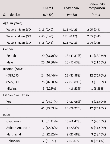
EEG data were collected in the third wave of a longitudinal study, while behavioral data were collected at all three waves. At Wave 3, participants were 54 toddlers (ages 2.51–4.03 years; M age = 3.16 years; see Table 1) and their primary caregivers, including 38 children in foster care and 16 community comparison children with no history of foster care placement. Descriptive statistics regarding age at Wave 1 (ages 1.51–3.01; M age = 2.13 years) and Wave 2 (ages 2.00–3.82 years; M age = 2.68 years) for children from whom EEG data were collected at Wave 3 are presented in Table 1. Prior to the first wave of data collection, foster toddlers entering a new foster care placement in one of two urban areas in the Pacific Northwest were recruited via referrals from the county public child welfare agency. Children and their caregivers completed laboratory assessments at approximately 1 month (Wave 1), 6 months (Wave 2), and 12 months (Wave 3) following placement. Aspects of these data, particularly related to behavioral and neuroendocrine differences, are reported elsewhere (DePasquale et al., Reference DePasquale, Olson, Desjardins, Bruce, Pears, Gunnar and Fisher2019; Olson, Kim, Bruce, & Fisher, Reference Olson, Kim, Bruce and Fisher2019; Perry, DePasquale, Fisher, & Gunnar, Reference Perry, DePasquale, Fisher and Gunnar2019).
Child demographic characteristics by group

The community comparison group (ages 2.60–3.67 years at Wave 3; M age = 3.04 years) included children with no history of involvement with the child welfare system who had lived with at least one biological parent throughout their life. Children and caregivers in the community comparison group were recruited via flyers, and laboratory assessments were completed at equivalent intervals to those in the foster care group. To obtain a low-income sample, an inclusion criterion for children in the community comparison group was to be eligible for free or reduced-price school lunches, and caregivers could not have completed more than one year of college. Seventy-five percent of families in the community comparison sample reported an annual income of less than $25,000 at Wave 3 (see Table 1). Caregivers in both groups were fluent in English, and children could not have any significant physical or developmental disorders that would make it difficult to complete the laboratory assessments. Children in foster care were granted permission to participate by the assigned child welfare caseworker (their legal guardian), and informed consent was obtained from foster caregivers in the foster care group (for their own participation) and from biological caregivers in the community comparison group (for the participation of themselves and their children). Institutional Review Board (IRB) approval for the study was obtained both from the university's IRB and from the state's Public Health IRB, which reviews all research involving children in foster care.
As noted, EEG data were collected at the third wave of data collection, which included 87 children. Thirty-three children did not have sufficient EEG data to calculate asymmetry due to refusal (n = 18) or experimental error (n = 15) and were therefore excluded from all analyses. Accordingly, the final sample included 54 children (38 children in foster care and 16 community comparison children). Sample sizes range from 49 to 54 for analyses due to differences in EEG and questionnaire collection. Table 1 describes child demographics.
Procedure
Data were collected at a laboratory session and via phone interview. In the phone interview, caregivers completed a demographics questionnaire. At the laboratory session, children were fitted with a Lycra Electro-Cap and asked to look at a book with fluorescent stars while EEG was collected. EEG was collected for a total of five minutes, comprised of alternating 30-second epochs during which the child's eyes were open and closed. Owing to difficulties separating eyes-open and eyes-closed conditions with toddlers, EEG data across these two conditions were collapsed for analyses.
Measures
Child Behavior Checklist (CBCL) for ages 1½–5 years
Caregivers completed the CBCL at all three waves. The CBCL (Achenbach & Rescorla, Reference Achenbach and Rescorla2001) is a caregiver report questionnaire regarding child problem behaviors. Items include a list of 100 behaviors, and caregivers indicate whether it is “not true,” “somewhat or sometimes true,” or “very true or often true” that each behavior describes the child. Responses can be combined to produce a total score, as well as scores on multiple subscales, including a subscale reflecting internalizing problem behaviors and a subscale reflecting externalizing problem behaviors.
Children's Behavior Questionnaire (CBQ) – short form
Caregivers completed the CBQ at Wave 3. The CBQ is a 94-item questionnaire asking caregivers to report on their child's responses to various situations using a 7-point Likert-type scale (Putnam & Rothbart, Reference Putnam and Rothbart2006). It yields scores on 15 subscales reflecting dimensions of temperament.
EEG collection and data reduction
Hardware and software by James Long Company (Caroga Lake, NY) was used to record EEG. EEG data were collected using a Lycra Electro-Cap with 32 tin electrodes using the International 10–20 Placement System (AFz, Cz, Fp1, Fp2, F7, F3, Fz, F4, F8, FC5, FCz, FC6, T7, T8, C4, C3, CP5, CP6, P7, P3, Pz, P4, P8, PO7, PO8, O1, Oz, O2, M1, M2, HEOG, VEOG) and sampled at 500 Hz. Eye movements were measured by placing one electrode under the right eye (VEOG) and another electrode horizontally to the right eye (HEOG). Online EEG was collected with the vertex (Cz) as the reference and an anterior midline site (AFz) as the ground electrode. After the child was fitted with the cap, the scalp was gently abraded and electrolytic gel was applied to each electrode site. Impedances were reduced to approximately 10 Ω (depending on child tolerance) prior to beginning data collection of the resting EEG.
All processing and analysis of the EEG was performed using the EEGLAB Toolbox (Delorme & Makeig, Reference Delorme and Makeig2004). First, the EEG was re-referenced to the average of the mastoid electrodes. Next, a bandpass filter between .1 (high pass) and 30 Hz (low pass) was applied to the continuous EEG. Second, eye channels were removed, and bad channels were identified and interpolated using spherical spline interpolation (Perrin, Pernier, Bertrand, & Echallier, Reference Perrin, Pernier, Bertrand and Echallier1989).
Continuous portions of the EEG data were rejected using spectrum thresholding in EEGLAB (i.e., pop_rejcont function). First, contiguous data epochs were extracted in half-second epochs and power was calculated (1 to 40 hz). Next, epochs were rejected if spectral thresholding exceeded 10 db across at least two epochs. Then, remaining sections of continuous EEG were rejected using visual inspection. Finally, independent component analysis (ICA) was conducted, and components containing eye-blinks and/or horizontal saccades were removed. Continuous EEG containing both eyes-open and eyes-closed conditions were collapsed for analyses. Participants had an average of 302.26 seconds (SD = 67.55) of artifact-free EEG data. The amount of artifact-free data increased with age, F(1, 51) = 9.73, p = .003, but did not significantly differ by group, t(52) = −0.08, p = .934. Age did not significantly differ between groups at Wave 1 (t(52) = 0.91, p = .367), Wave 2 (t(50) = 1.26, p = .212), or Wave 3 (t(51) = 1.40, p = .167). Similarly, gender did not differ between groups, χ2(1, N = 54) = 1.30, p = .254, correcting for continuity. The linear models revealed that at Wave 3, age was not significantly related to frontal, central, or parietal alpha asymmetry, ps > .30. Accordingly, age was not included in main analyses.
The alpha frequency band (6–9 Hz) was exported following previous literature for toddlers (Marshall, Bar-Haim, & Fox, Reference Marshall, Bar-Haim and Fox2002; McLaughlin et al., Reference McLaughlin, Fox, Zeanah and Nelson2011). Power spectral density was estimated using Welch's method, with a Hamming window with 50% overlap. We calculated absolute power by taking the natural logarithm of alpha power at each electrode location. Frontal asymmetry was calculated by taking the difference between the natural logarithm of alpha power in the right frontal electrode (F4) and left frontal electrode (F3):

Asymmetry was calculated in a parallel fashion for central and parietal regions:


Given the inverse relationship between alpha power and cortical activation, positive values indicate greater left (relative to right) activation, and negative values indicate greater right activation (Allen, Coan, & Nazarian, Reference Allen, Coan and Nazarian2004). There were no outliers greater than three standard deviations above or below the mean absolute power at any of the six electrode sites.
Data analysis
Preliminary analysis
Differences in variance
First, we tested whether variability in asymmetry differed by group at each of the three electrode locations. Specifically, we conducted F tests for equality of variance in alpha EEG asymmetry for frontal, central, and parietal locations.
Main analyses
Data analysis proceeded across two stages:
Absolute power by group, location, and hemisphere
To investigate group differences in absolute power and asymmetry, we ran a mixed analysis of variance (ANOVA) predicting absolute power (i.e., the natural logarithm of alpha power) based on group, location, and hemisphere. Specifically, to test whether there were main effects of group, location, or hemisphere on absolute power, we ran an ANOVA with location (frontal, central, parietal) and hemisphere (left, right) as within-subjects factors and group (foster care, community comparison) as a between-subjects factor. Asymmetry was examined as statistical interactions between group and hemisphere. Simple main and interaction effects were followed using Bonferroni correction. Significance values were adjusted using Greenhouse–Geisser corrections when appropriate.
Alpha asymmetry and behavioral measures
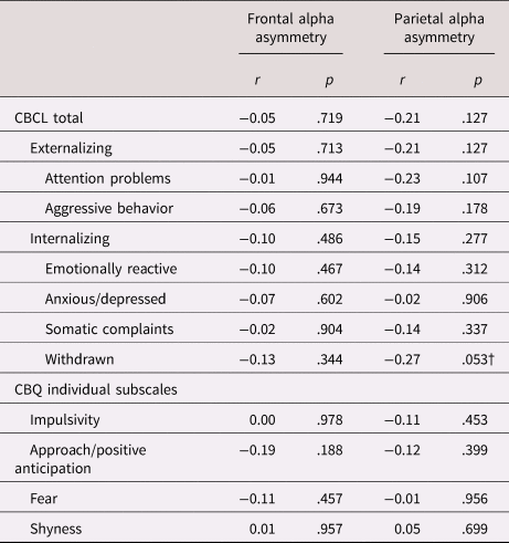
To investigate the relationship between EEG asymmetry and behavioral measures, we ran a series of bivariate correlation tests. First, we calculated the Pearson's r correlation coefficients to estimate the relationship between raw scores on each of the CBCL subscales included in the CBCL internalizing and CBCL externalizing subscales, as well as the CBCL total score, and frontal alpha asymmetry at Wave 3. Approach-related subscales included the CBCL externalizing subscale, and each of its constituent subscales: attention problems and aggressive behavior. Avoidance-related subscales included the CBCL internalizing subscale, and each of its constituent subscales: emotionally reactive, anxious/depressed, somatic complaints, and withdrawn. Pearson's r correlation coefficients were also calculated for the relationship between frontal alpha asymmetry at Wave 3 and each of the following CBQ subscales: impulsivity (approach-related), approach/positive anticipation (approach-related), fear (avoidance-related), and shyness (avoidance-related).
To investigate whether behavioral measures predicted later EEG asymmetry, parallel analyses were conducted to examine the relationship between CBCL scores at Wave 1 and frontal asymmetry at Wave 3, as well as CBCL scores at Wave 2 and frontal asymmetry at Wave 3. Finally, all correlation tests were repeated for the parietal location post hoc due to significant group differences observed in parietal asymmetry in the first stage of analyses.
Results
Preliminary analysis
Differences in variance
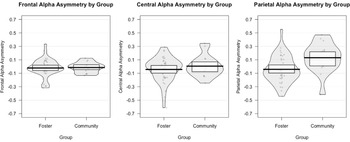
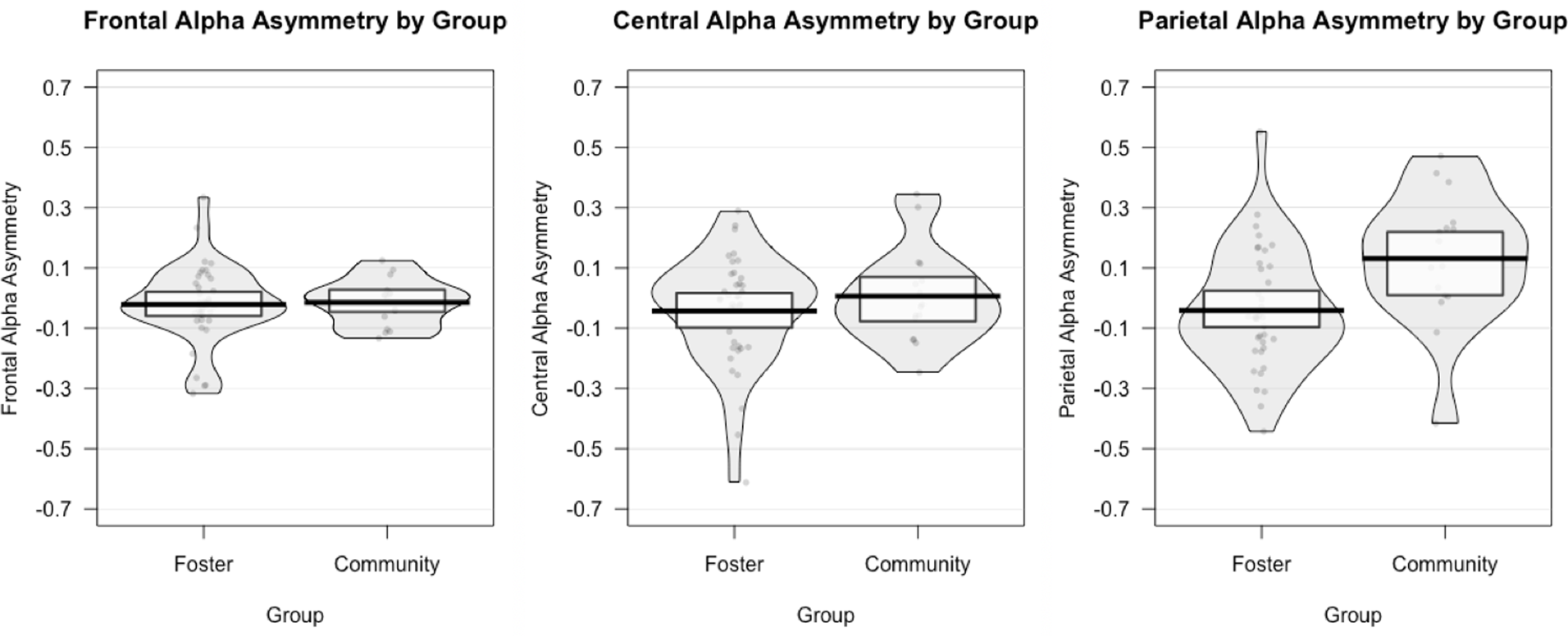
Notably, we observed a greater variance in frontal alpha asymmetry in the foster care group compared to the community comparison group, F(37, 15) = 2.94, p = .028 (see Figure 1). Variance did not significantly differ between groups for central alpha asymmetry, F(37, 15) = 1.37, p = .518, or parietal alpha asymmetry, F(37, 15) = 0.82, p = .601 (see Figure 1).
Variability in alpha electroencephalogram (EEG) asymmetry by location and group. An F test for equality of variance revealed that there was significantly greater variance in frontal alpha asymmetry in the foster care group relative to the community comparison group, p < .05. No significant group differences in variance were observed for central or parietal asymmetry, ps > .05.
Main analyses
Absolute power by group, location, and hemisphere
Main effect of location
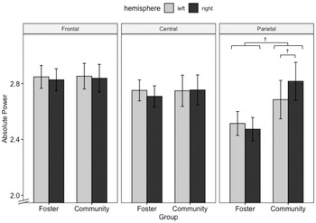
The mixed analysis of variance revealed a significant main effect of location, F(2, 104) = 13.57, p < .001, $\eta _p^2$ = .207, such that absolute power decreased from anterior to posterior regions (see Figure 2). No significant main effects of group, p = .478, or hemisphere, p = .869, were observed.
Mean absolute power (i.e., natural logarithm of alpha power) in each hemisphere by group and location. Lower absolute power reflects higher activation. Error bars represent standard error of the mean. †marginally significant, p < .10.

Group by location interaction
The main effect of location was qualified by an interaction between group and location, F(2, 104) = 5.50, p = .012, $\eta _p^2$ = .096, which was driven by a marginally significant difference in power between groups at the parietal location, p = .066, such that less absolute power was observed at parietal electrodes for the children in the foster care group (M = 2.49, SD = 0.52) as compared to children in the community comparison group (M = 2.75, SD = 0.54; see Figure 2). No group differences in power were observed for the frontal or central locations, ps > .90.
Group by hemisphere interaction
There was also a significant two-way interaction between group and hemisphere, F(1, 52) = 4.60, p = .037, $\eta _p^2$ = .081, indicating that the foster care group exhibited greater asymmetry across all scalp locations (i.e., frontal, central, parietal) as compared to the community group. This interaction was primarily driven by a greater marginally significant asymmetry (i.e., differences in power between hemispheres) in the foster care group, p = .066, relative to the community comparison group, p = .212, across all electrode locations.
Group by location by hemisphere interaction
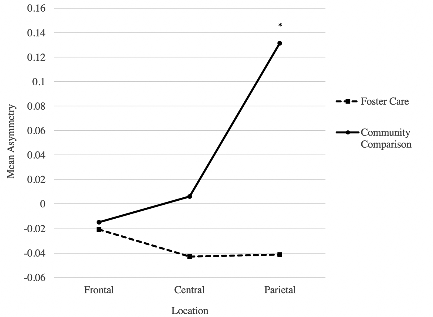
All main effects and two-way interactions were qualified by a significant three-way interaction between group, location, and hemisphere, F(2, 104) = 3.65, p = .035, $\eta _p^2$ = .066. Follow-up tests of simple interactions indicated that there was a statistically significant interaction between group and hemisphere at the parietal location, p = .021, indicating that parietal asymmetry values were significantly lower in the foster care group (M = −0.041, SD = 0.20) as compared to the comparison group (M = 0.131, SD = 0.22; see Table 2 for asymmetry values at each location and hemisphere). Specifically, at the parietal location, children in the community comparison group exhibited marginally greater absolute power in the right (M = 2.82, SD = 0.54) relative to left (M = 2.69, SD = 0.55) hemisphere, p = .062, while children in the foster care group exhibited no differences in absolute power between the right (M = 2.47, SD = 0.52) and left (M = 2.51, SD = 0.53) hemispheres, p = .422. No group differences were observed for frontal alpha asymmetry or central alpha asymmetry, ps > .90 (see Figure 3).
Alpha electroencephalogram (EEG) asymmetry across electrode locations by group. Values below zero indicate greater right relative to left alpha asymmetry. *p < .05.

Alpha electroencephalogram (EEG) asymmetry by location and group

Alpha asymmetry and behavioral measures
Frontal alpha asymmetry and behavioral measures
Frontal alpha asymmetry was not significantly correlated with the CBCL total score on any of the CBCL or CBQ subscales examined at Wave 3, ps > .10 (see Table 3). Similarly, CBCL scores at Wave 1 were not significantly correlated with frontal alpha asymmetry at Wave 3, ps > .20, nor were CBCL scores at Wave 2 significantly correlated with frontal alpha asymmetry at Wave 3, ps > .10.
Pearson's r correlations between alpha asymmetry and approach/avoidance-related Child Behavior Checklist (CBCL) and Children's Behavior Questionnaire (CBQ) subscales at Wave 3

†marginally significant, p < .10
Note: Higher asymmetry values reflect less right alpha electroencephalogram (EEG) asymmetry.
Parietal alpha asymmetry and behavioral measures
The correlation between toddlers’ scores on the withdrawn subscale of the CBCL at Wave 3 and parietal alpha asymmetry was marginally significant, r(52) = −0.27, p = .053. No other CBCL or CBQ scores at Wave 3 were significantly correlated with parietal alpha asymmetry, ps > .10 (see Table 3). CBCL scores at Waves 1 and 2 were not significantly correlated with parietal alpha asymmetry at Wave 3, ps > .10, with the exception of the CBCL attention problems subscale at Wave 1, which was significantly correlated with parietal alpha asymmetry at Wave 3, r(53) = −0.28, p = .043.
Discussion
The present study examined alpha electroencephalogram (EEG) asymmetry (i.e., differences in absolute power between hemispheres) and absolute power in a sample of toddlers in the United States foster care system and a community comparison low-income sample. As noted previously, children in foster care experience disrupted caregiving and are also likely to be exposed to neglect and other forms of maltreatment (U.S. Department of Health and Human Services, 2018). Given this fact, and in light of past research linking psychosocial deprivation and right frontal alpha EEG asymmetry (e.g., McLaughlin et al., Reference McLaughlin, Fox, Zeanah and Nelson2011), we predicted that toddlers in foster care would exhibit greater right frontal alpha EEG asymmetry relative to toddlers with no history of being in foster care. In addition, we explored whether there were more general differences in power between groups.
We observed significantly greater right asymmetry across scalp locations among toddlers in foster care relative to community comparison toddlers. Overall, the observed group differences in asymmetry across locations highlight potential neurobiological differences between children in foster care and community comparison children emerging as early as toddlerhood. The differences are especially noteworthy because the community comparison sample was comprised of children with very low family income. This suggests that the differences between groups were not the result of lack of access to economic resources but rather the nature of the caregiving experiences (including disruptions) to which the foster children were exposed.
Notably, the group differences we observed were primarily driven by differences in parietal alpha asymmetry rather than frontal. The finding that group differences in asymmetry were driven by differences at the parietal location was surprising in light of previous research that has observed a relationship between psychosocial deprivation, a form of maltreatment, and right frontal alpha asymmetry in children and adolescents (e.g., Miskovic et al., Reference Miskovic, Schmidt, Georgiades, Boyle and MacMillan2009). However, the observed differences in parietal asymmetry are consistent with the findings of Curtis and Cicchetti (Reference Curtis and Cicchetti2007), who observed greater right parietal alpha asymmetry among children with a history of maltreatment relative to children without a history of maltreatment.
While our hypotheses focused on asymmetry, or differences in power between hemispheres, we also observed some evidence of differences in absolute power between the foster care group and community comparison group. Specifically, children in the community comparison group exhibited marginally more absolute alpha power (across hemispheres) in the parietal region than children in the foster care group. Previous studies have likewise observed differences in absolute alpha power among young children with varying levels of exposure to adversity, generally finding decreased power among children exposed to greater psychosocial adversity. Marshall, Fox, and the BEIP Core Group (Reference Marshall and Fox2004) found that relative to toddlers with no history of institutionalization, toddlers who were institutionalized exhibited lower absolute alpha power across locations. This difference was primarily driven by group differences in the frontal and temporal regions rather than parietal regions; however, a follow-up study found that at eight years old, children who were institutionalized or placed in foster care after age two exhibited lower absolute alpha power in the parietal location (as well as other regions) compared to children who had never been institutionalized or who were placed in high quality foster care before age two (Vanderwert, Marshall, Nelson, Zeanah, & Fox, Reference Vanderwert, Marshall, Nelson, Zeanah and Fox2010).
We also observed significantly greater variability in frontal asymmetry in the foster care group. Although this is an exploratory finding that warrants further investigation, and may reflect normative individual differences, it may also be consistent with research that has observed greater day-level variability in diurnal cortisol levels in foster children relative to community controls (Fisher, Stoolmiller, Gunnar, & Burraston, Reference Fisher, Stoolmiller, Gunnar and Burraston2007). Together, these findings may indicate that as a group, foster children are more heterogeneous than children without a history of foster care in ways that impact neurobiological outcomes. Variations in early experiences of children in foster care may be considerable. For example, length of time in foster care, number of placement changes, specific maltreatment experiences, and other measures of adversity might be associated with distinct patterns of frontal alpha asymmetry that are obscured when results are collapsed across foster children. While the sample size and data available for the present study did not allow for such analyses, future research should consider individual differences between children in foster care when examining neurobiological outcomes.
One possible explanation for the finding that group differences were driven by parietal rather than frontal asymmetry is that group differences in frontal asymmetry are being obscured by changes in asymmetry across development. McLaughlin and colleagues (Reference McLaughlin, Fox, Zeanah and Nelson2011) examined developmental trends in frontal alpha asymmetry among children with and without a history of institutionalization. Though the two groups showed distinct trajectories across the first eight years of life, frontal asymmetry was fairly similar between groups around the age range of the present sample. Accordingly, it is possible that group differences would emerge if frontal alpha EEG asymmetry were examined at a slightly earlier or slightly later point in development.
Another possible explanation is that much of the research examining the relation between maltreatment and alpha EEG asymmetry in children has focused on children with a history of institutionalization (e.g., Frenkel et al., Reference Frenkel, Koss, Donzella, Frenn, Lamm, Fox and Gunnar2017; McLaughlin et al., Reference McLaughlin, Fox, Zeanah and Nelson2011). Although neglect is common in both children who have been institutionalized and children in foster care, foster children without a history of institutionalization constitute a distinct group from post-institutionalized children. Specifically, neglect in the context of foster care may be characterized by a lack of predictability rather than a complete lack of responsive care. In addition, many children in foster care experience other forms of maltreatment, including physical abuse and sexual abuse (U.S. Department of Health and Human Services, 2019). Maltreatment experiences characterized by deprivation, such as physical neglect, and maltreatment experiences characterized by threat, such as physical abuse, may affect neurobiological functioning through distinct pathways and be associated with distinct outcomes (McLaughlin, Sheridan, & Lambert, Reference McLaughlin, Sheridan and Lambert2014; Sheridan & McLaughlin, Reference Sheridan and McLaughlin2014). Consistent with the idea that different early experiences may be associated with distinct neurobiological pathways, Perry and colleagues (Reference Perry, DePasquale, Fisher and Gunnar2019) observed different patterns of diurnal cortisol production between foster children and post-institutionalized children. Thus, differences in early experiences may also help explain why our findings regarding frontal alpha asymmetry in foster children differ from past findings with post-institutionalized populations. It is also noteworthy that not all studies have observed elevated right frontal asymmetry in children with a history of institutionalization (e.g., Frenkel et al., Reference Frenkel, Koss, Donzella, Frenn, Lamm, Fox and Gunnar2017).
As noted previously, this is not the first report of this finding in the literature; Curtis and Cicchetti (Reference Curtis and Cicchetti2007) reported that children with a history of maltreatment showed greater right parietal alpha asymmetry than children without a history of maltreatment. Notably, Curtis and Cicchetti's (Reference Curtis and Cicchetti2007) sample included children between the ages of 6 and 12; the present study suggests that differences in parietal alpha asymmetry may emerge as early as toddlerhood. More recently, Meiers, Nooner, De Bellis, Debnath, and Tang (Reference Meiers, Nooner, De Bellis, Debnath and Tang2020) examined the relationship between child maltreatment history and trauma symptoms, parietal alpha asymmetry, and problem behaviors in early adolescence. Interestingly, they found that parietal asymmetry moderated the relationship between trauma symptoms and problem behaviors, such that for adolescents with greater right parietal asymmetry, there was a stronger positive relationship between trauma symptoms and problem behaviors. While further research is needed, this finding raises the question of whether children in foster care who show greater right parietal asymmetry might be at particular risk of developing later behavioral problems. Further research should further investigate the relationship between early experiences (e.g., maltreatment) and parietal alpha asymmetry, as well as behavioral correlates of parietal alpha asymmetry. The fact that children in the foster care group showed relatively greater right parietal asymmetry compared to the community comparison group highlights the need for future research to examine the developmental trajectories of parietal alpha asymmetry across development. Given that the prefrontal cortex is slow to develop in early childhood relative to other brain regions (Gogtay et al., Reference Gogtay, Giedd, Lusk, Hayashi, Greenstein, Vaituzis and Thompson2004), it is possible that differences in alpha asymmetry might emerge in posterior areas before frontal areas over the course of development.
It is also important to acknowledge that we observed little indication of a relationship between frontal alpha EEG asymmetry (at Wave 3) and caregiver-reported behavior at any wave. While this finding is inconsistent with some previous work (e.g., Smith & Bell, Reference Smith and Bell2010), it is not unprecedented. For example, a recent meta-analysis by Peltola and colleagues (Reference Peltola, Bakermans-Kranenburg, Alink, Huffmeijer, Biro and van IJzendoorn2014) similarly did not find a significant relationship between right frontal alpha asymmetry and internalizing problems or between left frontal alpha asymmetry and externalizing problems among children with psychosocial risk factors, including a history of maltreatment.
Given observed group differences in parietal asymmetry, we also examined the relationship between parietal alpha EEG asymmetry (at Wave 3) and caregiver-reported behavior at all three waves. While parietal asymmetry (at Wave 3) was not significantly related to caregiver-reported behavior at any wave, we observed a weak – but marginally significant – correlation between parietal alpha asymmetry and caregiver-reported child withdrawal behavior at Wave 3, such that greater right parietal alpha asymmetry was associated with greater withdrawal behavior. Interestingly, this pattern is inconsistent with previous findings observing a relationship between decreased right posterior asymmetry and low positive emotionality in children (e.g., Hayden et al., Reference Hayden, Shankman, Olino, Durbin, Tenke, Bruder and Klein2008; Shankman et al., Reference Shankman, Tenke, Bruder, Durbin, Hayden and Klein2005), warranting further investigation.
It is possible that behavioral patterns related to alpha EEG asymmetry emerge at different developmental time points. While we assessed whether caregiver-reported behavior at earlier time points reflected later alpha EEG asymmetry, we did not assess whether alpha asymmetry among children in the present sample might predict later behavior, as EEG was collected at the final wave. Supporting this possibility, McLaughlin and colleagues (Reference McLaughlin, Fox, Zeanah and Nelson2011) found that patterns of right frontal alpha asymmetry in early childhood were related to children's internalizing problems at 4.5 years. Similarly, Hayden and colleagues (Reference Hayden, Shankman, Olino, Durbin, Tenke, Bruder and Klein2008) found that less right parietal asymmetry at ages 5–6 was associated with low positive emotionality later in childhood. Interestingly, some research suggests that parietal asymmetry is not a stable trait marker among children (Müller, Kühn-Popp, Meinhardt, Sodian, & Paulus, Reference Müller, Kühn-Popp, Meinhardt, Sodian and Paulus2015), further emphasizing the need to explore behavioral correlates of parietal alpha asymmetry across development.
This is certainly not the first observation in the literature of neurobiological measures not being readily connected to observable changes in behavioral functioning following early life stress and disrupted caregiving. For example, although there has been extensive replication of alterations in cortisol production in maltreated samples (e.g., Bernard et al., Reference Bernard, Frost, Bennett and Lindhiem2017; Cicchetti & Rogosch, Reference Cicchetti and Rogosch2001; Fisher, Gunnar, Chamberlain, & Reid, Reference Fisher, Gunnar, Chamberlain and Reid2000), associations between cortisol levels and behavioral disruptions in these studies have been reported much less frequently (Flannery, Beauchamp, & Fisher, Reference Flannery, Beauchamp and Fisher2017). It is important not to discount the presence of biological changes in the absence of behavior changes, as they may still be important sequelae of disrupted early caregiving. Work on this topic might also benefit from more detailed behavioral assessments, as the present study relied solely on caregiver report measures.
Limitations and conclusions
The present study had several limitations. First, we had limited information regarding the early experiences of children in each group. While we intended to code child welfare records to obtain information regarding children's maltreatment experiences, changes in state regulations for research within the child welfare system made it impossible to access the necessary case files. Understanding whether there is a relationship between specific dimensions of early adverse experiences (e.g., deprivation, threat) is an important avenue for future research. Second, we were only able to collect EEG from a subset of participants in the larger study. Large data loss is common in developmental research, particularly – in our experience – with high-risk samples. Our recruitment of a high-risk sample of toddlers is a strength, as it is rare to obtain neurobiological measures from toddlers in foster care. A third and related limitation is our small sample size, particularly in the community comparison group. A fourth limitation is that the research is cross-sectional, which prevented us from being able to detect group differences in developmental trajectories or examine the relationship between alpha asymmetry and behavior at different points in development. Fifth, although collapsing across experimental conditions (i.e., eyes-open and eyes-closed) maximized the number of epochs from which we could use data, it might be considered a limitation. Similar studies examining alpha EEG asymmetry in children and adolescents have generally focused on one condition (e.g., Curtis & Cicchetti, Reference Curtis and Cicchetti2007; McLaughlin et al., Reference McLaughlin, Fox, Zeanah and Nelson2011) or combined data across conditions to produce a composite measure (e.g., Miskovic et al., Reference Miskovic, Schmidt, Georgiades, Boyle and MacMillan2009).
Despite these limitations, the present study sheds light on alpha EEG asymmetry and potential behavioral correlates in a population for which alpha EEG asymmetry has not been extensively studied: toddlers in the United States foster care system. Our findings point to the need to investigate alpha EEG asymmetry among foster children, and the relationship between alpha EEG asymmetry and behavioral measures, at different points in development. Future research should also differentiate between different aspects of adversity experienced by children in the foster care system when investigating the relationship between early adversity and alpha EEG asymmetry, and when considering alpha asymmetry as a potential mechanism by which early experiences affect behavior. Ultimately, further understanding the neurobiological mechanisms by which early adversity affects child outcomes has the potential to play a key role in informing prevention and intervention efforts to support children in the foster care system.
Acknowledgments
The present study would not have been possible without the generous long-term mentorship and collegiality provided by Megan Gunnar to the senior author. In addition, the authors thank the members of the Early Experience, Stress Neurobiology, and Development Network for their guidance in the design and execution of the study. We also are grateful to Kristen Greenley, Dr. Jacqueline Bruce, Dr. Katherine Pears, Dr. Nathan Fox, and Dr. Leslie Roos for their contributions to the conceptualization and execution of this study. We would also like to acknowledge and thank the families who participated in this research study.
Funding Statement
This research was supported by grants P50 DA035763, P50 MH078105, P50 DA048756, and F32HD097921.
Conflicts of Interest
None.