


Introduction
The mammary gland plays a key role in the performance of lactating swine and piglets, and minimizing the impacts of oxidative stress in the mammary gland tissue has been the focus of several studies (Kim and Wu, Reference Kim and Wu2009; Dallanora et al., Reference Dallanora, Walter, Marcon, Saremba, Bernardi, Wentz and Bortolozzo2016; Zhu et al., Reference Zhu, Guo, Gao, Wang, Chen, Ma and Jiang2017; Holanda et al., Reference Holanda, Marcolla, Guimarães, Neves, Hausman, Duarte, Abreu and Saraiva2019; Gao et al., Reference Gao, Wen, Guo, Wang, Ban, Yang, Wu and Jiang2020). The gland develops the capacity to secrete milk, which supports the growth of piglets. A high metabolic load in the mammary gland tissue results in the increased production of reactive oxygen species (ROS), especially those of mitochondrial origin (Berchieri-Ronchi et al., Reference Berchieri-Ronchi, Kim, Zhao, Correa, Yeum and Ferreira2011), which can potentially cause cellular damage to breast tissue and may impair milk production. In addition, there are other challenges related to the anatomical and physiological changes that can occur in the glands depending on their position.
The amino acid L-arginine has been the subject of research due to its multiple functions in animal metabolism, serving as a substrate for protein synthesis and as a precursor for the synthesis of important biological molecules (Wu et al., Reference Wu, Knabe and Kim2004). L-arginine supplementation in the diet can increase the production of nitric oxide and polyamines (Mateo et al., Reference Mateo, Wu, Moon, Carroll and Kim2008), which, in turn, stimulate cell proliferation and regulate lactogenesis (Oka and Perry, Reference Oka and Perry1974). Polyamines are also important for the regulation of gene expression, signal transduction, and protein and DNA synthesis (Igarashi and Kashiwagi, Reference Igarashi and Kashiwagi2000). Nitric oxide and polyamines stimulate cell growth, tissue remodeling, angiogenesis and vasodilation (Wu et al., Reference Wu, Bazer, Davis, Kim, Li, Rhoads, Satterfield, Smith, Spencer and Yin2009). Increased blood flow in the mammary gland increases nutrient availability for tissue and cellular maintenance. As L-arginine enhances blood flow in the mammary gland via nitric oxide, it may increase the availability of nutrients for milk synthesis, thus increasing milk production and improving milk quality (Kim and Wu, Reference Kim and Wu2009). This is extremely important for the current swine industry, in which sows, due to their reproductive characteristics and the intense selection pressure of breeding programs, need to increase milk production to meet the nutritional demand of a large litter.
The uptake of arginine by the mammary gland is greater than its concentration in milk due to the high demand for this amino acid for tissue metabolism (Kim and Wu, Reference Kim and Wu2009). Arginine and its metabolic products perform numerous functions in the body, such as protein synthesis, cell proliferation, hormone secretion, apoptosis regulation, immune response, tissue repair and blood flow. Therefore, arginine is considered a conditionally essential amino acid in the lactation phase (Tan et al., Reference Tan, Yin, Kong, Li, Li, Gao, Li, Huang and Wu2010). Studies on arginine supplementation for lactating sows are scarce, and the effects of arginine supplementation on the mammary gland are not clear. Thus, determining the effects of supplementation only in the lactation phase on the growth performance of pigs and on the mammary gland tissue is highly important since it can generate productive gains in swine production. Furthermore, the effects of the position of the mammary gland on the performance of piglets are not yet completely clear, and this information could help professionals in decision-making within the production system.
Therefore, the objective of this study was to evaluate the influence of supplementation with 10g L-arginine/kg on the performance of lactating sows and their litters by evaluating the milk amino acid composition, serum hormone levels, mammary tissue redox status indicators, mammary tissue histomorphology, and the expression of genes related to mammary gland metabolism; in addition to the effect of the suckling position in the mammary gland on piglet performance.
Materials and methods
Animals, location, and facilities
The study was conducted on a commercial farm, Fazenda São Paulo, located in the municipality of Oliveira, Minas Gerais, Brazil. Twenty-four culled commercial hybrid lineage multiparous lactating sows with parity 4 to 7 were used in this study. The sows were inseminated with the same batch of semen and distributed into two treatment groups depending on body weight to ensure uniformity across groups. The sows were transferred and housed in farrowing rooms at 109 days of gestation. The farrowing rooms were equipped with farrowing cages containing a concealer and lamp to keep the piglets warm. All of the cages had a semiautomatic feeder and a water bottle-type drink holder. In the internal environment of the farrowing sheds, thermohygrometers were installed at the mid-height of the pigs, and measurements were performed using these devices to monitor the temperature and relative humidity of the air in the facilities.
The average minimum and maximum temperatures recorded daily in the farrowing unit were 15.1 ± 1.87 °C and 28.1 ± 2.50 °C, respectively, and the average daily minimum and maximum relative humidity were 47.9 ± 4.50% and 73.7 ± 5.70%, respectively.
Housing of animals, experimental design and diets
The sows were housed in the farrowing unit from 109 days of gestation until the 23rd day of weaning. The day of farrowing was considered day zero (D0). A completely randomized design (CRD) was used in a 2 × 3 factorial arrangement, with two diets (control and arginine) and three positions in the mammary gland (thoracic, abdominal and inguinal), totaling six treatment groups. Moreover, the effects of arginine on the sow performance variables and plasma hormone concentrations were evaluated. Each dietary treatment consisted of 12 replicates, with each replicate equivalent to one experimental unit, which was considered a sow and its litter. For the distribution of sows in the experimental units of each dietary treatment, the following factors were considered: farrowing order, number of piglets born, and genetics. Subsequently, within 48 hours after farrowing, the litters were equalized to obtain at least 13 piglets per sow.
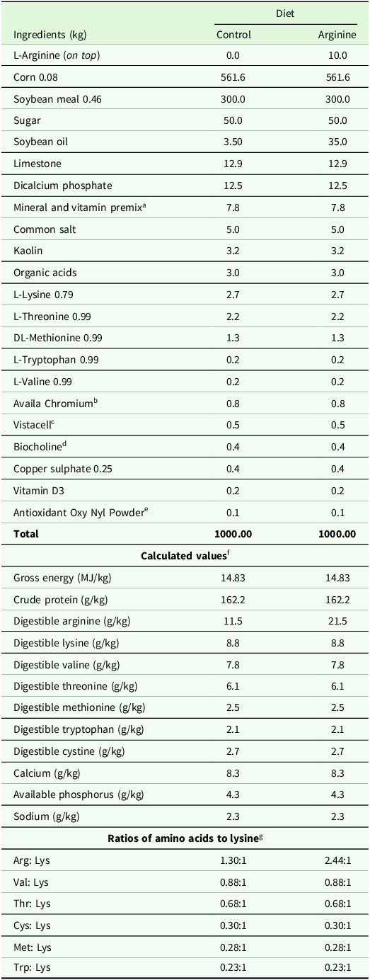
The experiment included two diets, one with L-arginine supplementation and the other without L-arginine supplementation. In the diet with L-arginine, 10 g L-arginine/kg of feed was added on top at the time of feeding. The feed used was provided by a commercial farm. Samples of the diets were collected and sent to the CBO Análises Laboratoriais (Campinas, Brazil) to determine the crude protein concentration by the Dumas method and amino acid concentration by HPLC (Hagen et al., Reference Hagen, Augustin, Grings and Tassinari1993). Additionally, enzymatic analysis was performed to determine the tryptophan concentration (Lucas and Sotelo, Reference Lucas and Sotelo1980) (Table 1).
Composition, calculated values and ratios of amino acids to lysine in the diets supplied to the sows during the experimental period (3–23 days of lactation)

a Assurance levels per kg of premix: folic acid 562.50 mg; pantothenic acid 2812.50 mg; biotin 63.28 mg; carnitine 6250.00 mg; copper 2109.00 mg; choline 75.00 g; chromium 56.25 mg; iron 14.06 g; phytase 70000.00 U; iodine 140.63 mg; manganese 7031.00 mg; niacin 4218.00 mg; selenium 63.28 mg; tryptophan 40.00 g; valine 120.00 g; vitamin A 1406250.00 IU; vitamin B1 210.00 mg; vitamin B12 4218.00 mcg; vitamin B2 703.00 mg; vitamin B6 421.00 mg; vitamin D3 281000.00 U; vitamin E 7734.38 U; vitamin K3 351.56 mg; zinc 16.88 g.
b Guarantee levels per kg of product: chromium methionine complex 1000 ppm.
c Guarantee levels per g of product: Saccharomyces cerevisiae 2.0 × 10¹⁰ CFU.
d Guarantee levels per kg of product: 16 mg of phosphatidylcholine.
e Product content: calcium carbonate, BHA, ethoxyquin, citric acid and phosphoric acid (the concentrations are not available from the manufacturer).
f Digestible values were calculated according to Rostagno et al. (Reference Rostagno, Albino, Hannas, Donzele, Sakomura, Perazzo, Saraiva, Abreu, Rodrigues, Oliveira, Barreto and Brito2017).
g The values of amino acid ratios relative to lysine were estimated considering the purity of the ingredients used in the diet formulation.
Experimental procedure
The females were weighed at 109 days of gestation and at weaning (23rd day of lactation). The diets were supplied at a fixed amount of 7.5 kg/day during the 22-day period of lactation in both treatment groups. Thus, the females treated with L-arginine supplementation received 75 g of L-arginine per day, which was divided into three feeding times according to the management of the farm (07:00 h, 13:00 h and 16:00 h). The feed leftovers were removed from the feeders and weighed in the morning of each day to calculate the daily feed intake. The piglets did not receive creep feeding during the lactation phase. Lactating sows and piglets had free access to water throughout the experimental period. Approximately 12 hours after farrowing, colostrum ingestion was guaranteed, and subsequently, these piglets were equalized to obtain at least 13 piglets per sow up to 48 hours after birth.
Performance of sows and piglets
All the piglets were weighed on the third day after farrowing and were uniform among the sows of the same treatment, maintaining at least 13 piglets per sow. The variables analysed to investigate the performance of the sows were weight change between 109 days of gestation and weaning (23rd day of lactation), litter weight at birth, litter weight at weaning, and sow feed intake. The piglets were weighed individually on the 3rd, 14th, and 23rd days of age, and then, on the 24th day, they were sent to the nursery sector. We evaluated weight gain from 3 to 14 days, from 14 to 23 days and from 3 to 23 days, in addition to the standard deviation and coefficient of variation of initial weight (at three days of age) and weaning weight (at 23 days of age).
The piglets received ear tags on the third day of the experiment for individual identification. To identify the suckling position of the piglets, they were marked with three different colours according to the position they occupied in the mammary complex. The markings were checked for accuracy on a daily basis. To confirm the suckling position, the piglets that remained in the same position more than 0.7 of the time were considered for the suckling position and performance evaluation.
Hormonal blood analysis and milk composition of sows
On the 3rd and 14th days postpartum, in the morning, blood was collected from the anterior vena cava of the sows using a pipe with a hypodermic needle (Injury ® 40×1.6). An eight-hour fasting period was used before blood collection. Blood was collected from each sow in two tubes: one siliconized tube without anticoagulant and one tube with EDTA.
After collection, the samples were immediately homogenized and placed in a Styrofoam™ container with ice. Within 20 minutes of collection, the samples were centrifuged (Eppendorf® 5702 R centrifuge) at 1800 ×g for 12 minutes to obtain the plasma and serum. The plasma and serum samples were pipetted into new tubes and stored in a freezer at –20 °C until further analysis. The levels of progesterone and prolactin were quantified in the serum samples.
Milk samples were also collected on the 3rd and 14th days postpartum. First, the teats of the females were cleaned with running water and subsequently dried with paper towels. The piglets were housed in the retractor so that they did not have contact with the mother for 30 minutes prior to collection. To collect the milk, 1.5 ml of oxytocin was intravenously administered in one of the branches (lateral, intermediate or medial) of the caudal auricular vein of the matrix. Approximately 40 ml of milk was collected in a Falcon tube from the mammary glands in the thoracic, abdominal and inguinal positions, for a total volume of approximately 120 ml. The samples were stored in a freezer at –20 °C and then freeze-dried for analysis of the amino acid composition.
The samples collected on the third day of lactation were used to determine the concentrations of total amino acids after lyophilization. A Biochrom 30+ AA analyser (Biochrom 30+ AA Analyser; Biochrom, Cambridge, UK) was used for ion exchange chromatography after hydrolysis in 6 M HCl for 23 hours. The sulfur contained in the amino acids was released by oxidation in a solution of hydrogen peroxide and performic acid for 16 h before hydrolysis.
Collection of tissue samples
The day after weaning, in the morning, 14 females were slaughtered in a commercial slaughterhouse located on the farm where the field experiment was performed. After stunning and bleeding, the entire breast complex was removed to collect tissue samples. The mammary gland tissue samples were obtained from two pairs of glands at each position (thoracic, abdominal and inguinal) based on size uniformity among all of the glands.
Samples from the most central portion of the mammary glands 4 cm caudal to the teat nipples were collected. For the evaluation of enzymes related to redox status and gene expression, samples 0.25 cm3 in size were placed in sterile cryogenic tubes, immediately frozen in liquid nitrogen, and subsequently stored at –80 °C.
For histological analysis, 1.0 cm3 samples were obtained from the same glandular region and fixed in 0.1 buffered formalin solution. The tissue fragments were transferred to a 0.7 ethanol solution for storage at room temperature 24 hours after sampling.
Analysis of redox status parameters
Total antioxidant capacity (TAC) and enzymatic activity were evaluated in the tissue homogenates using commercial kits. To quantify the total antioxidant capacity of the tissue, the Antioxidant Assay Kit (Cayman Chemical, Lansing, MI, USA) was used. For protein carbonyl levels, the Protein Carbonyl Colormetric Assay Kit was used, and glutathione peroxidase activity was determined using the Glutathione Peroxidase Assay Kit. Finally, thiobarbituric acid reactive substances (TBARS) were detected with a TBARS Assay Kit. All kits were obtained from Cayman Chemical.
Analysis of gene expression in mammary gland tissue
According to the manufacturer’s recommendations, total RNA was extracted using the Qiagen RNeasy Lipid Tissue Mini Kit (Hilden, Germany). The RNA was stained with GelRed (Biotium, Hayward, CA) and visualized with an E-Gel Imager Camera Hood (Life Technologies, Neve Yamin, Israel).
Subsequently, the absorbance was quantified at 260/280 nm and 260/230 nm using a DeNovix DS-11 instrument (Wilmington, DE, USA). Complementary deoxyribonucleic acid (cDNA) was synthesized using a High-Capacity cDNA Reverse Transcription Kit (Applied Biosystems, Foster City, CA, USA).
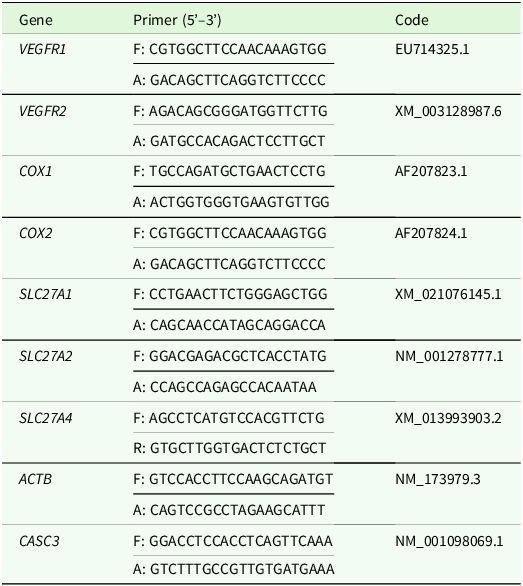
All primers used for the target and reference genes are described in Table 2. The reference genes selected for the assay were β-actin (ACTB) and cancer susceptibility candidate 3 (CASC3). All of the primers used were designed using the online program Primer3Plus based on sequences published and indexed in the GenBank database of the National Center for Biotechnology Information (NCBI) public platform. Afterward, the primers were commercially synthesized by Invitrogen (Carlsbad, CA, USA) and diluted in a solution of pooled samples to determine the respective efficiencies and regression coefficients (R2).
Sequences (5’ to 3’) of primers used in real-time polymerase chain reaction (PCR)

Abbreviations: VEGFR1, vascular endothelial growth factor receptor 1; VEGFR2, vascular endothelial growth factor receptor 2; COX1 and COX2 - cyclo-oxygenase enzymes; SLC27A1, SLC27A2 and SLC27A4 - fatty acid transport proteins; ACTB - actin beta; CASC3 - cancer susceptibility candidate 3.
Real-time quantitative polymerase chain reaction (RT–qPCR) was performed using the SYBR Green detection system (Applied Biosystems, USA) in an Eppendorf Reaplex thermocycler (Eppendorf, Hamburg, Germany). The relative expression levels were calculated according to the method described by Pfaffl (Reference Pfaffl2001).
Histological analyses of mammary gland tissue
Tissue samples were subsequently embedded in paraffin and sectioned at a thickness of 5.0 μm using a microtome (RM 2255; Leica Biosystems, Nussloch, Germany). The slides were stained with hematoxylin-eosin solution and qualitatively analysed under a light microscope (BX53; Olympus, Tokyo, Japan).
A total of 12 digital images of each glandular unit (n = 3) from all of the sows were randomly obtained using Cell Sens Dimensions 1.6 software (Olympus, Tokyo, Japan) and a light microscope equipped with a 1.3 MP CMOS digital camera. Bio-CAM (Takachiho, Japan). For the analysis of capillaries, 40× magnification was used, and for the evaluation of alveoli, parenchymal tissue, connective tissue, adipocytes and blood vessels, 10× magnification was used. After the images were captured, the variables were analysed using ImageJ software (v.1.50i; National Institutes of Health, Bethesda, USA). The quantification of adipocytes, blood vessels, and parenchymal and adipose tissue was performed by histological evaluation of the parameters analysed. A total of 18 histological fields (120,000 µm2 each field) per animal were counted with six fields per gland. The proportions of parenchymal tissue, connective tissue, adipocytes and blood vessels were evaluated by point count, accounting for 10,584 points per animal and 3,528 points per mammary gland. Each histological image has 588 points in a known area of 120,000 µm². The proportion of points in each component was calculated by the following formula: volumetric proportion = number of points in each component/3,528.
Statistical analysis
The statistical analyses were performed using Statistical Analysis System (SAS) software (SAS Institute, Inc., Cary, NC, USA, Version 9.3). To analyse the normality of the data using the PROC UNIVARIATE procedure, the Shapiro—Wilk test was performed, and when the data did not follow this distribution, transformation was performed using the PROC RANK tool in SAS. Tukey’s test was used to compare the means of the different treatment groups using the PROC MIXED procedure in SAS. P values below 0.05 were considered to indicate a significant difference; P values between 0.05 and 0.10 were considered a trend toward significance.
The statistical model used was:
 $$Y_{ikl} = \mu + D_{i} + P_{k} + DP_{ik} + e_{ikl},$$
in which Y ikl = represents the observation on experimental unit i, supplemented or not with L-arginine (D i ) and mammary gland position k (P k ), on repetition l; μ = the overall mean; D i = L-arginine supplementation effect (i = with or without); P k = effect of mammary gland position (k = 1, 2, 3); DP ik = interaction of the L-arginine supplementation i and the mammary gland position k; and e ikl = the residues corresponding to each observation, which, by hypothesis, has a normal distribution with zero mean and sigma squared variance.
Results
Performance of sows and piglets and litter quality
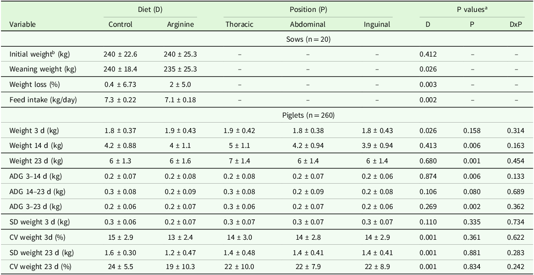
Statistical analysis revealed no significant interaction effects between diet and suckling position on piglet performance or litter quality (P > 0.05). The females administered the L-arginine-supplemented diet exhibited a lower feed intake (P = 0.002). There was an effect of diet on the performance parameters of the sows; those that received arginine supplementation exhibited significant weight loss during the period between 109 days of gestation and weaning (23rd day of lactation) (P = 0.003) and a lower weight at weaning (P = 0.026) (Table 3).
Main effects of diet and suckling position on performance of sows and piglets and litter quality (means ± SD)

a Tukey test at 5% significance level; values between 5 and 10% were considered a trend toward significance. Control: basal diet without added L-arginine; Arginine: control diet with the addition of L-arginine at 10 g/kg on top.
b Initial weight at 109 days of gestation.
Abbreviations: ADG, average daily weight gain; SD, standard deviation; CV, coefficient of variation.
The mean initial weight (third day) was greater (P = 0.026) in piglets from sows that received L-arginine supplementation. However, for the weight of the piglets at 14 and 23 days, L-arginine supplementation had no effect (P > 0.05), nor did it affect weight gain during the different periods. Regarding the variables related to litter quality, the standard deviation of the initial weight and the coefficient of variation of the initial and weaning weights of the piglets from sows that received the arginine supplement were lower (P = 0.001) than those of the piglets from sows that received the control diet (Table 3).
Suckling position significantly affected piglet weight at 14 (P = 0.006) and 23 days (P = 0.001) and daily weight gain at 14 (P = 0.006) and 23 days (P = 0.002). The piglets that suckled on the teats in the thoracic position had higher mean values than those that suckled on the teats in the inguinal position and in the abdominal position. Moreover, suckling position tended to affect daily weight gain from 14 to 23 days (P < 0.10), which was similar to the effects of suckling position on other variables (Table 3).
Sow milk amino acid composition
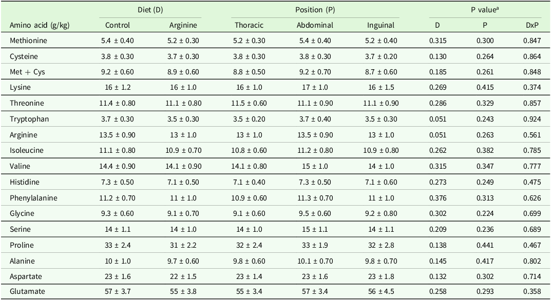
The milk composition was not significantly affected by diet or milk collection position (P > 0.05). However, there was a trend toward a reduction in arginine and tryptophan levels (P < 0.10) in the milk of sows fed a diet supplemented with L-arginine (Table 4).
Main effects of diet and mammary gland position on amino acid composition of sow milk collected on the 14th day of lactation (means ± standard deviation)

a Tukey test at 5% significance level; values between 5 and 10% were considered a trend toward significance. Control: basal diet without added L-arginine; Arginine: control diet with the addition of L-arginine at 10 g/kg on top.
Abbreviations: Met + Cys - methionine plus cysteine.
Hormonal levels in the plasma of sows
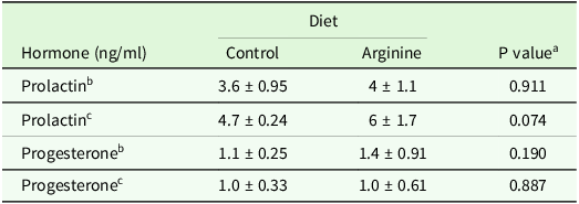
The L-arginine supplementation had no significant effect (P > 0.05) on the plasma levels of prolactin or progesterone measured on 3 and 14 days of lactation (Table 5). However, there was a trend toward increasing prolactin levels on the 14th day of lactation (P < 0.10) for the sows fed a diet supplemented with L-arginine.
Plasma prolactin and progesterone concentrations of lactating sows according to dietary treatment (means ± standard deviation)

a Tukey test at 5% significance level; values between 5 and 10% were considered a trend toward significance. Control: basal diet without added L-arginine; Arginine: control diet with the addition of L-arginine at 10 g/kg on top.
b Harvest was performed on the 3rd day of lactation.
c harvest was performed on the 14th day of lactation.
Redox status parameters of the mammary gland tissue
There was no significant interaction effect between diet and gland position (P > 0.05) on any of the redox status variables. The anatomical position of the mammary gland had an almost significant effect on the levels of protein carbonyls (P = 0.049). Moreover, there was a trend toward a reduction in glutathione peroxidase activity (P < 0.10) only in the mammary gland tissue of sows fed a diet supplemented with L-arginine (Table 6).
Main effects of diet and gland position on analyses of the redox status of the mammary gland tissue of sows (means ± standard deviation)

a Tukey test at 5% significance level; values between 5 and 10% were considered a trend toward significance. Control: basal diet without added L-arginine; Arginine: control diet with the addition of L-arginine at 10 g/kg on top.
Abbreviations: TBARS - thiobarbituric acid reactive substances, expressed in micromolar (μM) of malonic dialdehyde (MDA) per kilogram (kg) of tissue; CP - carbonyl protein (nmol/ml); GPx - glutathione peroxidase activity; TAC - total antioxidant capacity.
Gene expression in mammary gland tissue
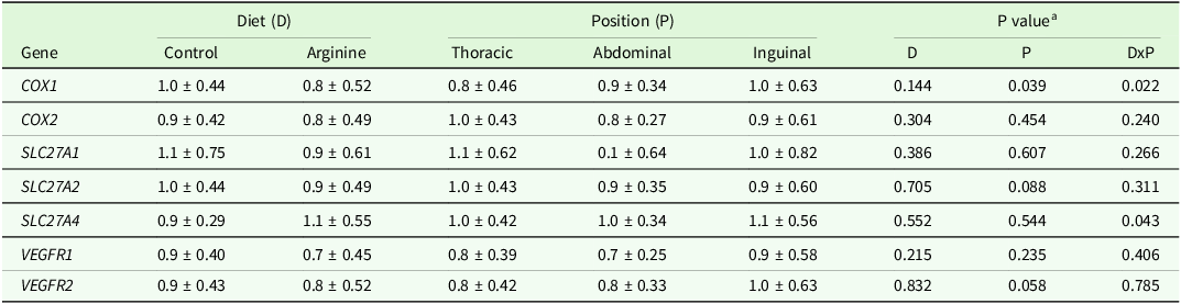
There was no single effect of diet (P > 0.05) on the gene expression variables. There was only a trend toward an effect of gland position (P < 0.10) on the expression of the SLC27A2 and VEGFR2 genes, with the thoracic and inguinal positions tending to have the highest values (Table 7). There was an interaction effect between diet and mammary gland position on COX1 (P = 0.022) and SLC27A4 (P = 0.043) gene expression in the samples from the thoracic and abdominal positions, but the expression of these genes did not significantly differ from that in females that received the control diet, regardless of gland position. With respect to the SLC27A4 gene, samples collected from the thoracic and inguinal positions had higher means than samples collected from the abdominal position, regardless of dietary treatment (Table 8).
Main effects of diet and mammary gland position on expression of genes related to mammary gland tissue metabolism (means ± standard deviation)

a Tukey test at 5% significance level; values between 5 and 10% were considered a trend toward significance. Control: basal diet without added L-arginine; Arginine: control diet with the addition of L-arginine at 10 g/kg on top.
Abbreviations: COX1 and COX2 - cyclo-oxygenase enzymes; SLC27A1, SLC27A2 and SLC27A4 - fatty acid transport proteins; VEGFR1 - vascular endothelial growth factor receptor 1; VEGFR2 - vascular endothelial growth factor receptor 2.
Slicing of the means of gene expression variables with significant interaction between diet and mammary gland position (means ± standard deviation)

Control: basal diet without added L-arginine; Arginine: control diet with the addition of L-arginine at 10 g/kg on top.
P values of the interaction between diet and mammary gland position: COX1 = 0.022; SLC27A4 = 0.043.
Abbreviations: COX1, cyclo-oxygenase enzymes; SLC27A4, fatty acid transport proteins.
Histology of mammary gland tissue
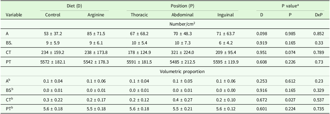
Histological analysis revealed no interaction between diet and mammary gland position (P > 0.05). The position of the mammary gland affected only the volumetric proportion of the connective tissue, with the highest value observed in the abdominal mammary glands in relation to the thoracic and inguinal mammary glands; these two parameters did not differ from each other. There was a trend toward an increase in the number of adipocytes in the breast tissue (P < 0.10) of sows fed a diet supplemented with L-arginine. Regarding the position of the gland, the tissue in the abdominal position tended to have a greater amount of connective tissue (P < 0.10) (Table 9).
Main effects of diet and mammary gland position on histomorphometric variables of the mammary gland of sows at 23 days of lactation (means ± standard deviation)

a Tukey test at 5% significance level; values between 5 and 10% were considered a trend toward significance. Control: basal diet without added L-arginine; Arginine: control diet with the addition of L-arginine at 10 g/kg on top.
b Area set for counting 120000 μm².
Abbreviations: A, adipocyte; BS., blood vessel; CT, connective tissue; PT, parenchymal tissue.
Discussion
The mammary gland in pregnant swine becomes enlarged close to parturition, accompanied by a large increase in cell proliferation and general tissue metabolism, leading to an increase in energy and amino acid requirements (Zhu et al., Reference Zhu, Guo, Gao, Wang, Chen, Ma and Jiang2017). The amino acid arginine is highly important in all stages of production, but particularly during lactation, as previously reported in the literature (Dallanora et al., Reference Dallanora, Walter, Marcon, Saremba, Bernardi, Wentz and Bortolozzo2016; Zhu et al., Reference Zhu, Guo, Gao, Wang, Chen, Ma and Jiang2017; Holanda et al., Reference Holanda, Marcolla, Guimarães, Neves, Hausman, Duarte, Abreu and Saraiva2019).
It should be noted that the temperature observed during the experimental period was above the thermoneutrality range for females, which, according to Black et al., (Reference Black, Mullan, Lorschy and Giles1993), is 16 to 22 °C, indicating that the sows were subjected to heat stress. However, the feed intake observed in the present study for both diets was within the expected range for lactating females.
A reduction in feed intake was observed during lactation when the females were supplemented with L-arginine. Laspiur and Trottier (Reference Laspiur and Trottier2001) found similar results. This reduction may be because arginine acts as a secretagogue of some hormones, such as growth hormone (GH), prolactin, and insulin. With the increase in the plasma concentrations of these hormones, there is greater uptake and use of nutrients for the metabolic processes of growth and lactation. Thus, with increasing nutrient utilization efficiency, females require less feed to meet their nutritional requirements, which explains the lower feed intake in sows supplemented with L-arginine. However, Zhu et al. (Reference Zhu, Guo, Gao, Wang, Chen, Ma and Jiang2017) reported no effect of L-arginine supplementation on the feed intake of lactating sows.
Supplementation with L-arginine promoted greater weight loss in sows during lactation, and consequently, they had lower body weights at weaning. The greater weight loss in sows supplemented with L-arginine may be related to a possible increase in milk production since this amino acid influences the endocrine system of pigs, especially the secretion of GH and prolactin (Mateo et al., Reference Mateo, Wu, Bazer, Park, Shinzato and Kim2007; Wu et al., Reference Wu, Bazer, Davis, Kim, Li, Rhoads, Satterfield, Smith, Spencer and Yin2009; Zhu et al., Reference Zhu, Guo, Gao, Wang, Chen, Ma and Jiang2017). An increase in GH secretion in plasma may stimulate the release of IGF-I through the GH-IGF-I axis, further increasing its concentration in plasma. Thus, IGF-I contributes to mammary differentiation and lactogenesis and, like prolactin, also has a galactopoietic effect in pigs (Zhu et al., Reference Zhu, Guo, Gao, Wang, Chen, Ma and Jiang2017; Gao et al., Reference Gao, Wen, Guo, Wang, Ban, Yang, Wu and Jiang2020). Therefore, L-arginine supplementation in lactating females may increase milk production so that there may be greater mobilization of body tissues, such as adipose tissue and muscle, to support milk synthesis, and thus greater weight loss may occur during lactation. In the present study, milk production was not measured. However, weight loss in both treatment groups can be considered normal because the loss was less than 0.08 of the female’s body weight (Gao et al., Reference Gao, Wen, Guo, Wang, Ban, Yang, Wu and Jiang2020) and thus did not compromise performance in the next reproductive cycle. The sows fed a supplemented diet exhibited higher nutrient utilization efficiency, as although their food consumption was lower, the performance of the piglets did not differ from that of the piglets from sows fed the control diet.
Piglets from sows fed a diet supplemented with L-arginine during lactation had greater initial weights. However, this difference in piglet weight was not related to L-arginine supplementation since the experimental diets were supplied only at the beginning of lactation. During pregnancy, there was no supplementation with this amino acid.
Regarding the dispersion measures, the initial weight of piglets from sows fed a diet supplemented with L-arginine had lower coefficients of variation (CV), and the weight at weaning also had lower CV and standard deviation values. This indicates that sows that received the diet with 10 g L-arginine/kg weaned piglets with more uniform weight (i.e., piglets that weighed around the average weight). The greater uniformity in the weight of the piglets at weaning may be related to a possible increase in milk production and nutrient content in sows fed a diet supplemented with L-arginine, which may benefit low-weight piglets with increased suckling capacity and reduce competition for teats among lighter piglets. Similar results were reported by Quesnel et al. (Reference Quesnel, Quiniou, Roy, Lottin, Boulot and Gondret2014) and Moreira et al. (Reference Moreira, Mendes, Palencia, Lemes, Roque, Kutschenko, Ferreira and Abreu2020), but supplementation was administered during pregnancy.
During suckling, the piglets that occupied the teats in the thoracic position generally performed better than those that occupied the teats in the inguinal position, which, in turn, presented a performance equivalent to that of the piglet that occupied the mammary glands in the abdominal position. A possible explanation for this finding is that, according to Kasanen and Algers (Reference Kasanen and Algers2002), female grunts may attract piglets to anterior/cranial teats (i.e., to the pairs of teats closest to the head). Thus, with competition for teats during suckling, the heaviest piglets tend to occupy the anterior and middle teats, while the lighter piglets tend to occupy the posterior teats (Moreira et al., Reference Moreira, Mendes, Palencia, Lemes, Roque, Kutschenko, Ferreira and Abreu2020). In the present study, this behavior was observed in the field and confirmed through the data. In addition, the fact that heavier piglets occupy the anterior and middle positions probably leads to greater production and secretion of milk in these regions, as there is a stronger and more active stimulation of the mammary glands (Skok et al., Reference Skok, Brus and Škorjanc2007). According to Špinka and Algers (Reference Špinka and Algers1995), the amount of milk sucked is directly related to the intensity and duration of teat massage. Furthermore, Skok et al. (Reference Skok, Brus and Škorjanc2007) reported a positive correlation between the body mass growth of piglets and gland milk production during different phases of lactation, which confirms that heavier piglets stimulate greater milk production in the positions they occupy. Thus, in addition to already tending to have greater weight at the beginning of lactation, piglets occupying the anterior and middle positions tend to exhibit greater growth during lactation because they stimulate more milk production from the mammary glands in these positions.
The availability of nutrients is another factor that contributes to the difference in the performance of piglets suckling on mammary glands at different positions. Blood flow differs between the mammary glands in different positions, as it tends to decrease in the direction of anterior to the posterior (Skok et al., Reference Skok, Brus and Škorjanc2007). In the same study, the milk consumption of piglets was evaluated in relation to the different positions of the mammary gland, and it was observed that, during the first week of lactation, the piglets that were positioned in the anterior and middle parts of the mammary complex consumed, on average, 0.42 more milk than did the piglets positioned in the rear. During the second week of lactation, the difference decreased to 0.34, and in the third and fourth weeks, the differences were 0.24 and 0.30, respectively (Skok et al., Reference Skok, Brus and Škorjanc2007).
L-arginine supplementation in lactating females may increase the milk amino acid content, as, according to Mateo et al. (Reference Mateo, Wu, Moon, Carroll and Kim2008), arginine may increase the uptake and use of amino acids by the mammary gland during lactation for the synthesis of proteins, peptides and other components of milk. This increased uptake may be because arginine has a secretagogue effect on insulin and GH, which in turn increases the uptake and use of amino acids by the mammary gland for the production of milk proteins (Kim et al., Reference Kim, McPherson and Wu2004; Laspiur et al., Reference Laspiur, Farmer, Kerr, Zanella and Trottier2006). However, in the present study, supplementation with 10g L-arginine/kg in lactating females did not affect the amino acid composition in milk at 14 days of lactation. Similar results were obtained by Dallanora et al. (Reference Dallanora, Walter, Marcon, Saremba, Bernardi, Wentz and Bortolozzo2016), who reported no change in the amino acid composition of milk at 10 and 20 days of lactation after the inclusion of L-arginine at 10g/kg in the diet of sows during lactation. These results indicate that, despite the physiological evidence that corroborates the hypothesis that arginine can increase the amino acid content in milk, supplementation with 10g L-arginine/kg did not alter the amino acid composition of milk in this study.
Regarding the effect of L-arginine supplementation on hormone levels, Mateo et al. (Reference Mateo, Wu, Bazer, Park, Shinzato and Kim2007) and Wu et al. (Reference Wu, Bazer, Davis, Kim, Li, Rhoads, Satterfield, Smith, Spencer and Yin2009) reported that this amino acid influences the endocrine system, mainly through its secretagogue effect, which increases the synthesis and secretion of several hormones, including prolactin. Thus, arginine supplementation tends to increase blood prolactin levels in lactating sows. However, in the present study, there were no changes in the plasma levels of prolactin or progesterone at 4 or 14 days of lactation after the inclusion of L-arginine in the diet of lactating females. Moreover, Gao et al. (Reference Gao, Wen, Guo, Wang, Ban, Yang, Wu and Jiang2020) evaluated the effect of supplementation with increasing levels of arginine and observed an increase in serum prolactin levels at 14 days of lactation with supplementation with 4g L-arginine/kg. Similarly, Zhu et al. (Reference Zhu, Guo, Gao, Wang, Chen, Ma and Jiang2017) reported an increase in the serum prolactin concentration with supplementation with 10g L-arginine/kg at 14 days of lactation. In both studies (Zhu et al., Reference Zhu, Guo, Gao, Wang, Chen, Ma and Jiang2017; Gao et al., Reference Gao, Wen, Guo, Wang, Ban, Yang, Wu and Jiang2020), a greater number of lactating sows was used than in the present study, which may have contributed to the divergence between the results.
L-arginine can attenuate oxidative stress in mammary gland tissue through several mechanisms, such as restoring glutathione production, stimulating the production of nitric oxide in the liver, and regulating antioxidant defence systems (Echeverría et al., Reference Echeverría, Valenzuela, Bustamante, Álvarez, Ortiz, Soto-Alarcon, Muñoz, Corbari and Videla2018). There are also reports of L-arginine improving the antioxidant capacity of tissues such as the liver and skeletal muscle (Zhang et al., Reference Zhang, Jin, Wang, Loor and Wang2020). In the present study, L-arginine supplementation did not affect the parameters related to the redox status in the mammary gland. However, with respect to the anatomical position of the mammary gland, a lower level of protein carbonyl was observed in the mammary glands in the thoracic position. Protein carbonyls are a marker of protein oxidation; the lower the concentration of a metabolite is, the lower the protein oxidation in that tissue and vice versa. However, this evaluation of only one indicator of oxidative stress is insufficient to conclude that redox status of mammary gland tissue is affected by its particular position.
Cyclooxygenases (COXs) are enzymes that catalyse the conversion of arachidonic acid into prostaglandin (PGE2), which plays an important role in various mammalian tissues. In the present study, the gene expression of the two main isoforms was evaluated: COX1, which is involved in homeostatic regulation and is required and expressed in almost all animal cells, and COX2, which is activated in inflammatory processes. In the literature, studies on the role of cyclooxygenases and PGE2 metabolic enzymes in mammary gland tissue in physiological conditions and during lactation are scarce. Upregulated COX2 expression is associated with pathological conditions, such as tissue damage, and COX2 expression is low in normal breast tissue. A modest increase is observed during breast involution. COX1 expression levels and total prostaglandin E2 (PGE2) levels increase with cell proliferation during lactogenesis. PGE2 regulates multiple biological processes under normal and pathological conditions and plays an important role in the physiology of epithelial cells (Chandrasekharan et al., Reference Chandrasekharan, Foley, Jania, Clark, Audoly and Koller2005).
Compared to the control treatment, L-arginine supplementation downregulated COX1 expression. The tissues in the thoracic and abdominal positions had lower means than those in the inguinal position. According to Holanda et al. (Reference Holanda, Marcolla, Guimarães, Neves, Hausman, Duarte, Abreu and Saraiva2019), higher COX1 gene expression indicates greater prostanoid metabolite synthesis, which favours events that promote cell differentiation, proliferation, angiogenesis, and antiapoptotic mechanisms in the tissue, in addition to prolonging periods of tissue cell growth and differentiation. In the study by Holanda et al. (Reference Holanda, Marcolla, Guimarães, Neves, Hausman, Duarte, Abreu and Saraiva2019), no effects were observed in the inguinal tissue, which may indicate greater metabolism of L-arginine by nonoxidative pathways, to the detriment of the formation of nitric oxide (NO) through the oxidative pathway (eNOS nitric oxide synthase) in the inguinal mammary glands. The decrease in COX1 expression levels in the tissue may be related to the increased formation of NO, which contributes to vasodilation, as well as to the synthesis of COX1-dependent PGI2 (Holanda et al., Reference Holanda, Marcolla, Guimarães, Neves, Hausman, Duarte, Abreu and Saraiva2019). Thus, the observed downregulation may be related to the balance of metabolites that act on blood vessels, since blood flow is mediated by several endothelial systems, including NO, prostaglandins (PGI 2 and PGE 2), and a family of endothelium-derived hyperpolarizing factors (EDHFs) (Chandrasekharan et al., Reference Chandrasekharan, Foley, Jania, Clark, Audoly and Koller2005). L-arginine can regulate the inflammatory response not only by acting as a precursor of NO but also by promoting the proliferation of polyamines (Satriano, Reference Satriano2003) in a way that potentially leads to a reduction in COX1 expression levels and, consequently, the production of prostaglandins in the cell. However, the possible relationships between these factors and mammary gland position need to be clarified.
The expression of VEGFR1 and VEGFR2 was evaluated. VEGF, through its receptors, regulates NO synthesis and blood flow in endothelial cells (Shibuya, Reference Shibuya2006). VEGF-related genes are indispensable for activating angiogenic pathways, including those mediated by angiopoietins (Eklund and Olsen, Reference Eklund and Olsen2006). In breast tissue, vascular density progressively increases during pregnancy, reaches its maximum, and progressively decreases in the final trimester and during lactation and involution. In a study conducted on the mammary glands of rodents, the transcript levels of VEGFR1 and VEGFR2 increased 2–4 times during lactation (Han et al., Reference Han, Li, Wang, Zhu, Wang, Guo, Lu, Wang and Yang2010). The largest increase in VEGF and VEGFRs 1 and 2 was observed during lactation and was directly related to the marked increase in the permeability of capillaries that transport nutrients from the blood to the alveolar epithelium during lactation. Thus, VEGF acts on capillary permeability during lactation in addition to inducing morphological changes in endothelial cells, including the appearance of vesicular vacuoles (Feng et al., Reference Feng, Nagy, Pyne, Hammel, Dvorak and Dvorak1999). However, in the present study, no difference was observed between the two groups. Similarly, Holanda et al. (Reference Holanda, Marcolla, Guimarães, Neves, Hausman, Duarte, Abreu and Saraiva2019) did not observe changes in VEGF expression in the mammary gland tissue of lactating females fed a diet supplemented with L-arginine.
The expression of SLC27 family genes related to lipid transport is naturally upregulated in the lactational phase due to the production and secretion of milk lipids, which increases the need for lipid transport into epithelial cells (Han et al., Reference Han, Li, Wang, Zhu, Wang, Guo, Lu, Wang and Yang2010; McManaman, Reference McManaman2014).
In the present study, SLC27A4 expression levels increased with L-arginine supplementation, and the thoracic and inguinal glands exhibited upregulated SLC27A4 expression. Arginine participates in the activation of the lipoprotein lipase enzyme, which releases glycerol and fatty acids from triglycerides for the synthesis of milk fat (Holanda et al., Reference Holanda, Marcolla, Guimarães, Neves, Hausman, Duarte, Abreu and Saraiva2019). This enzyme is expressed at high levels in the lactating mammary gland (Mateo et al., Reference Mateo, Wu, Moon, Carroll and Kim2008). This fact may explain the higher expression levels of the transport protein-encoding gene in the samples, as more transporters are required after the hydrolysis of plasma triglycerides. The upregulated expression of SLC27A4 in the inguinal glands may be a feature of tissue adaptation to optimize the uptake of fatty acids and glycerol since blood flow in this position is demonstrably lower than that in other positions (Paula et al., Reference Paula, Magalhães, Romão, Ferreira Junior, Moreira, Barcelos and Ribeiro2019).
According to the histological evaluations of the breast tissue, L-arginine supplementation did not significantly change the morphology of this tissue. Li et al. (Reference Li, Bazer, Johnson, Burghardt, Erikson, Frank, Spencer, Shinzato and Wu2010) and Wu et al. (Reference Wu, Bazer, Satterfield, Li, Wang, Johnson, Burghardt, Dai, Wang and Wu2013) reported that arginine improves tissue vascularization, especially in mammary gland tissue. However, in the present study, the proportion of blood vessels was not affected by L-arginine supplementation. Moreover, Holanda et al. (Reference Holanda, Marcolla, Guimarães, Neves, Hausman, Duarte, Abreu and Saraiva2019) also observed an increase in the number and proportion of blood vessels in the mammary tissue of females fed a diet supplemented with L-arginine during lactation. There are some differences among the studies that may be responsible for the varying results, such as the fact that Holanda et al. (Reference Holanda, Marcolla, Guimarães, Neves, Hausman, Duarte, Abreu and Saraiva2019) evaluated the mammary tissues of primiparous females. In the present study, multiparous females were used, with parity 4–7, which may have influenced the effect on the tissue. In the present study, a greater percentage of connective tissue was observed in the samples from the glands in the abdominal position than in those from the other two positions. This result may be indicative of the lower productivity of the glands in this position; however, the other results observed in this study, such as the performance of piglets at weaning, do not corroborate this observation. Another justification would be that a gland in that position was not stimulated during lactation, but this was not observed in the field. This is the first study to evaluate the effect of L-arginine supplementation on the mammary glands in different positions; however, additional studies are needed to clarify these results.
It is extremely important to understand the effects of L-arginine supplementation in different positions of the mammary gland, in addition to understanding the particularities of each position, since supplementation with the amino acid arginine, as well as with other functional nutrients, can have different effects according to the position of the mammary gland. This information contributes to the acquisition of knowledge about the metabolism of mammary tissue and helps in the assessment of the feasibility of using functional nutrients in the diet of lactating sows, such as arginine, also assisting in decision-making by nutritionists working in the swine industry.
Conclusion
Supplementation with 10g L-arginine/kg to sows during lactation improves litter quality by increasing weaning weight uniformity. Furthermore, this supplementation modulates the expression of the COX1 and SLC27A4 genes in mammary gland tissue. Regarding the position of the mammary gland, piglets that occupy the thoracic and abdominal positions perform better.
Acknowledgements
The authors would like to thank Fazenda São Paulo (Oliveira – MG, Brazil) for technical support and for providing the location, facilities and animals for this study.
Author contributions
MFSAM, TOS and MLTA conceived and designed the study. MFSAM, TOS and JVLB conducted data gathering. FLC and RHRM performed statistical analyses. FLC, TRSG, JMOJ, RHRM and MLTA wrote the article.
Funding statement
The authors acknowledge the financial support of the Conselho Nacional de Desenvolvimento Científico e Tecnológico (CNPq), Coordenação de Aperfeiçoamento de Pessoal de Nível Superior (CAPES), Fundação de Amparo à Pesquisa do Estado de Minas Gerais (FAPEMIG), and Instituto Nacional de Ciência e Tecnologia de Ciência Animal (INCT-CA).
Competing interests
The authors declare there are no conflicts of interest.
Ethical standards
The methods and protocols used in the experiment were approved by the Ethics Committee on Animal Use (CEUA) of the Federal University of Lavras, under protocol number 048/2019.