Mastitis is still ranked among the main production diseases in dairy herds of developed countries (Miller et al. Reference Miller, Bartlett, Lance, Anderson and Heider1993; Rajala-Schultz & Gröhn, Reference Rajala-Schultz and Gröhn1999). Especially intramammary infections with Escherichia coli very often lead to an acute mastitis with severe clinical effects (Bannermann et al. Reference Bannermann, Paape, Lee, Zhao, Hope and Rainard2004). Enormous economic losses during current and/or subsequent lactations are consequences (Seegers et al. Reference Seegers, Fourichon and Beaudeau2003). The identification of the inflammatory event at an early stage is not only a basic demand of milk hygiene and an essential precondition for a successful therapy (Grunert, Reference Grunert and Rosenberger1990) but also an important factor for the limitation of economic impacts due to yield losses (De Mol & Ouweltjes, Reference De Mol and Ouweltjes2001). Effects of metabolic activities of the mammary gland on mammary blood flow seem to be evident (Lough et al. Reference Lough, Beede and Wilcox1990). Clinical examination, California mastitis test (CMT) and bacteriological examinations (BE) are the most common non-invasive methods for diagnosing mastitis. In this present study a new non-invasive and quantitative method was used for examining the pudendoepigastric trunk by transrectal colour Doppler sonography (TCDS). The investigated vessel section is the only section of the inguinally running external pudic artery, the major supplier of the mammary gland of cows, which is transrectally accessible (Budras & Wünsche, Reference Budras, Wünsche, Budras and Wünsche2002). Earlier studies have shown that TCDS is an established non-invasive method for detecting physiological (Bollwein et al. Reference Bollwein, Baumgartner and Stolla2002; Krueger et al. Reference Krueger, Koerte, Tsousis, Herzog, Flachowsky and Bollwein2009) or pathological (Rauch et al. Reference Rauch, Krueger, Miyamoto and Bollwein2008) changes of genital organs in cattle. The objectives of this preliminary investigation were to evaluate the feasibility of TCDS for determining blood flow of the pudendoepigastric trunk in cows with experimentally induced Esch. coli mastitis.
Materials and Methods
Animals
The study included a homogeneous group of five primiparous Holstein dairy cows, 120–180 days in milk, not pregnant and with an average milk yield (MY) of 28·6±3·6 kg/d. None of the animals had ever shown any signs of mastitis before. Weekly regular milk tests during a 3-week adaptation phase made by staff at the Clinic for Ruminants and the Milchprüfring Bayern e.V. (Registered Association of Milk Quality Control in Bavaria) guaranteed all udder quarters were clinically healthy, somatic cell count (SCC) was <50 000 cells/ml and milk samples were bacteriologically negative at the beginning of the trial. Milk was always sampled before regular milking. Bacteriological examinations were performed from foremilk samples; SCC was determined in 10 ml whole milk after each udder quarter had been milked separately with a quarter milker (WestfaliaSurge, Bönen, Germany).
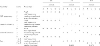
During the adaptation phase rectal temperatures (RT), MY and CMT were documented twice a day. Also, the clinical status of the animals was defined twice daily by a clinical score (CS) with a change-scale from 0 (Nothing Abnormal Detected) over 1 (mild impairment) and 2 (moderate impairment) to 3 (severe impairment) quantifying milk appearance, udder consistency after palpation and general condition of the animals (Table 1). During the investigation phase, consisting of the methodological and the experimental parts of the study, all of these parameters (BE, RT, MY, CMT, SCC and CS) were documented at the times of measurements (establishment of the method: oestrus/dioestrus; Esch. coli mastitis model: 0/12/24 h). During the adaptation phase as well as during the investigation phase all animals were kept under constant conditions at the Clinic for Ruminants, Ludwig-Maximilians-University Munich, Germany. The trial was conducted under the approval and supervision of the Ethics Committee of the regional government in Munich, Germany (No 55.2-1-54-2531-108-05).
Clinical scores of five cows (A, B, C, D, E) using three parameters to assess the clinical status in the experimental part of the study. Individual scores and the sum of the scores of each animal at different times are shown. Also shown is the Median (Minimum/Maximum) per time of these sums. (NAD=Nothing Abnormal Detected)

Stage of the oestrus cycle
For comparable hormonal conditions, all cows were synchronized by two injections of (+)-cloprostenol (Dalmazin®, Selectavet, Weyarn, Germany) at 12-d intervals. For an accurate determination of oestrus and dioestrus, palpations and ultrasound scanning of the ovaries as well as clinic-established and cattle-adapted laboratory techniques for detecting sexual steroids were used (Prakash et al. Reference Prakash, Meyer, Schallenberger and van de Wiel1987).
Scanning procedure
All TCDS measurements were carried out using an ultrasound unit (Hitachi EUB 8500, Hitachi Medical Systems GmbH, Wiesbaden, Germany) equipped with a 7·5 MHz linear rectal transducer (Hitachi EUP-033J; Hitachi Medical Systems GmbH). The range of the Doppler gate was adapted to the diameter of the pudendoepigastric trunk. Measurements were shown on-line at the display together with the ultrasonic image. The ‘frozen’ B-mode/moving Doppler signals were saved on hard disk and later recorded on DVD for analysis. The transverse section of the blood vessel was recorded in the B-mode as a ‘frozen’ image. All evaluations were made afterwards using a specific ultrasound image viewer program (US Image Viewer, PCS-UV 1; 2002, 2004; Hitachi Medical Corporation).
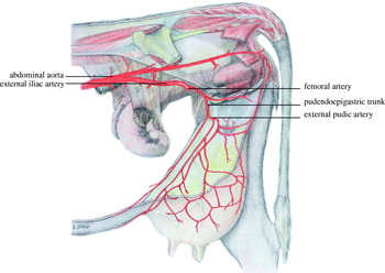
The blood vessels of interest were found transrectally in the following manner (Fig. 1). By holding the face of the ultrasound transducer dorsally it was easy to identify the abdominal aorta. The external iliac artery was found at the level of the branching of the abdominal aorta. By following the external iliac artery and passing the femoral artery the pudendoepigastric trunk was located cranioventral of the cranial edge of the osseous pelvis.
Anatomical location of the pudendoepigastric trunk in cows (based upon Budras & Wünsche Reference Budras, Wünsche, Budras and Wünsche2002).

Measurements were always performed by the same person in the same quiet room to which the animals had been conditioned during their adaptation phase. To study mammary blood flow, the left and the right pudendoepigastric trunk were examined. After correction for the specific angle at which the measurements had been taken, all waveforms of the blood flow were obtained at an interrogation angle between the Doppler ultrasound beam and the flow direction from 20 to 50 degrees. Both the left and the right pudendoepigastric trunk were scanned to determine the blood flow expressed by the blood flow volume (BFV, l/min). To calculate the BFV the area of the pudendoepigastric trunk was sized three times (Bollwein et al. Reference Bollwein, Weber, Woschée and Stolla2004) by circumscribing the vessel internally with the cursor of the PC and then taking the average of all measurements.
Establishment of the method (methodological part of the study)
Precision of TCDS was verified in the clinically healthy animals. For that purpose all scanning was done before feeding time. Measurements were carried out within 2 h before and after milking during oestrus and dioestrus; thus, four measurements were taken per animal. For the intra-observer precision three separate measurements on each side were taken with an interval of 1–10 min. From each of the three measurements two consecutive Doppler waveforms with identical systolic and end-diastolic amplitudes were analysed and averaged (Bollwein et al. Reference Bollwein, Maierl, Mayer and Stolla1998; Bollwein et al. Reference Bollwein, Meyer, Maierl, Weber, Baumgartner and Stolla2000; Bollwein et al. Reference Bollwein, Weber, Woschée and Stolla2004). Pre- and post-milking recordings of the left and the right pudendoepigastric trunk were made during oestrus as well as during dioestrus.
Escherichia coli mastitis model (experimental part of the study)
Three days after the second injection of (+)-cloprostenol the animals were inoculated with pathogens during oestrus. Twenty international units of oxytocin (Veyx-Pharma, Schwarzenborn, Germany) were applied intravenously and the udder was stripped completely. The teats were cleaned and disinfected with 70% ethanol and 500 CFU Esch. coli strain 1303 were administered intracisternally in the right rear quarter through the teat canal. Two millilitres of 0·9% sterile pyrogen-free saline without bacteria was inoculated into the left rear quarter as placebo (Petzl et al. Reference Petzl, Zerbe, Günther, Yang, Seyfert, Nürnberg and Schuberth2008). Following administration of Esch. coli 1303 to a single udder quarter, all inoculated animals suffered from acute mastitis. Major clinical symptoms, such as udder swelling, milk appearance and general condition, were typical for an acute Esch. coli mastitis (Table 1). All milk samples from the inoculated quarters were positive for Esch. coli bacteria, whereas neighbouring quarters remained bacteriologically negative during the observation period. Measurements by TCDS of the left and the right pudendoepigastric trunk were performed during this experimental part at time 0 (immediately before inoculation), 12 and 24 h post infectionem (p.i.), always before milking.
Statistical methods
Statistical analyses were performed using the statistics program NCSS (NCSS, PASS, & GESS, Kaysville, USA, www.ncss.com), SPSS (version 15.0, SPSS, Chicago, USA, www.spss.com), SAS (version 9.1, SAS, Cary, USA, www.sas.com) and Excel (Microsoft R Office Excel 2003 (11.8033.8036) SP2).
Establishment of the method (methodological part of the study):
Ranges, means, standard errors and differences were calculated for all data measured on the left and the right pudendoepigastric trunk, before and after milking, during oestrus and dioestrus. Intra-observer precision, meaning reproducibility, was determined by the coefficient of variation (CV) and the intraclass correlation coefficient (ICC) in accordance with previous studies (Bollwein et al. Reference Bollwein, Meyer, Maierl, Weber, Baumgartner and Stolla2000; Hollis et al. Reference Hollis, Mavrides, Campbell, Tekay and Thilaganathan2001). Differences between left and right, between pre- and post-milking, and between oestrus and dioestrus were compared using the non-parametric Wilcoxon signed-rank test (WSRT) owing to the limited number of animals and the non-normally distributed data (Bortz, Reference Bortz1993). Additionally, a repeated measures model (RMM) was calculated with the three individual values to confirm the results of the WSRT (SAS, ProcMixed). Results of P<0·05 were considered significant.
Esch. coli mastitis model (experimental part of the study):
For comparing the courses of BFV, RT, MY and SCC medians and quartiles were displayed. RMM was applied to examine the courses of these parameters. In addition, for comparisons between two different time points, the WSRT was applied again. Correlations between BFV and MY/SCC were evaluated using Spearman's rank correlation coefficient.
Results
Establishment of the method (methodological part of the study)
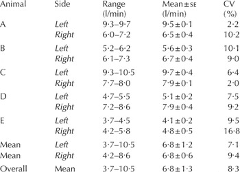
Intra-observer precision and the left-right comparison of the BFV of the left and the right pudendoepigastric trunk of five cows is shown in Table 2. The CV of the BFV was 7·1% (6·8±2·6 l/min) for the left and 9·4% (6·8±1·3 l/min) for the right pudendoepigastric trunk, an average of 8·3% (6·8±1·9 l/min). The ICC of the BFV was 0·99 (P<0·001) for the left and 0·75 (P=0·004) for the right vessel. The BFV of the left side did not differ significantly from the BFV of the right side (WSRT: P=0·893; RMM: P=0·410). The pre-post-milking comparison and the oestrus-dioestrus comparison are illustrated in Table 3. Values of BFV, means of left and means of right measurements did not differ significantly between pre- and post-milking (WSRT: left P=0·345; right P=0·345; RMM: left P=0·141; right P=0·536) or between oestrus and dioestrus (WSRT: left P=0·690; right P=0·893; RMM: left P=0·646; right P=0·829).
Intra-observer precision and left-right comparison of the blood flow volume (BFV; l/min) of the left and the right pudendoepigastric trunk of five cows (A, B, C, D, E) in the methodological part. Values describe ranges and means and se of three separate measurements on each side. Intra-observer precision is expressed by the coefficient of variation (CV), which is presented for each side of each individual cow (no significant difference in the precision between left and right, P=0·345)

Pre-post-milking comparison and oestrus-dioestrus comparison of the blood flow volume (BFV; l/min) of the left and the right pudendoepigastric trunk of five cows in the methodological part of the study. Values describe ranges and means and se of measurements of all animals on each side and differences between pre- and post-milking as well as between oestrus and dioestrus (measured only pre-milking)

Esch. coli mastitis model (experimental part of the study)
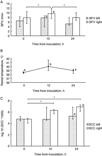
The BFV differed significantly in the course of the trial between the left and the right side (RMM: P<0·001). Significant differences of increasing BFV between 0 and 12 h p.i. (WSRT: P=0·043) and decreasing BFV between 12 and 24 h p.i. (WSRT P=0·043) were discovered for the pudendoepigastric trunk of the infected right side (Fig. 2A). No significant differences were detected for the BFV of the left non-infected side (P>0·05). In the left-right comparison a significant difference of BFV was observed at the time of 12 h p.i. (P=0·043).
24-h course of (A) the blood flow volume [BFV; l/min], (B) the rectal temperature [RT; °C] and (C) the somatic cell count [log10 (SCC*1000)] of five cows in the experimental part, expressed by medians and quartiles (first and third quartiles indicated by error bars) during an induced Escherichia coli mastitis. Different letters indicate a statistically significant difference (A, C) between the left and the right side or (B) between different times (P<0·05), * indicates a statistically significant difference (A, C) between different times (P<0·05).

A significant time effect in the rectal temperature was seen (RMM: P=0·006), whereby a significant increase of the RT could be detected between 0 and 12 h p.i. (WSRT: P=0·043; RMM: P=0·011) as well as a statistically significant decrease between 12 and 24 h p.i. (WSRT: P=0·043; RMM: P=0·037) (Fig. 2B).
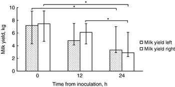
Between 12 and 24 h p.i. a significant decrease of MY was seen in the infected right udder half (P=0·042) (Fig. 3). Between 0 and 24 h p.i. both the left and right udder halves showed significant differences (WSRT: left P=0·043; right P=0·043; RMM: left P=0·007; right P=0·026). The left-right comparison showed no significant difference. Between 0 and 12 h p.i. the difference in the MY, which was decreasing, was correlated with the difference in the BFV of the right pudendoepigastric trunk, which was increasing (ρ=0·60; P=0·043).
24-h course of the milk yield (kg; average of the two left quarters v. average of the two right quarters) of five cows in the experimental part, expressed by medians and quartiles (first and third quartiles indicated by error bars) during an induced Escherichia coli mastitis. * indicates a statistically significant difference between different times (P<0·05).

In the RMM of the log 10 (SCC*1000) a significant time effect (P<0·001), side effect (P<0·001) and a significant time-side interaction effect (P=0·004) was found. SCC of the right udder quarters increased significantly between 0 and 12 h p.i. (WSRT: P=0·043; RMM: P<0·001) (Fig. 2C). Between 0 and 24 h p.i. the right udder half showed a significant increase in SCC (WSRT: P=0·043; RMM: P<0·001). A significant difference of the SCC was observed in the left-right comparison at the time of 24 h p.i. (WSRT: P=0·043; RMM: P=0·004). The correlation between BFV and SCC was examined by comparing the differences of the absolute values between different times. The difference of an increasing SCC correlated positively with the difference of an increasing BFV between 0 and 12 h p.i. (ρ=1·00; P=0·043) for the right side. Between 12 and 24 h p.i. the difference of an increasing SCC correlated negatively with the difference of a decreasing BFV (ρ=−0·60; P=0·043) for the right pudendoepigastric trunk.
Discussion
Establishment of the method (methodological part of the study)
In the beginning of this trial the intra-observer precision of this new method was necessarily assessed and possible dependences on disturbance variables (left/right; pre-/post-milking; oestrus/dioestrus) were tested. In the literature several methods of evaluating intra-observer precision are described (Lamb et al. Reference Lamb, Burton and Carlisle1999; Bollwein et al. Reference Bollwein, Meyer, Maierl, Weber, Baumgartner and Stolla2000; Schmucker et al. Reference Schmucker, Schatzmann, Budde, Gundel, Jäggin and Meier2000). In the present study measurements were taken three times on each side and precision was expressed by the CV and the ICC similarly to other studies (Bollwein et al. Reference Bollwein, Meyer, Maierl, Weber, Baumgartner and Stolla2000; Hollis et al. Reference Hollis, Mavrides, Campbell, Tekay and Thilaganathan2001). Intra-observer precision of repeatedly performed measurements in single animals was consistently good. The overall mean, using left and right measurements of all five animals, also supported this finding with a CV of 8·3%. It was demonstrated that TCDS of the pudendoepigastric trunk provides well reproducible and, thus, precise values. The method was precise, as it repeatedly measured the same values. However, a precise method does not necessarily mean a correct method. It is conceivable that the method measures precisely values which are a certain amount higher or lower than the real value. To evaluate the correctness of the method further investigations would be needed to compare TCDS with other methods, such as direct blood measurements. On the basis of former studies using Doppler sonography for different investigations, bilateral comparisons were drawn for information about relevancies and differences of the two scanning locations, the left and the right pudendoepigastric trunk (Christensen et al. Reference Christensen, Nielsen, Bauer and Hilden1989; Bollwein et al. Reference Bollwein, Maierl, Mayer and Stolla1998; Schmucker et al. Reference Schmucker, Schatzmann, Budde, Gundel, Jäggin and Meier2000; Bollwein et al. Reference Bollwein, Baumgartner and Stolla2002; Piccione et al. Reference Piccione, Arcigli, Fazio, Giudice and Caola2004a). We did not find a significant difference between measurements of the left and the right pudendoepigastric trunk; however, the animal number in the present study was limited. Yet, a power analysis using a sd of 0·5, which was found in the present study, calculated that a difference in the BFV of at least 1 l/min between left and right would have been identified with a power of 90% and alpha=0·05 using the five animals (calculated with NCSS/PASS). This indicates similar blood support of both udder halves and equates with almost equally distributed MY of the left and the right udder halves (data not shown) when kept under physiological conditions as in this trial. The results also corroborate findings of other authors using Doppler ultrasound for measurements of mammary blood flow velocities at different udder-associated vessels in lactating cows (Piccione et al. Reference Piccione, Arcigli, Fazio, Giudice and Caola2004a), goats (Christensen et al. Reference Christensen, Nielsen, Bauer and Hilden1989) and ewes (Piccione et al. Reference Piccione, Arcigli, Assenza, Percipalle and Caola2004b).
A significant difference between measurements before and after milking could not be discovered. Similar BFV observations are reported for mammary blood flow measurements in lactating goats (Christensen et al. Reference Christensen, Nielsen, Bauer and Hilden1989). Earlier studies using invasive methods at the external pudic artery in cows showed that positive changes of BFV are only measurable during milking. An abrupt and enormous increase of mammary blood flow with onset of milking followed by a very quick return of BFV within minutes to initial values at the end of milking has been described (Houvenaghel et al. Reference Houvenaghel, Peters and Verschooten1973; Davis & Collier, Reference Davis and Collier1985). This can be seen as an explanation for not detecting any differences between pre- and post-milking measurements within 2 h in the present trial.
In goats and cattle it is known that the oestrus cycle affects blood flow in certain blood vessels. In goats the mammary BFV is decreased the day before and on the day of oestrus, but no alteration is seen on any other days during the oestrus cycle (Burvenich, Reference Burvenich1980). In the present trial a significant difference of BFV between scannings during oestrus and dioestrus was not detected.
Esch. coli mastitis model (experimental part of the study)
Since observations of the early stage of an acute mastitis are not possible under field conditions a standardized and, thus, very well reproducible stringent Esch. coli mastitis model was used for this trial.
After pathogen inoculation a significant difference of BFV in the right infected udder half between 0 und 12 h p.i. was detected. The first change of BFV can be easily explained by the basic principles of inflammation. After contact with the pathogen, processes regulated by biochemical mediators provoke a dilatation of the arterioles, capillaries and venules. Increased hydrostatic pressure affects transudation and hyperaemia (Meurer, Reference Meurer1999). An increased volume of blood is moved into the mammary region. In an earlier study, measuring mammary blood flow (absolute integration values/unit of time; %) by an implanted electromagnetic flow probe after intramammary infusion of Esch. coli endotoxin two blood flow peaks were found (Dhondt et al. Reference Dhondt, Burvenich and Peeters1977). A first peak occurred at the third hour after infusion followed by a temporary return to the initial level and a second peak between the tenth and eleventh hour followed by a return to control levels between the thirteenth and fourteenth hour. Since, in our study, measurements were made at 0, 12 and 24 h p.i. detection of the described first blood flow peak was not possible. However, the described second peak could be entirely validated by TCDS.
Significant differences of simultaneously increasing RT and SCC in the infected quarter between 0 and 12 h p.i. also fit well with the inflammatory procress with systemic and cellular defence mechanisms during the acute phase. Thereby, the statistically positive correlation between the changes of the right SCC and the changes of the right BFV clarifies well the degree of acute inflammation and its effect on mammary blood flow. A significant difference of BFV between the right (infected) and the left (non-infected) side at 12 h p.i. is remarkable because it allows the exact identification of the infected udder half.
Although the decrease of the MY of the right infected udder half was not statistically significant, the change of the MY correlated positively with the change of the increasing right BFV. At this stage of experimental Esch. coli mastitis it is known that mammary tissue shuts down synthesis of milk constituents and favours the expression of antimicrobial effector molecules such as defensins (Vanselow et al. Reference Vanselow, Yang, Herrmann, Zerbe, Schuberth, Petzl, Tomek and Seyfert2006, Petzl et al. Reference Petzl, Zerbe, Günther, Yang, Seyfert, Nürnberg and Schuberth2008). This could explain the increase of the BFV up to 12 h p.i. due to proinflammatory actions, although MY is already decreasing. Between 12 and 24 h p.i. the BFV of the right pudendoepigastric trunk (infected side) decreased significantly accompanied by significantly decreasing RT and MY (right). These findings also follow the basic inflammatory principles: hours after the occurrence of inflammation the dilatation of arterioles and the arterial branch still continues, whereas the venous branch and small veins start to constrict. Hyperaemia and a slowdown of blood flow are the consequences (Meurer, Reference Meurer1999). A lower volume of blood is moved into the mammary region. After the acute phase of inflammation RT returns to a lower level. The significant decrease of MY of the right infected side can be explained by the ongoing progression of the systemic illness which influences, typically for Esch. coli mastitis, the MY of the left non-infected side (Burvenich et al. Reference Burvenich, Paape, Hoeben, Dosogne, Massart-Leen and Blum1999). A high correlation between decreasing MY of the uninfected gland and bacterial growth of the infected gland is known (Dosogne et al. Reference Dosogne, Burvenich, van Weren, Roets, Noordhuizen-Stassen and Goddeeris1997).
With this present study it could not only be shown that TCDS is a new non-invasive and precise method for evaluating the BFV of the pudendoepigastric trunk in healthy cows, but also that this technique was successfully used for detecting pathological mammary blood flow changes at an early stage in a stringent Esch. coli mastitis model. The discoveries of this study mark an important basis for further investigations of mammary blood flow, especially for experimental set-ups of infection models with udder pathogens and the associated detection of clinical, acute/chronic or subclinical mastitis by TCDS. Findings of this study provide significant information for bilateral comparisons and scanning times in mastitis infection models and should be seen as a helpful guideline for planned Doppler sonographic testings. It has to be noted that only initially udder-healthy animals with an equally distributed MY in both udder halves were examined in the present trial. Animals with pre-existing impairments of the udder and such with unequal milk distribution in the udder halves could have biased the results of the standardized study. This fact underlines the particular meaning and suitability of the findings described here with regard to well-defined mastitis infection models. Setting a ‘zero-standard’, meaning healthy-, left/right- and hour 0-status, of the single test animal before trial initiation is an absolute prerequisite for any examination.
Especially with a view to the severe problem of mastitis appearing worldwide and the need to detect it as early as possible, it is conceivable that TCDS may find its way from an experimental technique to a clinical application as another useful non-invasive and reliable tool for diagnosing pathological changes in the mammary gland of cows. However, more studies including some on the economical and logistical practicability of the method are needed.
This work was funded by the Deutsche Forschungsgemeinschaft (German Research Foundation; FOR 585, ZE 414/3-1). The authors thank Markus Reimann (Clinic for Ruminants with Ambulance Clinic and Herd Health Management, Ludwig-Maximilians-University Munich) and Antonia Goetze (Clinic for Cattle, University of Veterinary Medicine Hannover) for skilful assistance.