Introduction
Tonsillar squamous cell carcinoma represents 15–20 per cent of all oropharyngeal malignancies and is the most common oropharyngeal carcinoma type.Reference Galati, Myers and Johnson1 It ranks second in frequency to squamous cell carcinoma of the larynx, and is most common during the fifth and sixth decades of life.Reference Poulsen, Porceddu, Kingsley, Tripcony and Coman2 Tonsillar cancer is usually diagnosed at an advanced stage, at least the tumour 2 (T2) stage, with a diameter greater than 2 cm. In 60 per cent of cases, there is evidence for regional lymphatic channel involvement, attributed to the rich lymphatic supply in this anatomical region.Reference Guggenheimer, Verbin, Johnson, Horkowitz and Myers3
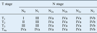
The tumour–node–metastasis (TNM) classification system of the American Joint Committee on Cancer is the official method of defining cancer progression and predicting prognosis, and is used worldwide for most tumours.Reference Takes, Rinaldo, Silver, Piccirillo, Haigentz and Suárez4 Based on different combinations of TNM levels, tumours are classified as stage I, II, III or IV. In this system, survival outcomes are considered to be similar within stages and different between stages. However, in oropharyngeal cancer, T1–4a and N0–2c tumours, which may exhibit very different biological behaviours, are classified as the same stage (IVa; see Table I).Reference Greene, Page, Fleming, Fritz, Balch and Haller5 Although patients with higher stage (e.g. IVa) tumours have worse prognoses, it is also clear that the prognoses of patients with tumours of the same stage may differ significantly. These differences may be explained by tumour biology or host- or environment-related factors not considered in the TNM system.Reference Takes, Rinaldo, Silver, Piccirillo, Haigentz and Suárez4
AJCC staging according to TNM classification*Reference Greene, Page, Fleming, Fritz, Balch and Haller5

*For cases without distant metastasis. AJCC = American Joint Committee on Cancer; N = node; T = tumour; I = stage I; II = stage II; III = stage III; IVa = stage IVa; IVb = stage IVb
Human papillomavirus (HPV) is considered a tumour biological factor in oropharyngeal cancer. Recent reports indicate that the tonsillar cancer incidence and proportion of HPV-positive tumours are increasing in parallel, suggesting that HPV infection may correlate with tonsillar cancer incidence.Reference Shiboski, Schmidt and Jordan6 Furthermore, because HPV-positive tonsillar cancers tend to have a better prognosis, HPV infection has been suggested as a prognostic biomarker.Reference Shiboski, Schmidt and Jordan6, Reference Hong, Dobbins, Lee, Jones, Harnett and Armstrong7
The objectives of this study were to identify clinical factors that can explain differences in treatment outcomes and to examine the value of HPV infection as a prognostic biomarker for stage IVa tonsillar carcinoma.
Materials and methods
Patients
Investigations were carried out in 116 stage IVa tonsillar squamous cell carcinoma patients treated with curative intent at the Korea University Medical Centre, Korea, between 1994 and 2009. Patient selection was based on the availability of clinicopathological data. Of these patients, 42 were excluded because of insufficient medical record data, a short follow-up period (less than 1 year) or the presence of a second primary cancer at another site. Fifteen patients who underwent cisplatin-based concurrent chemoradiation therapy were also excluded. Therefore, a total of 59 patients were included in the study; the median patient follow-up period was 80.5 months.
The analysis was performed according to the American Joint Committee on Cancer staging system.Reference Greene, Page, Fleming, Fritz, Balch and Haller5 Locoregional recurrences were determined by clinical and radiological parameters, and verified by biopsy. This study was approved by the institutional review board of Korea University Anam Hospital.
Treatment
Treatment modalities were chosen after discussion among surgical, medical, and radiation oncologists serving on the Head and Neck Tumour Board at our institution. Factors affecting the choice of treatment modality were patient preference, functional preservation and morbidity after specific treatments.
All patients underwent surgical treatment, followed by adjuvant therapies such as radiotherapy or combined chemoradiotherapy. For surgical resection of the primary tumour, lateral oropharyngectomy with or without reconstruction was performed, depending on the tumour's size and relationship to adjacent structures. Elective or therapeutic neck dissection was performed on patients suspected of having lymph node metastasis.
Clinical factors and tonsillar cancer reclassification
The clinical data collected from participants comprised gender, age, smoking exposure, American Joint Committee on Cancer T and N stage, tumour differentiation, resection margin, and nodal extracapsular spread.
Furthermore, tonsillar cancer limited to the tonsillar fossa (according to the final pathology results) was classified as ‘locally limited cancer’. Locally invasive tonsillar cancer, including invasion of the extrinsic muscles of the tongue, medial pterygoid or hard palate, was classified as ‘locally advanced cancer’. In addition, tonsillar cancer either without or with a single ipsilateral lymph node metastasis of 3 cm or less in diameter was classified as ‘early lymph node metastasis’. Tonsillar cancer with lymph node metastasis of more than 3 cm in diameter or with multiple lymph node metastases was classified as ‘advanced lymph node metastasis’. No patients had both locally limited cancer and early lymph node metastasis. Therefore, patients were classified into the following three groups: those with locally advanced cancer and early lymph node metastasis (Group 1), those with locally limited cancer and advanced lymph node metastasis (Group 2); and those with locally advanced cancer and advanced lymph node metastasis (Group 3).
Deoxyribonucleic acid extraction
Archived paraffin-embedded tissue samples were examined. Haematoxylin and eosin (H&E) stained sections were first reviewed to confirm the presence of sufficient tumour tissue. Of the 59 patients, paraffin-embedded tissue from 45 patients was available for HPV infection testing. For this, five consecutive 5-µm sections were cut from each paraffin block. Paraffin was removed by vortexing, incubation for 10 minutes in 1.0 ml xylene, and then washing twice with absolute ethanol. Deoxyribonucleic acid was then isolated using a QIAamp DNA mini kit (Qiagen, Valencia, California, USA) following the manufacturer's instructions.
Human papillomavirus genotyping
The commercially available PANArray HPV chip test (Panagene, Daejeon, Korea), a polymerase chain reaction based DNA microarray system, was used for HPV genotyping. The HPV-DNA chip contains 32 type-specific probes to 19 high-risk (i.e. 16, 18, 26, 31, 33, 35, 39, 45, 51, 52, 53, 56, 58, 59, 66, 68, 69, 70 and 73) and 13 low-risk (i.e. 6, 11, 32, 34, 40, 42, 43, 44, 54, 55, 62, 81 and 83) HPV subtypes. Analyses were performed according to the manufacturer's instructions.Reference Choi, Kim and Park8
p16 and p53 immunostaining and scoring
Three consecutive, 5-μm sections were cut from each paraffin-embedded block. One section from each specimen was routinely stained with H&E, and the other two sections were immunohistochemically stained with either monoclonal mouse anti-human p16INK4a antibody (MTM Laboratories, Heidelberg, Germany) or monoclonal mouse anti-human p53 antibody (DakoCytomation, Carpentaria, California, USA). Specific protein expression was revealed by developing the sections in a 3,3′-diaminobenzidine tetrahydrochloride (Sigma, St Louis, Missouri, USA) solution. For negative controls, non-immune serum immunoglobulin G or phosphate-buffered saline was used instead of the primary antibody. Sections were counterstained with haematoxylin and mounted. Absolute p16 expression was indicated by the presence of positively stained cells (Figure 1a); the p53 expression level was scored as the percentage of positively stained cells within a high-power field (0, less than 5 per cent; 1, 5–25 per cent; 2, 25–50 per cent; 3, 50–75 per cent; 4, 75–100 per cent; Figure 1b–d). Immunostaining was evaluated by a pathologist in at least 10 high-power fields at ×200 magnification. Moderately to intensely stained cells were counted by a recorder in a blinded manner.
Photomicrographs showing immunohistochemistry staining for p16 and p53 in tonsillar cancer tissue. (a) Strong, diffuse p16 expression in tonsillar cancer cells. (b–d) p53 expression in tonsillar cancer cells scored as 0 (b), 2 (c) and 4 (d). (×40)
Statistical analysis
Relationships between survival rate and various clinical factors, HPV positivity, and p16 and p53 expression were examined using Kaplan–Meier plots, log-rank tests and Cox regression models. Differences between groups were evaluated using the Pearson chi-square test and Fisher's exact test, as appropriate. For all tests, a probability value of less than 0.05 was accepted as statistically significant. All statistical analyses were conducted using SPSS software version 12.0 (SPSS Inc, Chicago, Illinois, USA).
Results
Patient characteristics and tumour risk factors
In all, 43 male and 16 female patients were included in the study. The age at diagnosis ranged from 29 to 78 years (overall mean 56.9 years); the mean ages for men and women were 57.8 and 54.5 years, respectively. Univariate analysis revealed no significant association between age or gender and the five-year overall survival rate or disease-free survival rate (data not shown; p > 0.05).
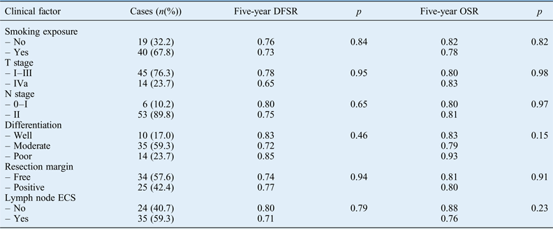
Similar analyses showed no association of smoking exposure, T stage, N stage, tumour differentiation, resection margin or extracapsular spread of lymph node with five-year overall survival rate or disease-free survival rate (p > 0.05; Table II).
Association of disease-free survival and overall survival rates with clinical factors*
*Univariate analysis. DFSR = disease-free survival rate; OSR = overall survival rate; ECS = extracapsular spread
Reclassification of tonsillar cancer cases
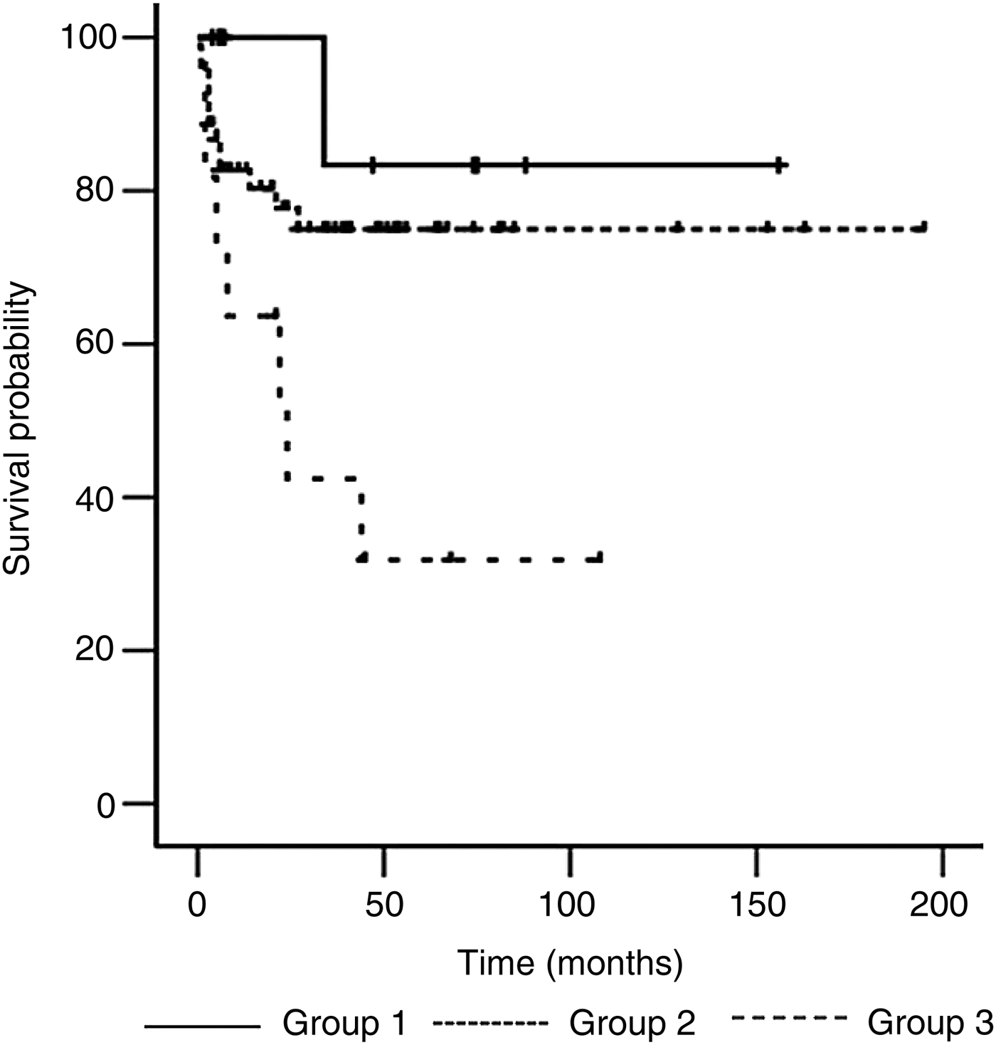
The overall survival rate was not significantly different among the three patient groups. The disease-free survival rate was highest for Group 1 patients (83.3 per cent), followed by Group 2 (74.9 per cent) and Group 3 (31.8 per cent, p = 0.01; Table II). As expected, the disease-free period was shortest for patients with ‘locally advanced and advanced lymph node metastasis’ (i.e. Group 3; Figure 2).
Kaplan–Meier plot showing significant differences in disease-free survival rates among the three study groups (p = 0.01). Group 1, locally advanced cancer and early lymph node metastasis; Group 2, locally limited cancer and advanced lymph node metastasis; Group 3, locally advanced cancer and advanced lymph node metastasis.
Human papillomavirus positivity and p16 and p53 expression
High-quality DNA was extracted from the tonsillar cancers of 45 patients: 23 patients (51 per cent) had HPV-positive tumours and 38 patients (84 per cent) had p16-expressing tumours. The HPV subtype present in all tumours was HPV16.
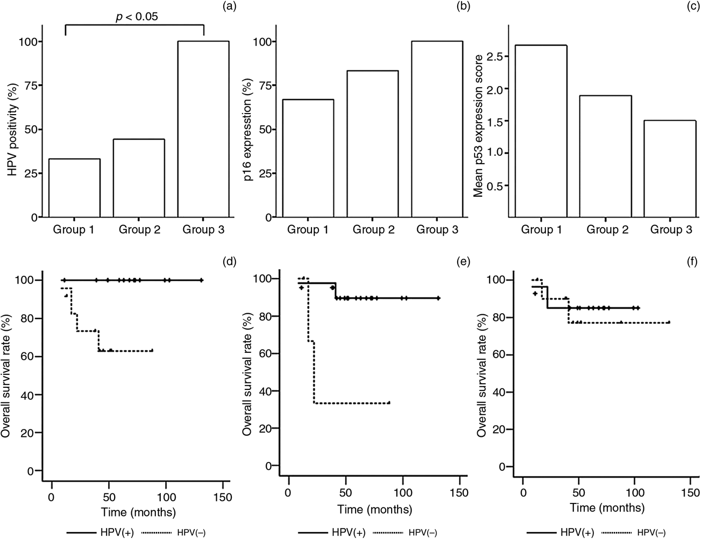
The distribution of HPV positivity and p16 expression differed among the three tonsillar cancer patient groups (Figure 3a and b). All Group 3 patients (100 per cent; with locally advanced tonsillar cancer and advanced lymph node metastasis) showed HPV positivity and p16 expression. In addition, the level of HPV positivity was significantly greater in tumours from Group 3 than from Group 1 patients (p= 0.04). In contrast, p53 expression was lowest in Group 3 patients, followed by those in Group 2 and then Group 1 (Figure 3c).
Graphs showing differences in tumour human papillomavirus (HPV) positivity, p16 expression, and p53 score among patient groups, and Kaplan–Meier curves showing overall survival in the three patient groups. The incidence of HPV-positive (a) and p16-positive (b) tumours, and the mean p53 expression score (c) in all patient groups. Overall survival rate by HPV positivity (d) p16 positivity (e) and p53 score (f) in the three patient groups. Group 1, locally advanced cancer and early lymph node metastasis; Group 2, locally limited cancer and advanced lymph node metastasis; Group 3, locally advanced cancer and advanced lymph node metastasis. p53 (+) = score higher than 1; p53 (−) = score of 0 or 1
The overall survival rate differed significantly according to HPV positivity and p16 expression; however, these variables were not associated with differences in the disease-free survival rate (Figure 3d and e). The five-year overall survival rates for patients with HPV-negative and HPV-positive tumours were 62.9 per cent and 100 per cent, respectively (p = 0.02). In addition, the five-year overall survival rates were 33.3 per cent and 89.6 per cent for patients with p16-negative and p16-positive tumours, respectively (p = 0.01). However, the five-year overall survival rates were not significantly different between those with p53-negative tumours (p53 score of 0 or 1) and those with p53-positive tumours (p53 score greater than 1; p > 0.05; Figure 3f).
Human papillomavirus positivity and smoking exposure
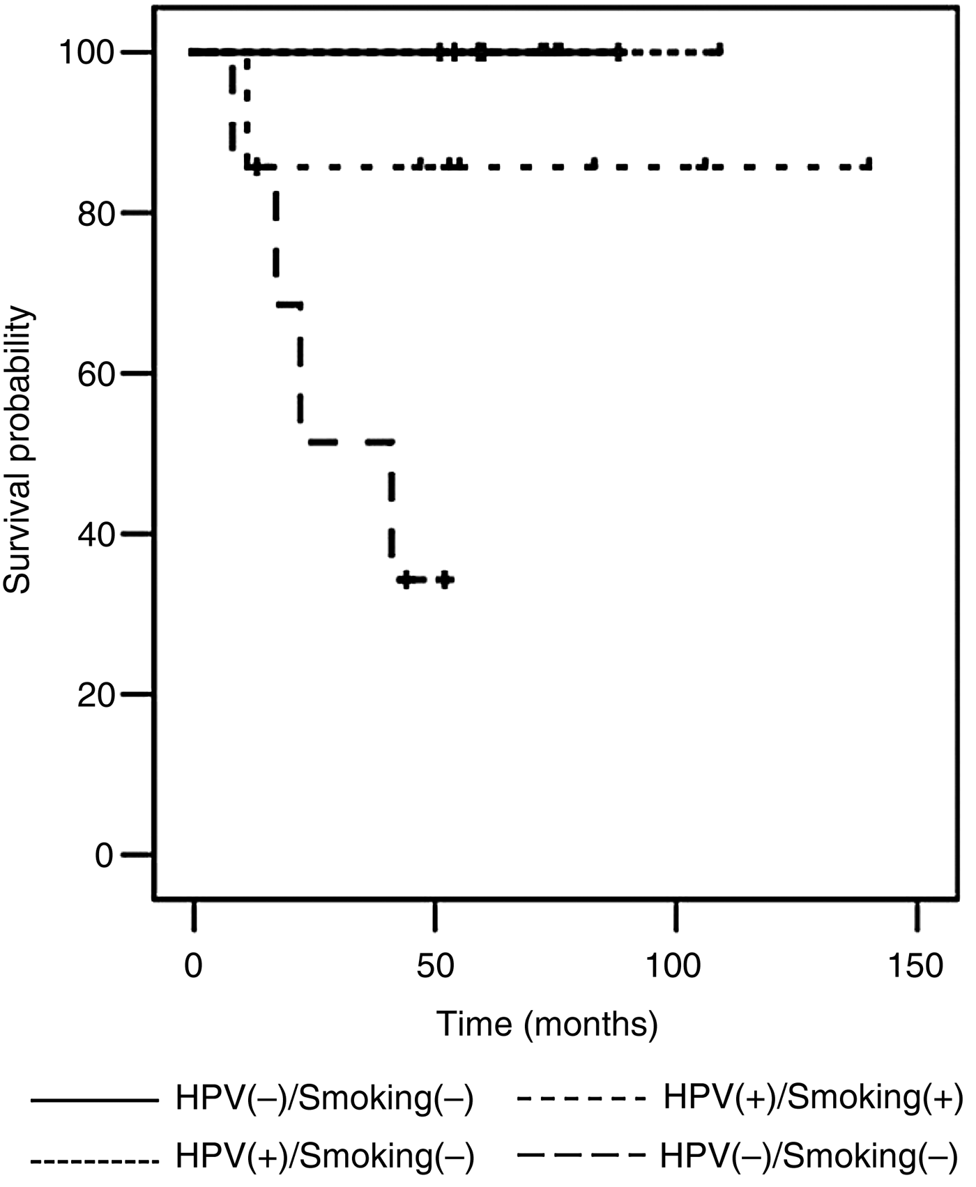
All patients were divided into four groups according to HPV positivity and smoking exposure at diagnosis: HPV-negative or HPV-positive, with or without smoking exposure. Although there was no difference among the four groups in disease-free survival rate, the overall survival rates were significantly reduced for groups exposed to smoking (p = 0.02; Figure 4). The five-year overall survival rate was 100 per cent in patients with no history of smoking exposure. However, for patients with a history of smoking exposure, the five-year overall survival rates were 85.7 per cent for those with HPV-positive tumours and 34.3 per cent for those with HPV-negative tumours.
Kaplan–Meier curves showing overall survival by human papillomavirus (HPV) positivity and smoking exposure.
Association between disease-free survival and overall survival rate after tonsillar cancer reclassification*
*Univariate analysis. †Group 1, locally advanced cancer and early lymph node metastasis; Group 2, locally limited cancer and advanced lymph node metastasis; Group 3, locally advanced cancer and advanced lymph node metastasis. DFSR = disease-free survival rate; OSR = overall survival rate
Discussion
Tonsillar squamous cell carcinoma is the most common oropharyngeal carcinoma. This cancer has considerable medical and social impacts on patients through its effects on critical functions of the oropharynx, including speech and swallowing.
Previous studies have suggested that the clinical stage at initial presentation is the most important prognostic factor, independent of other factors such as gender, age and tumour location.Reference Ildstad, Tollerud and Bigelow9 Generally, the survival rate of patients is similar within each TNM stage group, but markedly different between TNM stage groups.Reference Takes, Rinaldo, Silver, Piccirillo, Haigentz and Suárez4 However, tonsillar carcinomas with varying characteristics can be classified as the same TNM stage, especially American Joint Committee on Cancer stage IVa tumours. Because most patients with tonsillar carcinoma are diagnosed as stage IVa, it is very important to identify prognostic factors for the different treatment outcomes of patients with stage IVa tumours with various clinical characteristics.
• The biological behaviour of T1–4a and N0–2c tonsillar cancers classified as stage IVa may be notably different
• All stage IVa tonsillar cancers showed the same overall survival rate
• The disease-free survival rate inversely correlated with local invasiveness and nodal status
• Human papillomavirus (HPV) and p16 positivity was more common in locally advanced tonsillar carcinomas with advanced lymph node metastasis
• The five-year disease-specific survival rate was better for HPV-positive, p16-positive tumour patients
• HPV infection may be a useful biomarker for predicting treatment outcomes in stage VIa tumours
Prognosis for tonsillar carcinoma patients is significantly associated with the primary tumour and metastatic lymph node stages.Reference Ildstad, Tollerud and Bigelow9–Reference Perez, Patel, Chao, Simpson, Sessions and Spector11 However, because patients with stage I–IV cancers were included in most previous studies, it is difficult to identify prognostic differences within American Joint Committee on Cancer stage IVa, which consists of diverse tumour and lymph node stages. In the present study, no meaningful clinical factor was associated with overall survival rate. However, a univariate analysis revealed that the disease-free survival rate was significantly different among three patient groups classified according to local tumour invasiveness and lymph node metastasis.
This result suggests that the American Joint Committee on Cancer staging system is a well-designed prognostic system that predicts the same overall survival rate for all stage IVa tonsillar carcinoma patients. However, it also suggests that tumour recurrence is related to primary tumour invasiveness and the nodal status. Therefore, because prognosis can differ even among patients with stage IVa tumours, the treatment modality should take local invasiveness and nodal status into consideration.
Many recent studies have reported that oncogenic HPV infection may be significantly associated with prognosis for oropharyngeal carcinoma patients. Some authors have suggested that the combined biomarkers oncogenic HPV positivity and p53, pRb and p21 tumour suppressor expression may provide prognostic molecular classification superior to that of the TNM staging system for advanced tonsillar squamous cell carcinomas.Reference Chung, Lee, Horng, Jian, Cheng and Tsai12 Although the prognostic significance of HPV infection for head and neck cancers is not yet certain, recent studies have reported a better prognosis for patients with HPV-positive tumours than those with HPV-negative tumours.Reference Kuo, Hsiao, Lin, Kuo, Huang and Lin13, Reference Schwartz, Yueh, McDougall, Daling and Schwartz14 The association between HPV positivity and other risk factors, such as tumour size, tumour invasiveness and extent of lymph node metastasis, differed considerably between studies.Reference Schwartz, Yueh, McDougall, Daling and Schwartz14–Reference Klozar, Kratochvil, Salakova, Smahelova, Vesela and Hamsikova17 Hoffmann et al. reported that survival was not significantly associated with HPV positivity, even though HPV-positive tumours corresponded to higher nodal stages.Reference Hoffmann, Görögh, Gottschlich, Lohrey, Rittgen and Ambrosch15 However, these authors reported that patients with HPV-positive tumours had better survival rates than those with HPV-negative tumours at the same nodal stage. Strome et al. reported that HPV positivity was not associated with nodal status but was associated with a better prognosis.Reference Strome, Savva, Brissett, Gostout, Lewis and Clayton16 Furthermore, these authors reported that, after adjusting for age, HPV positivity was not associated with survival. However, Klozar et al. suggested that the survival of HPV-positive tumour patients was significantly better than that of HPV-negative tumour patients, irrespective of age.Reference Klozar, Kratochvil, Salakova, Smahelova, Vesela and Hamsikova17 In the present study, HPV positivity and p16 expression differed among the three patient groups classified according to local tumour invasiveness and lymph node metastasis. Although not all tumours were tested for HPV infection or p16 expression, all patients in Group 3 (with locally advanced cancer and advanced lymph node metastasis) were positive for both HPV infection and p16 expression. Additionally, HPV positivity differed significantly between Group 1 (with early nodal stage tumours) and Group 3 (with advanced nodal stage tumours) patients. The overall survival rate differed significantly between patients with HPV-positive, p16-positive and HPV-negative, p16-negative tumours classified as American Joint Committee on Cancer stage IVa tonsillar cancers.
Previous studies have suggested that HPV positivity and smoking exposure are significant prognostic factors for patients with oropharyngeal carcinoma.Reference Ang, Harris, Wheeler, Weber, Rosenthal and Nguyen-Tân18, Reference Gillison, Zhang, Jordan, Xiao, Westra and Trotti19 Similarly, of all HPV-positive patients in the present study, the overall survival rate was significantly lower in smokers. Furthermore, in smokers with HPV-negative tumours, the overall survival rate was reduced to 34.3 per cent.
Conclusion
Among stage IVa tonsillar cancers, locoregional tumour recurrence can differ according to local tumour invasiveness and nodal status. Although HPV infection may be associated with regional tonsillar cancer progression, the prognosis for patients with HPV-positive tumours was better than for patients with HPV-negative tumours. In addition, among patients with a history of smoking, the prognosis differed significantly according to HPV positivity. Therefore, HPV infection may be a useful biomarker for predicting different treatment outcomes in stage IVa patients with varying clinical characteristics.
Acknowledgements
This study was supported by a Grant-in-Aid award from the Korea University Research and Business Foundation.