INTRODUCTION
Common bean (Phaseolus vulgaris L.) and cowpea (Vigna unguiculata (L.) Walp) are two important legume crops that are commonly grown in sub-Saharan Africa for food, cash, animal feed and as soil improvers. The seeds of the two crops are a cheap source of protein for the people of this region and ease of storage leads to their preference over meat products by smallholder farmers (Msuku et al. Reference Msuku, Saka and Munthali2000), who mostly lack cooling facilities. It has been reported that the two crops also have medicinal properties (Kritzinger et al. Reference Kritzinger, Lall and Aveling2005; Wong et al. Reference Wong, Zhang, Wang and Ng2006; Jeng et al. Reference Jeng, Shih, Lai, Wu and Sung2009). Leaf extracts of cowpea (Kritzinger et al. Reference Kritzinger, Lall and Aveling2005) and seed extracts of both crops have antifungal activities against some phytopathogens (Ng Reference Ng2004), making them a potential source of agricultural fungicides.
Despite the importance of the two crops, farmers fail to realize maximum potential yields because of several limiting factors, among them the incidence and severity of diseases (Beaver et al. Reference Beaver, Rosas, Myers, Acosta, Kelly, Nchimbi-Msolla, Misangu, Bokosi, Temple, Arnaud-Santana and Coyne2003; Adebanjo & Bankole Reference Adebanjo and Bankole2004). Anthracnose is a common disease of legumes and is caused by Colletotrichum spp. Common bean anthracnose is caused by Colletotrichum lindemuthianum (Sacc. and Magn.) Bri. and Cav., whereas cowpea anthracnose is initiated by both Colletotrichum dematium (Fr.) Grove var. truncata and C. lindemuthianum. Anthracnose is a seed-borne disease and complete crop failure can be expected under heavy infection (Allen Reference Allen1983; Pastor-Corrales & Tu Reference Pastor-Corrales, Tu, Schwartz and Pastor-Corrales1989). The pathogen has also been reported to be associated with humans and animals, making its control imperative (Cano et al. Reference Cano, Guarro and Gené2004; Mendiratta et al. Reference Mendiratta, Thamke, Shukla and Narang2005). For example, C. dematium has been implicated in corneal ulcers in humans (Mendiratta et al. Reference Mendiratta, Thamke, Shukla and Narang2005).
The current major means of controlling anthracnose is the use of synthetic fungicides (Tian et al. Reference Tian, Zhu, Sun, Jiang, Wang and Zhang2007) through seed dressing and foliar application. However, several problems pertaining to the use of synthetic fungicides have been reported, including accumulation of harmful chemical residues in seeds, water and soil and also the induction of resistance in the pathogens (Falandysz Reference Falandysz2000; Shovan et al. Reference Shovan, Bhuiyan, Begum and Pervez2008). Therefore, there is a need for safer alternative fungicides.
A survey by Mwine et al. (Reference Mwine, Van Damme, Kamoga, Kudamba and Jumba2011) in the Masaka district, southern Uganda, reported that 34 plant species belonging to 18 families were used in traditional plant production for plant protection by smallholder farmers. Among these were Allium sativum (garlic) water extracts, used against field and storage pests, and Carica papaya (pawpaw) water extracts, used against ‘blight’. In a survey involving smallholder farmers in Zimbabwe, Sibanda et al. (Reference Sibanda, Dobson, Cooper, Manyangarirwa and Chiimba2000) found that many of the farmers had tried or heard about spraying with botanical extracts from plants such as Mexican Marigold (Tagetes spp.), garlic, chillies (Capsicum frutescens L.) and tobacco (Nicotiana tabacum L. and other spp.) against pathogens and pests. The present authors were unable to find any reported surveys on the use of plant extracts as seed treatments by smallholder farmers.
There are many plants that are toxic to Colletotrichum; however, research in this field has focused upon the anthracnose diseases of other vegetables and fruits (Raghavendra et al. Reference Raghavendra, Lokesh, Girisha, Govindappa and Prakash2006; Chowdhury et al. Reference Chowdhury, Rahim, Khalequzzaman, Humauan and Alam2007; Nduagu et al. Reference Nduagu, Ekefan and Nkwankiti2008; Mdee et al. Reference Mdee, Masoko and Ellof2009) and little has been done on the anthracnose of beans and cowpea (Amadioha Reference Amadioha2003; Shovan et al. Reference Shovan, Bhuiyan, Begum and Pervez2008; Akinbode & Ikotun Reference Akinbode and Ikotun2008). There is a need to search for plants that have fungicidal properties against Colletotrichum on legumes. The plant species A. sativum, Agapanthus, Syzygium, C. papaya, Chlorophytum and Ipomoea batatas have been reported to possess antifungal properties against other phytopathogens (Pretorius et al. Reference Pretorius, Zietsman and Eksteen2002; Taiga et al. Reference Taiga, Suleiman, Sule and Olufolaji2008; Tegegne et al. Reference Tegegne, Pretorius and Swart2008).
The present work therefore aimed to (1) evaluate plant extracts for their antifungal activities against C. dematium and C. lindemuthianum that could possibly lead to their use for the control of cowpea and bean anthracnose, (2) determine the minimum inhibitory concentrations (MICs) of different plant extracts extracted by different solvents and (3) evaluate the performance of some extracts as cowpea and bean seed treatments against C. dematium and C. lindemuthianum, respectively, in vivo in the greenhouse.
MATERIALS AND METHODS
Plant material
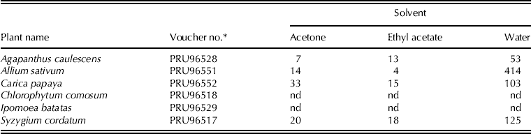
The plant materials used in the present study were Syzygium cordatum Hochst.ex Krauss. (fruits from a tree in the family Myrtaceae), Chlorophytum comosum (Thunb.) Jacq. cvar variegatum (whole plant, spider plant, herbaceous, family Asparagaceae), Agapanthus caulescens Spreng. (whole plant, herbaceous, family Amaryllidaceae), I. batatas (L.) Lam. (leaves, sweet potato, family Convolvulaceae), A. sativum L. (bulbs, garlic, family Amaryllidaceae) and C. papaya L. (leaves, pawpaw, family Caricaceae). Collections of S. cordatum, C. comosum and A. caulescens were made from the Manie van der Schijff Botanical Garden at the University of Pretoria, Pretoria, Republic of South Africa (RSA). The sweet potato leaves were collected from the University of Pretoria's Experimental Farm, whereas the garlic bulbs were bought at the Pretoria fresh market and the pawpaw leaves were collected from Durban in Kwazulu-Natal province, RSA. Samples of the collected plants were deposited in the University of Pretoria H.G.W.J. Schweickerdt Herbarium and voucher numbers were assigned (Table 1).
The quantities (g) of crude extracts harvested from 1 kg plant material using acetone, ethyl acetate and water as solvents
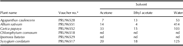
* Voucher number assigned by the University of Pretoria H.G.W.J. Schweickerdt Herbarium.
nd, not determined.
Preparation of the crude plant extracts
All the plant materials were air dried in the shade and ground to powder using a pestle and mortar. Sequential extractions were performed on 1 kg of each plant powder by soaking them in 2 litres of 27·06 moles acetone, 20·30 moles ethyl acetate and sterile distilled water (24 h in each). The plants were soaked in that order contingent to the polarity of the solvents. The organic solvents were removed using a Büchi Rotavapor (Model R-200, Switzerland) and water plant filtrates were concentrated to powder using a freeze drier (Edwards High Vacuum International, Sussex, England). Different quantities of crude extracts were harvested from different solvents as shown in Table 1. All the extracts were then stored in the fridge at 4 °C.
Collection of anthracnose infected plants and isolation of Collectotrichum spp.
Anthracnose-infected common bean and cowpea plants were collected from Middelburg (25°39′S and 029°45′E), Mpumalanga province, RSA and the Department of Agriculture experimental farm (25°46′S and 31°1′E), Nelspruit, Mpumalanga, RSA, respectively. Stems and leaves were surface disinfected in 0·15 mg/ml sodium hypochlorite for 3 min and rinsed three times in sterile distilled water, and then placed either on moist blotters in Petri dishes or on potato dextrose agar (PDA) under continuous ultraviolet light for 7 days at 24±2 °C. Colletotrichum spp. were morphologically identified using the Common Laboratory Seed Health Testing Methods for Detecting Fungi (Mathur & Kongsdal Reference Mathur and Kongsdal2003) and were sub-cultured and stored in a fridge (4 °C) until needed.
Antifungal bioassays
Agar infusion assay
The antifungal activity of the extracts was determined by adopting the technique described by Kritzinger et al. (Reference Kritzinger, Lall and Aveling2005) where appropriate amounts of each stock of crude extract was added to 100 ml of PDA before pouring into Petri dishes to yield final concentrations of 0·5, 1·0, 2·5 and 5·0 mg/ml. The amended PDA was poured into 65 mm Petri dishes with preset diametrical lines drawn on the bottom plate to identify the centre of the plate. Plugs (5 ml diameter) of C. lindemunthianum and C. dematium from 7-day-old fungal cultures were placed in the centre of the Petri dishes containing PDA amended with either water extracts, acetone extracts 0·05 ml/ml (v/v), ethyl acetate extracts 0·05 ml/ml (v/v), the fungicide fludioxonil+mefenoxam (as commercial product Celest® XL (25 gai/l) Syngenta SA, Midrand). The Petri dishes were then sealed with Parafilm® and incubated at 25 °C. Acetone, ethyl acetate and unamended PDA represented the negative controls. Celest® XL represented the positive control based on previous work (Aveling et al. Reference Aveling, Govender, Kandolo and Kritzinger2012). The treatments were arranged in a completely randomized design and each treatment was replicated four times. The experiment was repeated twice. Evaluation of Petri dishes was executed by measuring the diameter of growth inhibition zones after 3, 6 and 9 days of incubation (DAI). The formula of Tegegne et al. (Reference Tegegne, Pretorius and Swart2008) was used to calculate the proportional inhibition of mycelial growth:
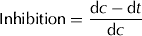 $${\rm Inhibition} = {\rm} \displaystyle{{{\rm d}c - {\rm d}t} \over {{\rm d}c}}$$
where dc is the average diameter of fungal colony of the negative control (PDA only) and dt is the average diameter of fungal colonies grown in the presence of plant extracts or standard fungicides.
The inhibition data were arc sine transformed and then analysed statistically using the Genstat computer package (VSN International 2008). The untransformed and transformed data led to the same conclusion, hence the untransformed analysis data have been presented.
Microtitre double-dilution assay
Malt extract broth was inoculated with Colletotrichum fungi and incubated for 5 days at 25 °C. The broth was adjusted to 0·2 optical density (thus 1×10⁵ and 1×10⁶ spores/ml for C. lindemuthianum and C. dematium, respectively) using an electrospectrometer just before inoculation.
In order to yield a series of 50 mg/ml solutions of extracts, 50 mg of each of the plant extracts were dissolved in 1000 μl of 121·7 mg/ml dimethyl sulphoxide (DMSO). The 96-well microtitre plates were used and 100 μl of the broth was added to all the wells as follows: 100 μl of stock solution (50 mg/ml) of acetone plant extracts were added to the first three wells of row A, ethyl acetate plant extracts were added to the wells 4–6 and water plant extracts were added to wells 7–9. Nutrient broth was added to well 10 as a negative (sterility) control, while Celest® XL (100 μl of 2·5 ml/ml (v/v)) was added to well 11 as a positive control. To well 12, 100 μl of 121·7 mg/ml DMSO was added as a negative control. A series of dilutions of plant extracts, Celest® XL and 121·7 mg/ml DMSO were carried out to row H.
Six-day-old malt broth cultures (100 μl) of Colletotrichum fungi were added to each well. The plates were divided into two sets; the first set (six plates) was inoculated with C. lindemuthianum and the other with C. dematium. The plates were then covered with lids, sealed with Parafilm® and incubated at 25 °C for 48 h. Thereafter, 40 μl of 0·2 mg/ml iodonitrotetrazolium (INT) chloride was added to all the wells, with the exception of columns 3, 6 and 9 as a growth indicator to determine the MIC values for the 18 plant extracts. The microtitre plates were incubated for 24 h at 25 °C and evaluated. The first clear wells from A to H were regarded as the MICs of extracts. The experiment was repeated three times and the final MIC for the extracts was calculated as described by Fawole et al. (Reference Fawole, Finnie and Van Staden2009).
In vivo antifungal test
The varieties used in the present study were the common bean variety Jenny, and IT93K5132 (cream coloured) and Pan 311 (brown coloured) cowpea varieties. Bean and cowpea seeds were soaked in sterile distilled water for 30 min. A small hole of 3 mm was made on each seed into the cotyledon using a sterile needle. The pricked seeds were then soaked in 1×10⁶Colletotrichum spore concentration made from a 10-day-old culture to which 20 drops of Tween 20 was added to yield 0·075 mg/ml Tween 20 solution. Bean and cowpea seed were soaked in C. lindemuthianum and C. dematium, respectively, for 4 h in the dark. The suspension was drained after 2 h and the seeds were placed under high humidity conditions by placing them between moist sterile paper towels in a tightly sealed container overnight, to allow fungal infection to take place. The seeds were then soaked in 150 ml of selected plant extracts diluted to 5 and 15 mg/ml for 24 h at 25 °C and kept in the dark. The inoculated seeds soaked in Celest® XL (25 gai/l) and non-inoculated seeds soaked in distilled water served as positive controls. The negative controls comprised inoculated seeds soaked in distilled water and 121·7 mg/ml DSMO, respectively. The seeds were then sown in pots 150 (diameter)×125 mm (depth) filled with a steam-sterilized mixture of sand and garden compost (Culterra (Pty) Ltd, Pretoria) in a ratio of 1:1 (m/m) at the rate of six seeds per pot. Each treatment was replicated four times and treatments were arranged in a randomized complete design. Water was applied daily in order to maintain soil moisture at field capacity. Each experiment was repeated twice. Data collected included anthracnose disease incidence and severity. The disease incidence was calculated by the formula below:
 $${\rm Disease}\;{\rm incidence} = \displaystyle{{{\rm No}{\rm.} \;{\rm of}\;{\rm infected}\;{\rm plants\;per\;pot}} \over {{\rm No}{\rm.} \;{\rm of}\;{\rm plants}\;{\rm per}\;{\rm pot}}}$$
The anthracnose disease severity scale of 1–9 (CIAT Reference Van Schoonhoven and Pastor-Corrales1987) was employed during the disease severity data collection. Arc sine transformation was performed on anthracnose disease severity data before statistical analysis. The Genstat Discovery statistical package (VSN International 2008) was used to analyse the transformed and untransformed disease data whose analysis was compared and gave similar results.
RESULTS
Agar infusion technique
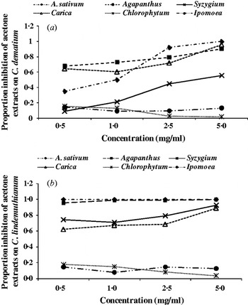
Figure 1a shows the inhibitory effect of acetone plant extracts on C. lindemuthianum at 6 DAI. Allium extract completely inhibited C. lindemuthianum at all concentrations and Agapanthus extract completely inhibited C. lindemuthianum at 5·0 mg/ml over the control. The inhibition increased as the concentrations of all extracts increased, with the exception of Chlorophytum and Ipomoea leaf extracts. In fact, Chlorophytum extracts stimulated fungal growth as the concentrations increased. Allium acetone extracts completely inhibited the growth of C. dematium at 5·0 mg/ml only, whereas Carica and Agapanthus extracts inhibited the growth of C. dematium compared with the control by 96 and 90%, respectively, at the highest concentration (Fig. 1b). Likewise, in C. lindemuthianum, the increase in Chlorophytum and Ipomoea extract concentrations promoted the growth of C. dematium.
The effect of acetone crude plant extracts on the colony growth of (a) C. lindemuthianum and (b) C. dematium at 6 days after inoculation.

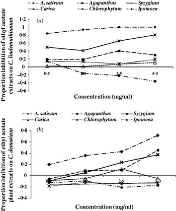
Allium ethyl acetate extracts completely inhibited the growth of C. lindemuthianum at 2·5 and 5·0 mg/ml (Fig. 2a), whereas Syzygium ethyl acetate extract inhibited growth by 80% at 5·0 mg/ml compared with the control. All other ethyl acetate plant extracts (Agapanthus, Carica, Chlorophytum and Ipomoea) failed to inhibit C. lindemuthianum by 50% or more at any concentration. Allium ethyl acetate extract inhibited growth of C. dematium by 87%, but the lower concentrations of extracts from this species inhibited growth by less than 50% compared with the control (Fig. 2b). Extracts of Ipomoea promoted the growth of C. lindemuthianum and C. dematium as concentrations increased.
The effect of ethyl acetate crude plant extracts on the colony growth of (a) C. lindemuthianum and (b) C. dematium at 6 days after inoculation.

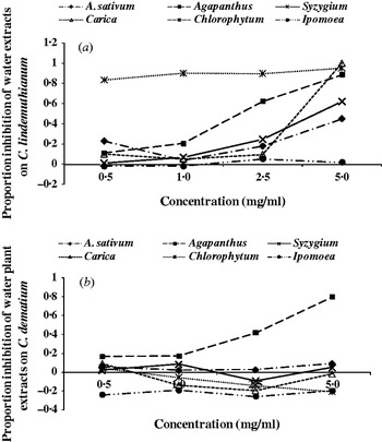
The Carica water extract completely inhibited the growth of C. lindemuthianum at 5·0 mg/ml followed by Chlorophytum and Agapanthus extracts that inhibited the growth by 95 and 89%, respectively (Fig. 3a). Agapanthus water extracts inhibited growth of C. dematium by 80%, whereas the remainder of the extracts failed to reach 50% inhibition (Fig. 3b). Ipomoea extracts of all the concentrations, promoted growth of C. dematium. The Chlorophytum water extracts promoted the growth of C. dematium.
The effect of water crude plant extracts on the colony growth of (a) C. lindemuthianum and (b) C. dematium at 6 days after inoculation.

Microtitre double-dilution method
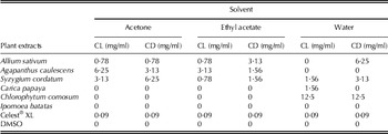
Table 2 shows the MIC extracted with acetone, ethyl acetate and water extracts from different plant species. All the Syzygium extracts were active against C. dematium and C. lindemuthianum. Allium extracts were also active against C. dematium and C. lindemuthianum; however, the water extract failed to inhibit the growth of C. lindemuthianum. Agapanthus acetone and ethyl acetate extracts were active against both fungi with MICs of 1·56, 3·13 and 6·25 mg/ml, respectively, but the range of concentrations of water extracts did not reveal a MIC. Carica and Chlorophytum water extracts were active against C. lindemuthianum, but their organic extracts were inactive against both C. dematium and C. lindemuthianum. All the Ipomoea extracts and DMSO were inactive against both C. dematium and C. lindemuthianum. Celest® XL had a MIC of 0·09 mg/ml, the highest inhibitory activity when compared with all the plant extracts.
The MIC of selected plants extracts on Colletotrichum dematium and Colletotrichum lindemuthianum

CL, colletotrichum lindemuthianum; CD, colletotrichum dematium; DMSO, dimethyl sulphoxide.
In vivo antifungal test
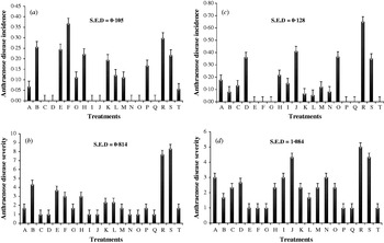
The plants from bean seeds treated with water extracts of Agapanthus (5 and 15 mg/ml), acetone extracts from Carica (15 mg/ml) and Syzygium (5 mg/ml) extracts showed no anthracnose disease incidence (Fig. 4a). The above extracts and 5 mg/ml water extracts from Allium had low anthracnose disease incidence and compared well with the non-inoculated control and Celest® XL. All the acetone plant extracts, 5 mg/ml Allium and Syzygium water extract-treated seeds gave bean plants that had significantly (P⩽0·05) lower anthracnose disease severity than other water treatments and compared well with Celest® XL and the non-inoculated control (Fig. 4b).
(a) Proportion of bean seedlings affected with anthracnose: disease incidence. (b) Anthracnose disease severity of bean seedlings from seeds treated with plant extracts. (c) Proportion of cowpea seedlings affected with anthracnose: disease incidence. (d) Anthracnose disease severity of cowpea seedlings from seeds treated with plant extracts. Treatments A–H=water extracts and I–P=acetone extracts (A=5 mg/ml garlic extracts; B=15 mg/ml garlic extracts; C=5 mg/ml Agapanthus extracts; D=15 mg/ml Agapanthus extracts; E=5 mg/ml Carica extracts; F=15 mg/ml Carica extracts; G=5 mg/ml Syzygium extracts; H=15 mg/ml Syzygium extracts; I=5 mg/ml garlic extracts; J=15 mg/ml garlic extracts; K=5 mg/ml Agapanthus extracts; L=15 mg/ml Agapanthus extracts; M=5 mg/ml Carica extracts; N=15 mg/ml Carica extracts; O=5 mg/ml Syzygium extracts; P=15 mg/ml Syzygium extracts). Q=non-inoculated control; R=inoculated control; S=DMSO and T=Celest® XL.

Water extracts from Allium, Carica and Syzygium and acetone extracts from Agapanthus, Carica and Syzygium showed no or the lowest cowpea anthracnose disease incidence (Fig. 4c) and compared well with the non-inoculated control and Celest® XL. All the plant extracts and the positive controls had low cowpea anthracnose disease severity and were not significantly different (P⩽0·05) from each other, except for the plants from seeds treated with (15 mg/ml) Allium acetone extract and the negative controls (Fig. 4d).
DISCUSSION
Acetone and ethyl acetate extracts from Allium compared well with Celest® XL by completely inhibiting C. lindemuthianum and C. dematium in the agar infusion technique. The Allium water extract inhibited the growth of C. dematium in the microtitre double-dilution technique. The performance of Allium water extracts on C. dematium is similar to the findings of Shovan et al. (Reference Shovan, Bhuiyan, Begum and Pervez2008), where Allium water extracts inhibited the growth of C. dematium.
Chlorophytum water extracts had higher inhibition values on C. lindemuthianum than acetone or ethyl acetate extracts and it can be speculated that the C. lindemuthianum antifungal chemical in C. comosum is only soluble in water and not in acetone or ethyl acetate. Chlorophytum water extracts failed to inhibit the growth of C. dematium, which contrasted with the findings of Raghavendra et al. (Reference Raghavendra, Lokesh, Girisha, Govindappa and Prakash2006) where aqueous extracts of Chlorophytum boriviliam reduced the incidence of C. dematium. The difference in results could be due to differences in species of Chlorophytum used in the two studies.
Carica leaf water extracts at 5·0 mg/ml performed well and was comparable to Celest® XL, by completely inhibiting the growth of C. lindemuthianum in the agar infusion technique, but failed to inhibit the growth of C. dematium. The failure of Carica water extracts to inhibit the growth of C. dematium could be due to differences in chemical or genetic composition between the two Colletotrichum spp. used in the present study. The performance of Carica water extracts on C. lindemuthianum was superior to that of organic extracts in both in vitro techniques used in the present study and these results are in agreement with the findings of Anibijuwon & Udeze (Reference Anibijuwon and Udeze2009), who reported that Carica leaf water extracts gave higher inhibition against bacteria than organic extracts. However, Carica water extracts failed to maintain superiority in vivo by permitting a higher incidence and severity of bean anthracnose disease than other extracts, but was one of the best treatments in recording no incidence of cowpea anthracnose disease.
All Syzygium extracts were active against both C. dematium and C. lindemuthianum in the microtitre double-dilution experiment and the in vivo experiment where they had the lowest cowpea and bean anthracnose disease incidence and severity.
Generally, there was an increase in inhibition of fungal colony growth by the other plant extracts as concentrations increased and this could have been due to the increased availability of the antifungal chemicals in the media. Cao & van Bruggen (Reference Cao and Van Bruggen2001) reported the increase in inhibition of the growth of Phytophthora infestans in media when A. sativum extract concentrations were increased. However, the growth of Colletotrichum increased with concentration of Ipomoea leaf extracts and it can be speculated that these leaves have some constituents that are required for growth of Colletotrichum. Morris & Ward (Reference Morris and Ward1992) found that some plant chemical compounds such as the isoflavone daidzein stimulated the germination of zoospores of Phytophthora sojae (Kaufman and Gerdemann) and Pythium irregulare (Buisman) on Glycine max (L.) Merrill soybean (G. max (L.) Merrill).
Acetone plant extracts showed the highest inhibitory activity among the solvent extracts in vitro and also gave good results in vivo. Masoko et al. (Reference Masoko, Picard and Eloff2005) also found that acetone extracts were superior to other extraction solvents such as hexane, dichloromethane and methanol. The sequential extracting method could have a contribution towards the poor performance of ethyl acetate extracts compared with acetone extracts because most of the organic soluble chemicals in plants had already be extracted in the acetone and little remained in the residues. The water extracts gave poor results in vitro, but performed well in vivo because lower concentrations (0·5–5 mg/ml) were used in vitro and higher concentration (5–15 mg/ml) in vivo. The water extract results could be due to the solubility of compounds in water as speculated by Masoko et al. (Reference Masoko, Picard and Eloff2005), who stated that water fails to extract non-polar active compounds in plant materials. This explains a need for higher plant water extracts concentration to achieve fungal toxic level.
The present study has contributed an additional list of plants that can potentially be used by smallholder farmers to control anthracnose diseases of common bean and cowpea. All the plant extracts except Carica water extracts were effective in reducing anthracnose disease severity. The anthracnose disease incidences in some seedlings from extracts and Celest® XL treated seeds could be due to the deep-seated infections that escaped treatment. Extracts from the plant species used in the present study can be adopted easily as seed treatments by farmers because the plants from which the extracts were made can be accessed easily around their homesteads and the seed treatment process is simple to follow. The smallholder farmers can therefore use A. sativum, A. caulescens and S. cordatum extracts to treat their common bean and cowpea seeds by soaking to avoid the yield losses due to anthracnose disease. The use of the above-stated plant extracts by farmers can also result in the reduction of pesticide-related illnesses suffered by farmers after using the synthetic fungicides (Ngowi et al. Reference Ngowi, Mbise, Ijani, London and Ajayi2007).
There is, however, a need to investigate further the performance of water and acetone A. sativum, A. caulescens, C. papaya and S. cordatum extracts in vivo as both seed treatments and foliar fungicides to control anthracnose disease of cowpea and common bean in the same way that A. sativum and C. papaya leaf extracts have been used as foliar fungicides to control pests and blight in Uganda (Mwine et al. Reference Mwine, Van Damme, Kamoga, Kudamba and Jumba2011).
We thank the Forum for Agricultural Research in Africa (FARA) and the African Development Bank for student bursary support. The National Research Foundation (NRF) for financial assistance and Syngenta SA (Pty) Ltd for providing Celest® XL.