Introduction
The development of the taxonomy of the family Hymenolepididae is one of the most complex and confusing among cestodes. This is associated with the presence of a large number of species in the family and, on the other hand, with considerable morphological uniformity of its representatives. Difficulties have been further confounded by the inconsistency and brevity of morphological descriptions for many taxa (Mariaux et al., Reference Mariaux, Tkach, Vasileva, Caira and Jensen2017). Due to the intense development of molecular systematics, the taxonomic position of many species, established originally based on morphological criteria, requires now a re-evaluation as has been repeatedly demonstrated among the related groups of hymenolepidids (e.g. Haukisalmi et al., Reference Haukisalmi, Hardman, Foronda, Feliu, Laakkonen, Niemimaa, Lehtonen and Henttonen2010; Neov et al., Reference Neov, Vasileva, Radoslavov, Hristov, Littlewood DT and Georgiev2019; Haas et al., Reference Haas, Hoberg, Cook, Henttonen, Makarikov, Gallagher, Dokuchaev and Galbreath2020).
Among an assemblage of problematic species requiring clarification of generic affinities, Rodentolepis (sensu lato) asymmetrica (Janicki, Reference Janicki1904) is currently recognized as a cestode with a broad distribution in the western Palaearctic as a parasite in various arvicoline rodents. It was originally described from the common vole Microtus arvalis (Pallas) (syn: Arvicola arvalis Pallas) from Central Europe (Switzerland) and attributed to the genus Hymenolepis Weinland, 1858 (Janicki, Reference Janicki1904, Reference Janicki1906). The original description was very brief and based on specimens without scoleces. The relative position of female gonads was regarded as the most important distinctive character of. Hymenolepis asymmetrica; in this species, the vitellarium is shifted to the antiporal side of proglottis in relation to the ovary.
Using this feature, Baer (Reference Baer1932) attributed his material collected from Chionomys nivalis (Martins) (syn: Microtus nivalis Martins) from Switzerland to H. asymmetrica; he was the first to describe the scolex. According to Baer (Reference Baer1932), the scolex of this cestode is armed with 20–22 rostellar hooks, 19.2 μm in length; an illustration of rostellar hook was provided but no other morphological characters were illustrated. Furthermore, Baer (Reference Baer1932) recognized Hymenolepis arvicolae Galli-Valerio, 1930 from M. arvalis from Switzerland as a junior synonym of H. asymmetrica.
Joyeux & Baer (Reference Joyeux and Baer1936) reported this species in a both M. arvalis and C. nivalis; the presented metrical data were nearly identical to those reported by Baer (Reference Baer1932), with the exception of the larger cirrus-sac (160–170 vs. 170–220).
Skrjabin & Matevosyan (Reference Skrjabin and Matevosyan1948) repeated the morphological data for H. asymmetrica as originally reported by Janicki (Reference Janicki1904, Reference Janicki1906) and Baer (Reference Baer1932).
Erhardová (Reference Erhardová1955) described Hymenolepis ampla Erhardová, Reference Erhardová1955 from C. nivalis from Slovakia. Baer & Tenora (Reference Baer and Tenora1970), based on morphological similarity, recognized this species as a synonym of H. asymmetrica.
Many authors provided descriptions and illustrations of the rostellar hooks and the anatomy of the proglottides of H. asymmetrica based on specimens from various arvicoline rodents from different regions of Europe (e.g. Žarnowski, Reference Žarnowski1955; Baer & Tenora, Reference Baer and Tenora1970; Tenora & Murai, Reference Tenora and Murai1972; Murai, Reference Murai1974; Genov, Reference Genov1984; Santalla et al., Reference Santalla, Casanova, Durand, Vaucher, Renaud and Feliu2002). All of these descriptions were in general agreement relative to shape, size and number of rostellar hooks as well as the antiporal position of the vitellarium in relation to the ovary.
Spassky (Reference Spassky1954) proposed the specificity to definitive hosts as the major criterion for the taxonomic classification of hymenolepidids from small mammals at the generic level. The genus Rodentolepis Spassky, Reference Spassky1954 was erected for various hymenolepidids from rodents with scolex armed with rostellar hooks, including R. asymmetrica.
Subsequently, the validity of the genus Rodentolepis was questioned and R. asymmetrica was transferred again to Hymenolepis (e.g. Erhardová, Reference Erhardová1955; Vaucher, Reference Vaucher1967; Baer & Tenora, Reference Baer and Tenora1970; Murai, Reference Murai1974; Genov, Reference Genov1984) or to Vampirolepis Spassky, Reference Spassky1954 (e.g. Schmidt, Reference Schmidt1986; Murai, Reference Murai1989). After recognition of the independent status of Rodentolepis by Czaplinski & Vaucher (Reference Czaplinski, Vaucher, Khalil, Jones and Bray1994), R. asymmetrica has been considered as member of this genus (e.g. Santalla et al., Reference Santalla, Casanova, Durand, Vaucher, Renaud and Feliu2002).
Recent phylogenetic studies exploring relationships among hymenolepidids from rodents, insectivores and bats assigned to the Rodentolepis clade have demonstrated that the genus Rodentolepis (sensu lato) is a non-monophyletic taxon requiring thorough revision. Species of Rodentolepis (sensu lato), for which molecular data are available, are consistent in exhibiting a very high level of phylogenetic divergence from the type-species Rodentolepis straminea (Goeze, 1792) (Haukisalmi et al., Reference Haukisalmi, Hardman, Foronda, Feliu, Laakkonen, Niemimaa, Lehtonen and Henttonen2010; Greiman & Tkach, Reference Greiman and Tkach2012; Makarikov et al., Reference Makarikov, Mel'nikova and Tkach2015; Neov et al., Reference Neov, Vasileva, Radoslavov, Hristov, Littlewood DT and Georgiev2019). Some linages previously attributed to Rodentolepis (sensu lato) were later recognized as the genera Pararodentolepis Makarikov & Gulyaev, Reference Makarikov and Gulyaev2009 and Nomadolepis Makarikov, Gulyaev & Krivopalov, Reference Makarikov, Gulyaev and Krivopalov2010 (Makarikov & Gulyaev, Reference Makarikov and Gulyaev2009; Makarikov et al., Reference Makarikov, Gulyaev and Krivopalov2010, Reference Makarikov, Mel'nikova and Tkach2015). Furthermore, it was shown that R. asymmetrica was not only phylogenetically unrelated to the type species of the genus but also was not included in the ‘Rodentolepis clade’ (Haukisalmi et al., Reference Haukisalmi, Hardman, Foronda, Feliu, Laakkonen, Niemimaa, Lehtonen and Henttonen2010; Neov et al., Reference Neov, Vasileva, Radoslavov, Hristov, Littlewood DT and Georgiev2019) and thus having an uncertain generic allocation. Incomplete morphological data in the original description and the brief subsequent redescriptions of R. asymmetrica have also hindered a clear picture of the taxonomy of this species.
Specimens of R. asymmetrica from voles Mi. arvalis, Mi. agrestis (Linnaeus) and Myodes glareolus (Schreber) from Eastern Europe (Belarus, Estonia, Latvia, Lithuania, Ukraine and north-western Russia) were collected for the present study. In addition, voucher specimens from voles in France, Hungary, Spain and Switzerland, attributed to R. asymmetrica by various researchers and deposited in the helminthological collection of the Geneva Museum of Natural History, Switzerland (MHNG), have been studied for comparison.
The aim of the present article is to complete a detailed morphological redescription of R. asymmetrica and to analyse its relationships with other hymenolepidids from mammals based on partial sequences of the nuclear ribosomal 28S rRNA gene. The generic allocation of this species is clarified based on integration of morphological criteria and molecular phylogenetic analysis.
Materials and methods
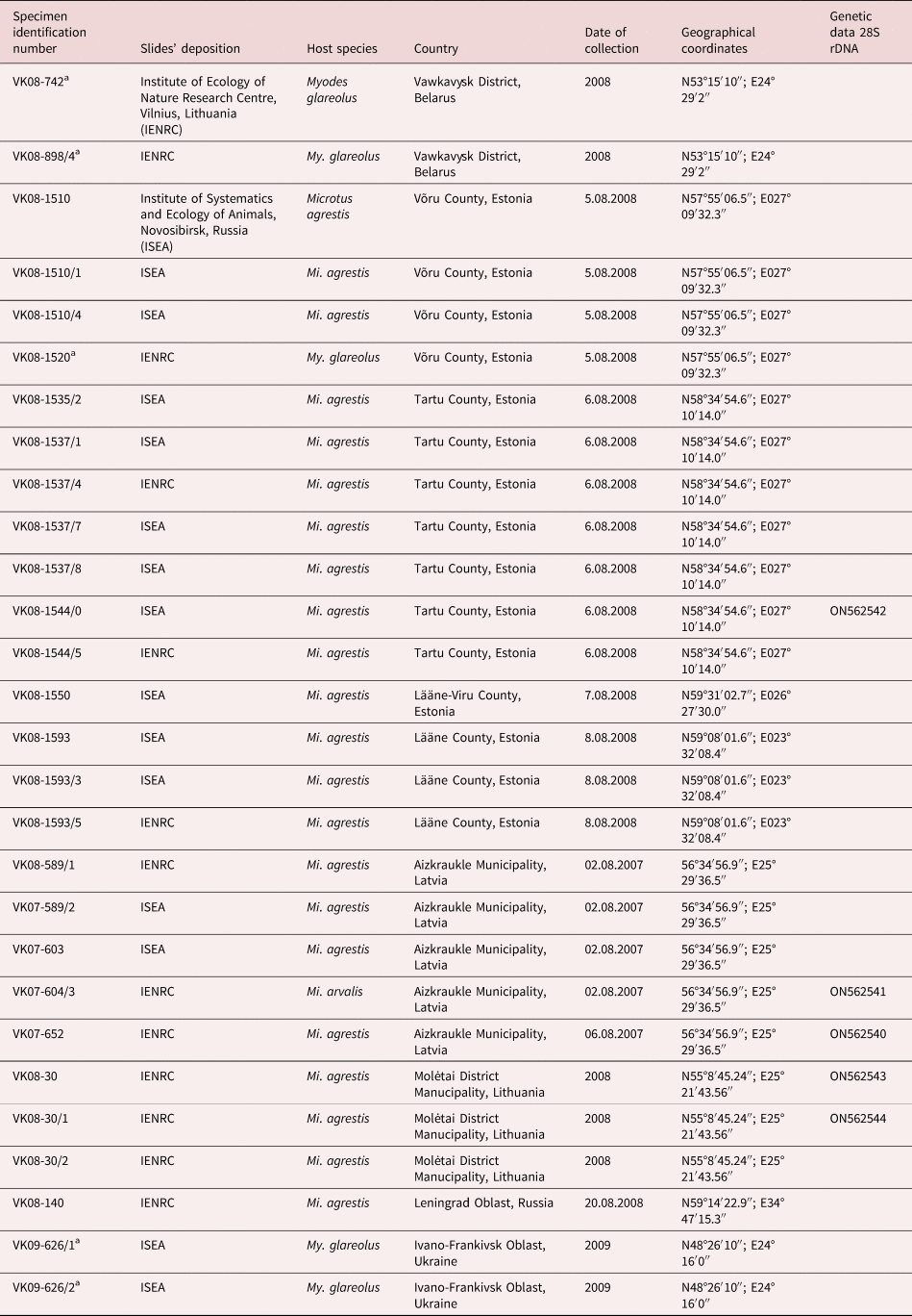
Mounted specimens of R. asymmetrica used in the present study and some tissues stored in ethanol were taken from the research collection of the late Dr Vytautas L. Kontrimavichus supported by the Lithuanian State Science and Studies Foundation. Cestodes from Mi. agrestis, Mi. arvalis and My. glareolus were assembled from field surveys during 2007–2009 conducted in Belarus, Estonia, Latvia, Lithuania, Ukraine and north-western Russia. Sample collection sites and their geographical coordinates are presented in table 1.
Specimens of Rodentolepis (sensu lato) asymmetrica examined in the current study, including vouchers used for morphological redescription and DNA extraction.

a Specimens of Rodentolepis (sensu lato) asymmetrica from Myodes glareolus from Belarus, Estonia and Ukraine used for morphological comparison but not included in the redescription.
Host specimens were dissected fresh. Cestodes were isolated, rinsed and relaxed in water, and preserved in 70% ethanol. Specimens were stained with Ehrlich's haematoxylin, dehydrated in an ethanol series, cleared in clove oil and mounted in Canada balsam. Some scoleces and fragments of strobila were mounted in Berlese's medium to facilitate detailed examination of the rostellar hooks, suckers, cirrus armature and structure of the eggs. Additional tissue was subsampled from some strobila and stored in 96% ethanol for molecular analyses. Specimens were studied using standard light and differential interference contrast microscopy. In the descriptions, measurements are given in micrometres except where otherwise stated; they are presented as the range followed by the mean and the number of the measurements (n) in parentheses. The voucher specimens of R. asymmetrica have been deposited in the collection of the Institute of Systematics and Ecology of Animals, Novosibirsk, Russia and the Institute of Ecology of Nature Research Centre, Vilnius, Lithuania. Mammalian taxonomy follows Musser & Carelton (Reference Musser, Carelton, Wilson and Reeder2005).
The type materials of R. asymmetrica are not available as those are not mentioned in the catalogues of the international collections of helminthes. With this regard, the following voucher specimens attributed to R. asymmetrica and deposited in the helminthological collection of the MHNG, were studied for comparison purposes:
(1) Vouchers of R. asymmetrica: MHNG-PLAT-17730 (C40/96), ex Microtus arvalis, Németbánya, Veszprém County, Hungary, 1973, collector E. Murai, description published by Murai (Reference Murai1974).
(2) Vouchers of R. asymmetrica: MHNG-PLAT-40927 (118/76-77), ex Chionomys nivalis, exact locality is not specified, collector F. Tenora.
(3) Vouchers of R. asymmetrica: MHNG-PLAT-40928 (118/78-80), ex Microtus subterraneus, exact locality is not specified, collector F. Tenora.
(4) Vouchers of R. asymmetrica: MHNG-PLAT-11779-11781 (C6/35-39), ex Microtus sp., Ramosch, Canton Grisons (Graubünden), Switzerland, 1971, collector A. Meylan, identified by C. Vaucher.
(5) Vouchers of R. asymmetrica: MHNG-PLAT-18413 (C61/54), ex Microtus agrestis, Allanche, Auvergne region, France, 1993, collector A. de Chambrier, identified by C. Vaucher.
(6) Vouchers of R. asymmetrica: MHNG-PLAT-18399, 18502-18503 (C61/90-96), MHNG-PLAT-18156, 18165 (C56/43-45), ex Microtus agrestis, Le Lieu, Le Brasus, Canton Vaud, Switzerland, 1993-1994, collection and identification C. Vaucher.
(7) Vouchers of R. asymmetrica: MHNG-PLAT-18362 (C60/07-10), ex C. nivalis, Col de Bretolet, Canton Valais, Switzerland, 1968, collection and identification C. Vaucher.
(8) Vouchers of R. asymmetrica: MHNG-PLAT-18363 (C60/11), 30549 (C99/38) ex Mi. agrestis, La Praz, Canton Vaud, Switzerland, 1967, 1994, collection and identification C. Vaucher.
(9) Vouchers of R. asymmetrica: MHNG-PLAT-18364 (C60/12-14), ex Mi. subterraneus, Col de Bretolet, Canton Valais, Switzerland, 1968, collection and identification C. Vaucher.
(10) Vouchers of R. asymmetrica: MHNG-PLAT-18365 (C60/15), ex Mi. arvalis, Col de Bretolet, Canton Valais, Switzerland, 1968, collection and identification C. Vaucher.
(11) Vouchers of R. asymmetrica: MHNG-PLAT-17613 (C40/29-30), ex Mi. agrestis, Valangin, Canton Neuchâtel, Switzerland, 1966, collector C. Vaucher.
(12) Vouchers of R. asymmetrica: MHNG-PLAT-39304 (C53/3-6), ex Myodes glareolus, La Chaux-de-Fonds, Canton Neuchâtel, Switzerland, 1967, collection and identification W. Reichenbach.
(13) Vouchers of R. asymmetrica: MHNG-PLAT-32861 (C107/078), ex My. glareolus, Moulis, Oriental Pyrenees, France, 1997, collection and identification C. Feliu, description published by Santalla et al. (Reference Santalla, Casanova, Durand, Vaucher, Renaud and Feliu2002).
(14) Vouchers of R. asymmetrica: MHNG-PLAT-32862 (C107/079), ex Microtus gerbei, Eugi, Navarra, Spain, 1996, collection and identification C. Feliu, description published by Santalla et al. (Reference Santalla, Casanova, Durand, Vaucher, Renaud and Feliu2002).
(15) Vouchers of R. asymmetrica: MHNG-PLAT-32863 (C107/80), ex Mi. arvalis, Val d'Aran, Cataluña, Spain, 1997, collection and identification C. Feliu, description published by Santalla et al. (Reference Santalla, Casanova, Durand, Vaucher, Renaud and Feliu2002).
(16) Vouchers of R. asymmetrica: MHNG-PLAT-30668, 39413 (C54/45, 59), ex Mi. subterraneus, Bex, Montricher, Canton Vaud, Switzerland, 1973, collector A. Meylan, identified by C. Vaucher.
(17) Vouchers of R. asymmetrica: MHNG-PLAT-30533 (C99/4), ex C. nivalis, Arolla, Canton Valais, Switzerland, 1997, collector C. Vaucher & F. Catzeflis, identified by C. Vaucher.
(18) Vouchers of R. asymmetrica: MHNG-PLAT-30536-30539 (C99/7-12), ex Mi. subterraneus, Microtus arvalis, Ain, France, 1998, collector C. Vaucher & A. Meylan, identified by C. Vaucher.
(19) Vouchers of R. asymmetrica: MHNG-PLAT-19075, 19078, 19162-19165, 19181, 19183-19184, 19205-19206, 19663 (C82/14-27, 31, 39, 41; C99/37), ex Mi. agrestis, Mi. arvalis, Le Lieu, Canton Vaud, Switzerland, 1993, 1994, collection and identification C. Vaucher.
(20) Vouchers of R. asymmetrica: MHNG-PLAT-30668-30669, 30672-30673 (C99/78-84), ex Microtus subterraneus, Mont Tendre, Canton Vaud, Switzerland, 1973, collector A. Meylan, identified by C. Vaucher.
(21) Vouchers of R. asymmetrica: MHNG-PLAT-19321 (C82/40), MHNG-PLAT-19328 (C99/94), ex Microtus subterraneus, Piora Valley, Canton Ticino, Switzerland, 1994, collection and identification C. Vaucher.
(22) Vouchers of R. asymmetrica: MHNG-PLAT-40937 (11/72-73), ex C. nivalis, Zaté (Val d'Hérens), Switzerland, 1931, collection and identification J. Baer, description published by Baer (Reference Baer1932).
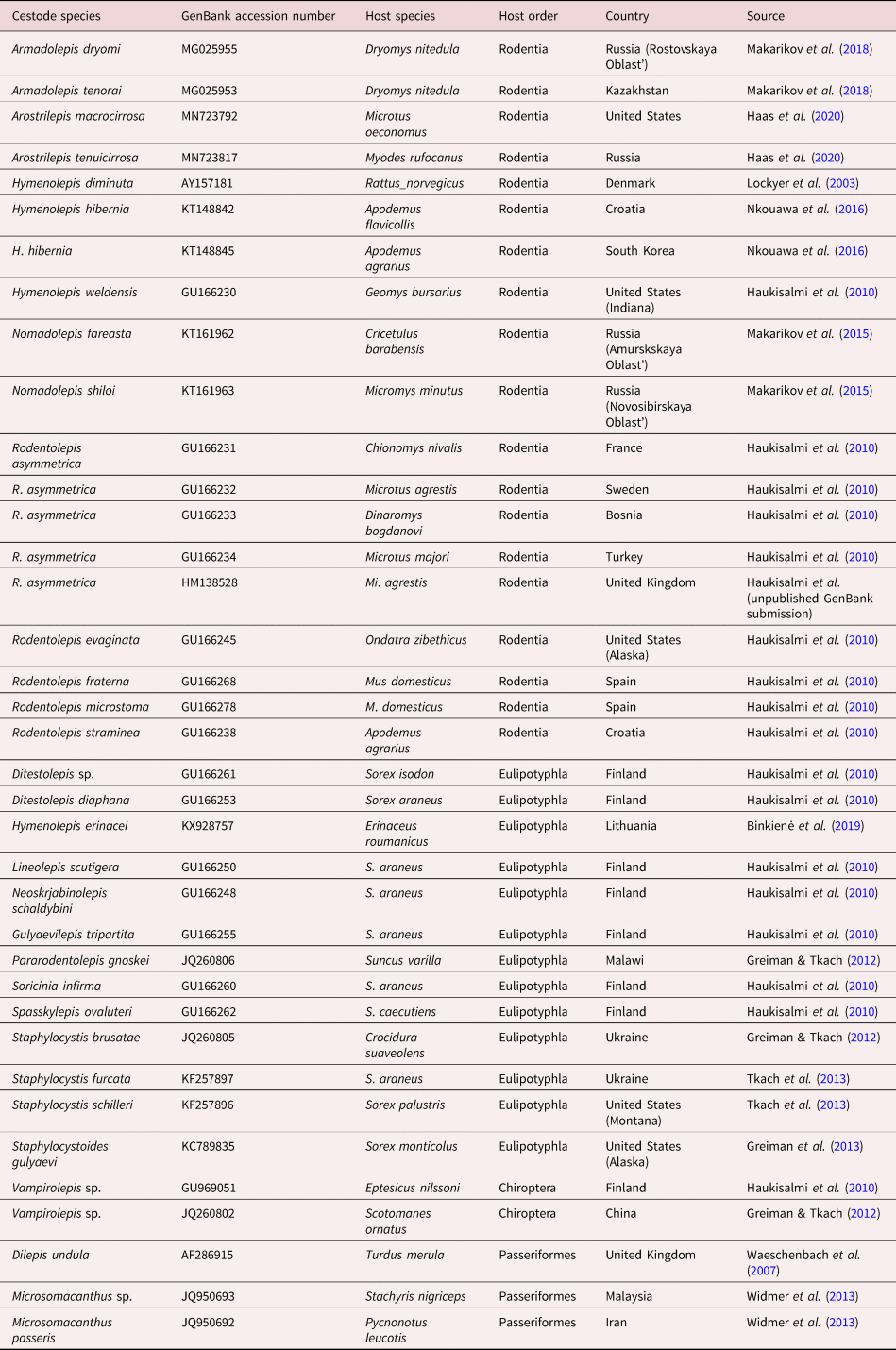
Genomic DNA for molecular phylogenetic analysis was extracted from specimens of Rodentolepis (sensu lato) asymmetrica collected from Estonia, Latvia and Lithuania. A fragment (1.5–2 mm long) of a single adult worm was used for each DNA extraction upon preliminary morphological identification. Scoleces and the remaining strobila have been mounted on slides. DNA was extracted using a standard phenol–chloroform protocol as described by Vainio et al. (Reference Vainio, Korhonen and Hantula1998) and Sambrook & Russell (Reference Sambrook and Russell2002). DNA fragments approximately 1080 base pairs (bp) long at the 5′ end of the nuclear large ribosomal subunit (28S) gene was amplified by polymerase chain reaction (PCR) on an Eppendorf EP Gradient thermal cycler using OneTaq Quick-load Mastermix from New England Biolabs (Ipswich, MA) according to the manufacturer's instructions. Forward primer 28S-5′ (5′-TAC CCG CTG AAC TTA AGC ATA T-3′) and reverse primer 28S-3′ (5′-CTC CTT GGT CCG TGT TTC AAG AC-3′) designed by Zehnder & Mariaux (Reference Zehnder and Mariaux1999) were used for amplification; the PCR protocol included 40 cycles with annealing temperature 53°C. Sequencing (from both sides) was carried out by Macrogen Inc. (Geumchun-gu Seoul 153-781, Korea) using the same primers. Sequences were aligned using BioEdit software, version 7.0.1. (Hall, Reference Hall1999). Pairwise comparisons of sequences of R. asymmetrica were calculated using MEGA X (Kumar et al., Reference Kumar, Stecher, Li, Knyaz and Tamura2018). To build a phylogenetic tree and reconstruct relationships between the R. asymmetrica and other hymenolepidids, we used maximum likelihood with a general time reversible model as distance substitution. For phylogenetic analyses, we used 5 newly obtained nucleotide sequences of R. asymmetrica and those of hymenolepidids published in previous studies outlined in table 2 (Lockyer et al., Reference Lockyer, Olson and Littlewood2003; Waeschenbach et al., Reference Waeschenbach, Webster, Bray and Littlewood2007; Haukisalmi et al., Reference Haukisalmi, Hardman, Foronda, Feliu, Laakkonen, Niemimaa, Lehtonen and Henttonen2010; Greiman & Tkach, Reference Greiman and Tkach2012; Tkach et al., Reference Tkach, Makarikov and Kinsella2013; Widmer et al., Reference Widmer, Georgiev and Mariaux2013; Makarikov et al., Reference Makarikov, Mel'nikova and Tkach2015; Nkouawa et al., Reference Nkouawa, Haukisalmi, Li, Nakao, Lavikainen, Chen, Henttonen and Ito2016; Binkienė et al., Reference Binkienė, Miliūtė and Stunžėnas2019; Haas et al., Reference Haas, Hoberg, Cook, Henttonen, Makarikov, Gallagher, Dokuchaev and Galbreath2020). Dilepis undula (Schrank, 1788) was used as an outgroup as proposed by Neov et al. (Reference Neov, Vasileva, Radoslavov, Hristov, Littlewood DT and Georgiev2019) for the phylogeny of Hymenolepididae. Bootstrap values were counted using MEGA as the percentage of 1000 replicates.
Published sequences of 28S rDNA of cestodes deposited in GenBank included in the present phylogenetic analysis.

Results
Morphological study of R. asymmetrica specimens revealed the presence of a set of unique morphological features of supraspecific level that distinguish this species from other genera of mammalian hymenolepidids, and this clearly indicated the need to erect a new genus for this cestode.
Family Hymenolepididae Perrier, 1987
Genus Kontrimavichusia n. g.
Diagnosis
Hymenolepididae of medium size. Development of proglottides gradual. Proglottides numerous, transversely elongate, craspedote. Rostellar apparatus well developed. Rhynchus armed with one row of small cricetoid-like hooks; number of rostellar hooks more than 10 and less 40. Suckers muscular, not prominent, armed along its entire surface with minute spines. Dorsal and ventral osmoregulatory canals located on same sagittal plane. Ventral canals connected by irregularly spaced transverse anastomoses. Genital pores unilateral, dextral; genital ducts pass dorsally to osmoregulatory canals. Testes, usually three, as exception four to five, situated in one row or antiporal testes lie on top of each other; poral testis separated from two antiporal testes by female gonads. Cirrus-sac does not reach median line of proglottis. Cirrus armed with minuscule spines. External and internal seminal vesicles present. Internal seminal vesicle with circular musculature. Ovary median, transversely elongate, fan-shaped. Vitellarium postovarian, median or slightly shifted to lateral side of proglottis, slightly lobed. Vagina with two distinct parts: copulatory part surrounded by circular musculature and covered externally by dense layer of intensely-stained cells; and conductive part thin-walled, clearly distinguishable from seminal receptacle. Uterus, initially transversely elongated stripe, situated dorsally to genital ducts and extending laterally beyond longitudinal osmoregulatory canals. Fully developed uterus labyrinthine, extending beyond osmoregulatory canals into both lateral fields, situated dorsally to osmoregulatory canals and genital ducts. Eggs numerous, subspherical, with thin outer coat. Embryophore subspherical, without polar filaments. Parasites of voles (Rodentia, Arvicolinae) in western Palaearctic.
Type species: Kontrimavichusia asymmetrica (Janicki, Reference Janicki1904) n. comb.
ZooBank registration: To comply with the regulations set out in Article 8.5 of the amended version of the International Code of Zoological Nomenclature (ICZN, 2012), details of the new genus have been submitted to ZooBank. The Life Science Identifier for Kontrimavichusia n. g. is urn:lsid:-zoobank.org:act:CCA8465A-4DDF-4500-B82B-D2B6CB8B928C.
Etymology: This species has been named in honour of the late Dr Vytautas L. Kontrimavichus, in recognition of his seminal and critical studies of parasites of vertebrates, helminth systematics, biogeography, ecology and evolution.
Remarks
Kontrimavichusia n. g. is most similar to other genera of hymenolepidids from mammals in having a scolex armed with rostellar hooks. These genera are Armadolepis Spassky, Reference Spassky1954, Arvicolepis Makarikov, Gulyaev & Chechulin, Reference Makarikov, Gulyaev, Chechulin and Alimov2005, Nomadolepis Makarikov, Gulyaev & Krivopalov, Reference Makarikov, Gulyaev and Krivopalov2010, Pararodentolepis Makarikov & Gulyaev, Reference Makarikov and Gulyaev2009, Relictolepis Gulyaev & Makarikov, 2007 and Rodentolepis Spassky, Reference Spassky1954 from rodents, Staphylocystis Villot, 1877 from shrews and Vampirolepis Spassky, Reference Spassky1954 from bats. The new genus is consistently distinguished from these taxa by a complex of unique characters such as the presence of armament on suckers, vagina subdivided into copulatory part and conductive part, copulatory part of vagina surrounded by circular musculature and enveloped in a dense sleeve of intensely-stained cells and by the presence of musculature on the internal seminal vesicle.
In specimens of Kontrimavichusia n. g., the apex of rostellum is invaginable and blades of retracted hooks are directed anteriorly. In contrast, all members of the Rodentolepis clade with an armed scolex, Armadolepis, Nomadolepis, Pararodentolepis, Rodentolepis, Staphylocystis and Vampirolepis, have a rostellum with non-invaginable apex; when rostellar apparatus is retracted, the blades of the hooks are directed posteriorly. Kontrimavichusia n. g. also differs from the majority of hymenolepidids from mammals by the presence of irregularly spaced transverse anastomoses between the ventral osmoregulatory canals; so far this character has also been noted in some species of Nomadolepis (Makarikov et al., Reference Makarikov, Gulyaev and Krivopalov2010).
Kontrimavichusia asymmetrica n. comb. differs from morphologically related taxa in uterine position and structure. When gravid, the fully-developed uterus is labyrinthine in shape, extending beyond the osmoregulatory canals into both lateral fields and is situated dorsally to osmoregulatory canals and genital ducts. These characters could be used as additional features to distinguish the new genus from other morphologically related genera from mammals. As exemplified, Rodentolepis (sensu stricto) and its type species R. straminea as well as species of Armadolepis, Nomadolepis, Relictolepis and Pararodentolepis have the uterus situated dorsally to the osmoregulatory canals and ventrally to genital ducts. The gravid uterus of these genera (Nomadolepis, Pararodentolepis, some species of Staphylocystis and Vampirolepis) is a bilobed sac not extending laterally beyond the osmoregulatory canals.
Kontrimavichusia asymmetrica (Janicki, Reference Janicki1904) n. comb.
Synonyms: Hymenolepis asymmetrica Janicki, Reference Janicki1904; Rodentolepis asymmetrica (Janicki, Reference Janicki1904) Spassky, Reference Spassky1954; Vampirolepis asymmetrica (Janicki, Reference Janicki1904) Schmidt, Reference Schmidt1986; Hymenolepis arvicolae Galli-Valerio, 1930; and Hymenolepis ampla Erhardová, Reference Erhardová1955.
Type host: Microtus arvalis (Pallas) (Rodentia: Cricetidae).
Other hosts: Chionomys nivalis (Martins), Microtus agrestis (Linnaeus, 1761), Mi. subterraneus (de Sélys-Longchamps, 1836), Myodes glareolus (Schreber) (Rodentia: Cricetidae).
Type locality: suburbs of Basel, Switzerland.
Redescription (figs 1–4)
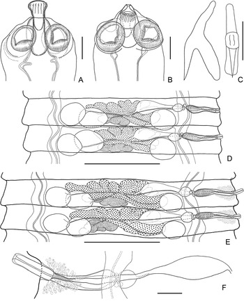
Kontrimavichusia asymmetrica (Janicki, Reference Janicki1904) n. comb. (A) voucher (VK08-1593/3, ex Microtus agrestis, Estonia), dorso-ventral view of scolex; (B) voucher (VK08-1537/8, ex M. agrestis, Estonia), dorso-ventral view of scolex; (C) voucher (VK08-1537/7, ex M. agrestis, Estonia), rostellar hooks in profile and frontal view (note narrow hook guard); (D) voucher (VK08-1535/2, ex M. agrestis, Estonia), male mature proglottides, dorsal view; (E) voucher (VK08-1535/2, ex M. agrestis, Estonia), hermaphroditic mature proglottis, dorsal view; and (F) voucher (VK08-1537/8, ex M. agrestis, Estonia), genital ducts, ventral view. Scale bars: (A, B) 100 μm; (C) 10 μm; (D, E) 600 μm; (F) 60 μm.

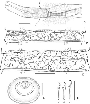
Kontrimavichusia asymmetrica (Janicki, Reference Janicki1904) n. comb. (A) voucher (VK08-1537/8, ex Microtus agrestis, Estonia), cirrus and vagina, ventral view; (B) voucher (VK08-1535/2, ex M. agrestis, Estonia), pregravid proglottis, showing appearance of uterine diverticula, ventral view; (C) voucher (VK08-1593/5, ex M. agrestis, Estonia), gravid proglottis, showing labyrinthine uterus, dorsal view; (D) voucher (VK08-1510, ex M. agrestis, Estonia), egg; and (E) voucher (VK08-1510, ex M. agrestis, Estonia), embryonic hooks (m, median; al, anterolateral; pl, postero-lateral);. Scale bars: (A) 30 μm; (B, C) 500 μm; (D) 20 μm; (E) 10 μm.

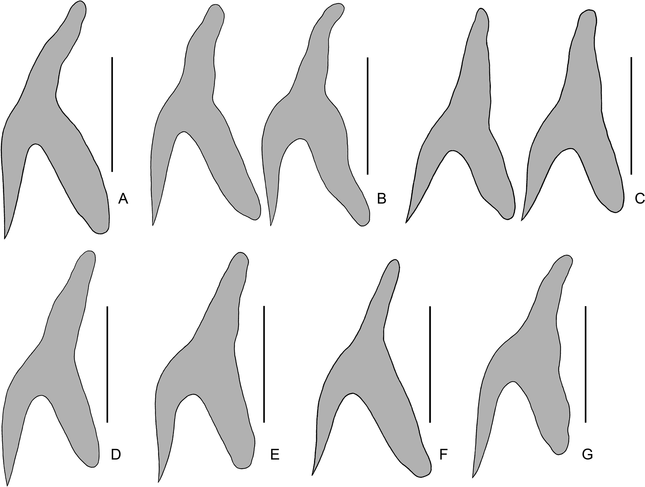
Kontrimavichusia asymmetrica (Janicki, Reference Janicki1904) n. comb. (A–G) rostellar hooks, showing variation in hooks shape between specimens. (A) voucher VK08-1537/7, ex Microtus agrestis, Estonia; (B) voucher MHNG-PLAT-18363 ex M. agrestis, Switzerland; (C) voucher MHNG-PLAT-40937 ex Chionomys nivalis, Switzerland; (D) voucher MHNG-PLAT-30668 ex M. subterraneus, Switzerland; (E) voucher MHNG-PLAT-11781, ex Microtus sp., Switzerland; (F) voucher 626/2, ex Myodes glareolus, Ukraine; and (G) voucher MHNG-PLAT-39304, ex Myodes glareolus, Switzerland. Scale bars: (A–G), 10 μm.

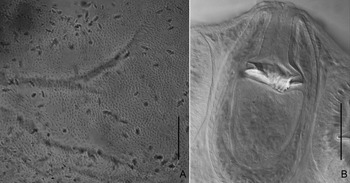
Kontrimavichusia asymmetrica (Janicki, Reference Janicki1904) n. comb. (A) sucker surface of the scolex mounted in Berlese's medium, showing presence of armature; (B) fully retracted rostellar apparatus, showing blades of hooks directed anteriorly. Scale bars: (A) 20 μm; (B) 50 μm.

(Based on 18 specimens from Microtus agrestis and Mi. arvalis from north-eastern Europe). Fully developed strobila 98–160 (122, n = 7) mm long, with maximum width 2.5–4.45 (3.1, n = 7) mm at level pregravid or gravid proglottides. Strobila flat, consisting of 780–940 craspedote proglottides. Scolex slightly compressed dorso-ventrally, 245–325 wide (280, n = 7), not clearly distinct from neck. Suckers small, thick-walled, rounded or oval, cup-shaped, 120–144 × 105–125 (134 × 111; n = 22), usually reaching lateral margins of scolex, armed with minute (less than 1 mm long) spines; spines covering entire sucker surface (figs 1A, B and 4A). Rostellar pouch 168–185 × 83–115 (175 × 100; n = 7), with muscular walls, its bottom reaching or slightly extending beyond level of posterior margin of suckers. Rostellum 113–135 × 42–81 (120 × 64; n = 7), sac-like, muscular, apex invaginable; when rostellar apparatus retracted, rostellar hooks with blades directed anteriorly (fig. 4B). Rhynchus 65–80 long and 45–84 wide, with well-developed circular musculature, armed with single crown of 18–23 (n = 12) rostellar hooks of cricetoid-like type (figs 1C and 3A–G). Rostellar hooks with relatively short handle and straight blade; axis of blade situated to the axis of guard at an acute angle; guard narrow in anterior surface; handle and blade slightly shorter or equal in length with guard. Hook measurements: total length 20–22.5 (21.1; n = 20); handle 7.5–9 (7.9; n = 20); blade 7.5–9 (8.1; n = 20); and guard 8–9.5 (8.7; n = 20). Neck 240–320 (n = 7), approximately equal in width with scolex (fig. 1A, B).
Ventral osmoregulatory canals 65–165 (118; n = 21) wide, connected by irregularly spaced transverse anastomoses (in 9–28% proglottides) (fig. 1D). Dorsal osmoregulatory canals very thin, 7–12 (9; n = 21) wide at level of hermaphroditic proglottides, usually situated directly dorsal (not shifted left or right) to ventral canals. Genital pores unilateral, dextral (fig. 1D, E). Genital ducts pass dorsally to both ventral and dorsal longitudinal osmoregulatory canals. Development of proglottides gradual, protandrous.
Mature proglottides 150–200 × 1450–1950 (176 × 1711; n = 18), transversely elongate, trapezoid (fig. 1D, E). Testes 3, relatively large, almost equal in size, 155–215 × 110–168 (183 × 138; n = 30), round or oval, most often situated in one row or, rarely, in triangle with flat angle (anterior antiporal testis shifted to lateral side of proglottis in relation to posterior antiporal testis), poral testis separated from two antiporal testes by female gonads. Number of testes usually constant, variation with four testes per proglottis infrequent. Cirrus-sac relatively short, 215–270 × 48–66 (243 × 54; n = 28), with thick muscular walls. Antiporal part of cirrus-sac reaching ventral osmoregulatory canal, rarely overlapping or slightly crossing it (fig. 1E, F). Genital atrium simple, cup-shaped, opens laterally, approximately in middle of lateral proglottis margin. Cirrus large, 105–166 × 25–36 (143 × 28; n = 32), cylindrical, armed with very small (up to 1.0–1.5 long), needle-shaped spines; distal part of fully-evaginated cirrus unarmed (fig. 2A). Internal seminal vesicle with circular musculature, ovoid, 126–164 × 42–56 (148 × 49; n = 30), occupying less than half of cirrus-sac length (fig. 1E, F). External seminal vesicle, 75–152 × 70–105 (104 × 82; n = 20), round or oval, clearly distinguishable from vas deferens, distinctly smaller than seminal receptacle.
Ovary 410–690 (543; n = 20) wide, median, transversely elongate, fan-shaped, irregularly lobed, ventral to male genital organs, occupying substantial part of median field, overlapping testes (fig. 1E). Vitellarium 70–105 × 192–276 (81 × 220 n = 20), postovarian, slightly shifted to lateral side of proglottis, slightly lobed. Vagina tubular, clearly distinct from seminal receptacle; ventral to cirrus-sac. Copulatory part of vagina 67–90 × 10–22 (76 × 15; n = 15), shorter than cirrus, thick-walled, surrounded by circular musculature and covered externally by dense layer of intensely stained cells; proximal part of vagina infundibular (figs 1F and 2A). Conductive part of vagina 145–190 × 9–18 (175 × 13; n = 15), thin-walled. Seminal receptacle relatively large, transversely elongate, 240–445 × 80–110 (346 × 92; n = 20).
Uterus appears as perforated, transversely-elongate stripe, situated dorsally to testes, genital ducts and osmoregulatory canals and extending laterally beyond longitudinal osmoregulatory canals. With proglottis development, uterus forms numerous diverticula on ventral side and becomes labyrinthine in terminal postmature proglottides. Testes persist in postmature proglottides; cirrus-sac and vagina persist in gravid proglottides (fig. 2B). Gravid proglottides transversely elongate, 320–480 × 2350–4450 (392 × 2966; n = 20). Fully developed uterus labyrinthine, occupying entire median field, extending bilaterally, dorsally, beyond longitudinal osmoregulatory canals (fig. 2C). Uterus contains numerous (up to 2000–2300) small eggs. Eggs 42–50 × 47–54, subspherical, with very thin outer coat (up to 0.8–1 thick); oncospheres 18–21 × 20–25 (fig. 2D). Embryophores very thin, 22–26 × 25–32, without polar filaments. Embryonic hooks small, antero-lateral hooks 9.0–9.5, much more robust than slender postero-lateral (9.0–9.5) and median (10.0–10.5) hooks (fig. 2E).
Molecular phylogenetic analysis
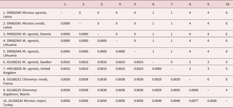
The length of the alignment after trimming was 975 nucleotides. All sequences of K. asymmetrica n. comb. from Eastern Europe including specimens from Mi. agrestis (GenBank: ON562541), and Mi. arvalis (GenBank: ON562540, ON562542–ON562544) obtained in the present study were identical and form one well-supported clade with those previously deposited in GenBank that were attributed to this species (table 2). It should be noted that the intraspecific differences between all available sequences of putative K. asymmetrica reach up to 1–6 bp (table 3).
Pairwise uncorrected genetic distances (below diagonal) and total nucleotide differences (above diagonal) in sequences of 28S rDNA between putative Kontrimavichusia asymmetrica specimens.

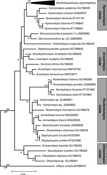
Kontrimavichusia n. g. is placed as the putative sister for species of Hymenolepis (sensu stricto), with the latter characterized by an unarmed scolex, rudimentary rostellar apparatus consisting of a partly reduced rostellar pouch and rhynchus and a fully reduced rostellum (fig. 5). Topology is consistent with that presented for these taxa in the analyses by Haukisalmi et al. (Reference Haukisalmi, Hardman, Foronda, Feliu, Laakkonen, Niemimaa, Lehtonen and Henttonen2010) and Neov et al. (Reference Neov, Vasileva, Radoslavov, Hristov, Littlewood DT and Georgiev2019). The monophyly of each of the two clades corresponding to Kontrimavichusia and Hymenolepis is well supported (1.0 and 0.99 posterior probability, respectively) but the support of the basal branching of the Hymenolepis clade is poorly supported. In the current analysis, the relationship of the Hymenolepis clade constituting Kontrimavichusia n. g. and Hymenolepis remains in a polytomy with certain species of Microsomacanthus from passerine birds and Rodentolepis (sensu lato) evaginata (Barker & Andrews, 1915) from muskrat.
Maximum likelihood phylogenetic tree of hymenolepidids based on analysis of partial sequences of the 28S rRNA gene. Bootstrap support given for maximum likelihood analysis based on 1000 replicates. Bootstrap support values lower than 70% are not shown.

Notably the majority of other hymenolepidids from mammals (Rodentia, Insectivora and Chiroptera), considered in our partial analyses, have variable levels of support and the overall topology is poorly resolved. These encompass taxa having scoleces armed with cricetoid or fraternoid hooks, including the type species of Rodentolepis (R. straminea) from the Rodentolepis clade sensu Haukisalmi et al. (Reference Haukisalmi, Hardman, Foronda, Feliu, Laakkonen, Niemimaa, Lehtonen and Henttonen2010). Although Kontrimavichusia n. g. and members of the Rodentolepis clade have rostellar hooks of similar shape, these taxa are not related. The closest to the Hymenolepis clade is the Arostrilepis clade, which would unite cestodes from rodents and insectivores having a fully reduced rostellar apparatus or normally well-developed armed rostellum with rostellar hooks (fig. 5). The Ditestolepis clade herein, including only cestodes with a rudimentary rostellar apparatus, a fully reduced rostellar pouch and partly reduced rostellum, forms the poorly supported as a sister-group for other hymenolepidids from mammals. Overall, instability in topology across these taxa may reflect limited taxon sampling and insufficient genetic diversity in a single 28S dataset.
Discussion
There have been a number of redescriptions of K. asymmetrica n. comb. based on specimens from various arvicoline hosts from multiple regions and field collections across Europe (Janicki, Reference Janicki1906; Baer, Reference Baer1932; Žarnowski, Reference Žarnowski1955; Baer & Tenora, Reference Baer and Tenora1970; Tenora & Murai, Reference Tenora and Murai1972; Murai, Reference Murai1974; Genov, Reference Genov1984; Santalla et al., Reference Santalla, Casanova, Durand, Vaucher, Renaud and Feliu2002). Although in general agreement, none of these contain complete information about the morphology of this species. Additionally, we documented several discrepancies in this range of published descriptions addressing some morphological features for this cestode. We address a variety of intraspecific morphological features and apparent host specificity of K. asymmetrica n. comb. based on specimens collected from voles from Eastern Europe, materials deposited at MHNG and published data outlined below. Furthermore, we discuss the taxonomic significance of supraspecific characteristics for the justification of Kontrimavichusia n. g.
Morphological analysis of specimens
Scolex
The morphology of the scolex has always been an essential characteristic in the division of cestodes at higher taxonomic levels (Mas-Coma & Galan-Puchades, Reference Mas-Coma and Galan-Puchades1991; Mariaux et al., Reference Mariaux, Tkach, Vasileva, Caira and Jensen2017).
Rostellum: Hymenolepidids with either an invaginable or non-invaginable apex of rostellum reflect a long-standing differentiation of ontogenetic development of metacestodes in relatively distant taxa and thus the morphology of rostellar apparatus can be used in generic and suprageneric taxonomy (Gulyaev, Reference Gulyaev2000). For instance, the orientation of retracted rostellar hooks is a major character for distinguishing of the family Gryporhynchidae from Dilepididae (Mariaux et al., Reference Mariaux, Tkach, Vasileva, Caira and Jensen2017). This feature clearly distinguishes Kontrimavichusia from members of the Rodentolepis clade, in which the apex of the rostellum is non-invaginable. In fig. 1B, the rostellar hooks of the specimen from Estonia show the blades directed posteriorly as the rostellar apparatus is not fully retracted. Other specimens from Eastern Europe, with a fully retracted rostellar apparatus and blades of hooks directed anteriorly, were mounted in Berlese's medium. Furthermore, in all specimens from MHNG (MHNG-PLAT 11779; MHNG-PLAT 18502 – two specimens; MHNG-PLAT 18165; MHNG-PLAT 19162–19164) having a fully retracted rostellar apparatus, the blades of hooks are also directed anteriorly (fig. 4B).
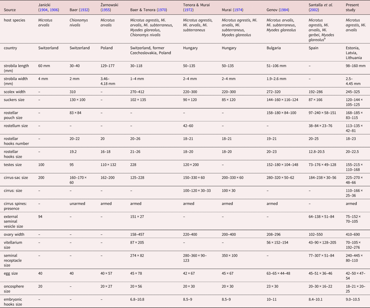
Rostellar hooks: The number, size and shape of rostellar hooks are traditionally among the most important characteristics for the identification of species with an armed scolex. Based on the published descriptions of K. asymmetrica, the number of hooks in this cestode varies from 18 to 26 (table 4). Material on this species from Eastern Europe is in the middle of this range. No distinct differences among specimens from different hosts and regions were noted. Also, an examination of voucher specimens of K. asymmetrica deposited in MHNG showed the most common number of rostellar hooks is in the range between 18 and 22 (18–3; 19–3; 20–5; 21–1; 22–3; 23–1; 23–1; 24–2; 25–1; 26–1).
Comparative morphometric data of Rodentolepis (sensu lato) asymmetrica by various authors and the present study (measurements in micrometres except where otherwise stated).

a Measurements by Santalla et al. (Reference Santalla, Casanova, Durand, Vaucher, Renaud and Feliu2002) are presented only from specimens originated from Microtus agrestis and Mi. arvalis.
The size of rostellar hooks usually varies between 18 and 23 micrometres. No distinct differences between specimens from different hosts and regions were noted (table 4). At the same time the hooks length presented by Santalla et al. (Reference Santalla, Casanova, Durand, Vaucher, Renaud and Feliu2002) is notably different from the specimens examined in our study and the data published by other authors, and extends intraspecific limits. Possible reasons for these differences can be both errors in measurements and, less likely, presence of more than one species in this material. In any case, these data from Santalla et al. (Reference Santalla, Casanova, Durand, Vaucher, Renaud and Feliu2002) cannot be used for subsequent comparisons. We had the opportunity to examine several vouchers of K. asymmetrica from Santalla et al. (Reference Santalla, Casanova, Durand, Vaucher, Renaud and Feliu2002) deposited at MHNG (MHNG-PLAT-32861–32863). There were no scoleces mounted in Berlese's medium and only one stained specimen retained the scolex, with the hooks in the crown laid very compact, which made it impossible to count and measure them correctly.
It should be noted that specimens attributable to K. asymmetrica examined in the present study demonstrate a relatively high variation in hook shape that has not been observed in other mammalian hymenolepidids (fig. 3). As it was described above, the most common hook shape of this species is the cricetoid type, characterized by a relatively short handle equal or shorter in length with the guard and straight, a blade shorter than former two structures (fig. 3A, B). There is also a variety of hook shape with guard shorter than blade and handle; however, this modification appears relatively rarely and is most likely intraspecific (fig. 3D, G). The dependence of the shape of the hooks on the species of the host was not noticed in the present study and the same specimen can have hooks with both aforementioned shapes. At the same time, the most peculiar shape of hooks was found in the material from Chionomys nivalis, which was used by Baer (Reference Baer1932) for the first description of the scolex morphology of this species (MHNG-PLAT-40937); hooks are characterized by a relatively short blade and guard, and both structures are shorter than the handle (fig. 3C). Such a modification is not found in cestodes from other hosts including the type. However, there is not enough data to state that if this shape of hook is associated with this host or not; in addition, other specimens from hosts of the genus Chionomys Miller (MHNG-PLAT-18362; MHNG-PLAT-30533) have hooks in similar shape to cestodes from Microtus spp.
Suckers: The suckers armed with minute hooks were previously noted in Hymenolepis myoxi (Rudolphi, 1819) sensu Baer (Reference Baer1932) from glirid rodents. This single character was used by Spassky (Reference Spassky1954) to erect the genus Armadolepis Spassky, Reference Spassky1954, although, this feature subsequently was rejected in this taxon (Makarikov, Reference Makarikov2017). Based on a current understanding of diversity, the presence of armature on suckers in Kontrimavichusia is a unique character, which is not known in any other hymenolepidids from mammals. There are also some hymenolepidids from birds, which have suckers armed with spines (e.g. Anatinella Spassky & Spasskaja, 1954; Diorchis Clerc, 1903; Echinolepis Spassky & Spasskaja, 1954; Gastrotaenia Wolffhügel, 1938; Skrjabinoparaksis Krotov, 1949). All these genera have completely different strobilar morphology and are unrelated, thus suggesting the independent origin of armament on suckers in different hymenolepidid taxa.
It should be noted that the spines on the suckers in Kontrimavichusia are usually poorly visible in stained specimens under light microscopy but become well distinguished on scoleces mounted in Berlese's medium (fig. 4A). The nature and chemical composition of these structures is not yet clear. In any case, it was repeatedly noted that the presence and pattern of the distribution of armature on suckers is a diagnostic character of genera (Skrjabin & Matevosyan, Reference Skrjabin and Matevosyan1948; Spassky, Reference Spassky1954; Mas-Coma & Galan-Puchades, Reference Mas-Coma and Galan-Puchades1991; Czaplinski & Vaucher, Reference Czaplinski, Vaucher, Khalil, Jones and Bray1994).
Excretory system
The presence of transverse anastomoses between the ventral osmoregulatory canals was indicated in the original description of K. asymmetrica: ‘Von den 2 Exkretionsgefäßpaaren ist das eine außerordentlich stark entwickelt; es bildet Queranastomosen’ (Janicki, Reference Janicki1904). Furthermore, this character has been later redescribed and illustrated by the author of this species: ‘Von den zwei Paaren der Exkretionsgefäße fällt das eine durch seine ungewöhnlich starke Entwicklung auf, namentlich in jüngeren Gliedern (vgl. Taf. XXII, fig. 42 und 43 Text fig. 11); es verläuft unter starker Schlängelung und bildet in einer jeden Proglottis Queranastomosen’ (Janicki, Reference Janicki1906). In all subsequent redescriptions of this species by other authors this feature was completely ignored (Baer, Reference Baer1932; Žarnowski, Reference Žarnowski1955; Baer & Tenora, Reference Baer and Tenora1970; Tenora & Murai, Reference Tenora and Murai1972; Murai, Reference Murai1974; Genov, Reference Genov1984; Santalla et al., Reference Santalla, Casanova, Durand, Vaucher, Renaud and Feliu2002). The ventral osmoregulatory canals connected with transverse anastomoses is among the generic characters of Hymenolepis (sensu stricto) (Makarikov & Tkach, Reference Makarikov and Tkach2013). In contrast to Hymenolepis, in Kontrimavichusia, transverse anastomoses are not located in every proglottis; those are randomly spaced throughout the strobila. Furthermore, in the proglottis anastomoses can pass terminally, medially or anteriorly, and in this, their irregularity is manifested. Irregular transverse anastomoses between ventral osmoregulatory canals also have been observed among some species of Nomadolepis and this feature is used among generic characters (Makarikov et al., Reference Makarikov, Gulyaev and Krivopalov2010), which can also be applied to the genus Kontrimavichusia.
Mature proglottides
Testes: No distinct variations in number and arrangement of the testes were observed in the material examined in the present study. Specimens are generally uniform in the distribution of testes, which usually are situated as noted in the redescription: in transverse line, one poral and two antiporal, separated by female gonads. At the same time, the following modifications in number and arrangement were noted: two poral testes and one antiporal testis (2–9 cases of 100 proglottides); triangular arrangement of the testes (1–8 cases of 100 proglottides); and four testes per proglottis (0–2 cases of 100 proglottides). The geometric arrangement of testes in hymenolepidids is used as a feature both for differentiation between species and between genera (Mas-Coma & Galan-Puchades, Reference Mas-Coma and Galan-Puchades1991; Czaplinski & Vaucher, Reference Czaplinski, Vaucher, Khalil, Jones and Bray1994). Since Kontrimavichusia is currently a monotypic genus, there is no possibility to assess its systematic significance for this taxon.
Cirrus: Baer (Reference Baer1932) gave initial information on the cirrus of this species and considered that this structure was unarmed (table 4). Subsequently, all the authors who redescribed the cirrus of K. asymmetrica were unanimous in reporting the presence of the armature (Žarnowski, Reference Žarnowski1955; Baer & Tenora, Reference Baer and Tenora1970; Tenora & Murai, Reference Tenora and Murai1972; Murai, Reference Murai1974; Genov, Reference Genov1984). We found that the armature of the cirrus is present although not evenly distributed; the distal part of the fully evaginated cirrus lacks spines (fig. 2A). Although in some groups of cestodes the patterns of spination are used to differentiate between species (Kornienko et al., Reference Kornienko, Gulyaev and Mel'nikova2006; Makarikov et al., Reference Makarikov, Galbreath and Hoberg2013), we believe that in this case this feature would not have taxonomic significance, since it is likely that the tiny spines in distal part of the cirrus may have been partially lost due to fixation and/or staining.
Vagina: A detailed description of the female genital ducts in K. asymmetrica was fully neglected as well as in the most other hymenolepidids from mammals. However, the position of the vagina in relation to the cirrus sac and its morphology are used for generic differentiation of avian hymenolepidids (Mas-Coma & Galan-Puchades, Reference Mas-Coma and Galan-Puchades1991). Similarly, we believe that peculiarities in vaginal structure of this species can be useful for exploring characters at the generic level. In Kontrimavichusia, this primarily refers to the division of the vagina into distinct parts; the copulatory part of the vagina is surrounded by circular musculature and covered externally by a dense layer of intensely stained cells, while the conductive part thin-walled is clearly distinguishable from the seminal receptacle (figs 1F and 2A). Structurally, this is markedly different from representatives of the Rodentolepis clade, in which the copulatory part of the vagina is simply organized without additional structures and the conductive part is indistinguishable (Makarikov et al., Reference Makarikov, Mel'nikova and Tkach2015, Reference Makarikov, Stakheev and Tkach2018).
Relative position of female gonads: The shifted position of the vitellarium to the lateral side of the proglottis in relation to the ovary was the key feature in initial differentiation of this species (Baer, Reference Baer1932; Joyeux & Baer, Reference Joyeux and Baer1936; Skrjabin & Matevosyan, Reference Skrjabin and Matevosyan1948). Apparently this character cannot be considered as reliable, since different groups of hymenolepidids also show a varying degree of shifting of the vitellarium to the antiporal side of the proglottis in relation to the ovary.
Gravid proglottides
Uterus: The shape of the uterus through different stages of strobilar development as well as its relative position to the other internal organs and extent of the fully developed uterus relative to the osmoregulatory canals are considered to be useful for generic classification of hymenolepidids (Mas-Coma & Galan-Puchades, Reference Mas-Coma and Galan-Puchades1991; Mariaux et al., Reference Mariaux, Tkach, Vasileva, Caira and Jensen2017). Characteristics of the uterus both in the original description as well as in its subsequent redescriptions for K. asymmetrica were incomplete. We stated that the shape and topography of the uterus in K. asymmetrica is more consistent with the genus Hymenolepis (sensu stricto) rather than with representatives of the Rodentolepis clade. The uterine morphology of K. asymmetrica and Hymenolepis has the following common features: the presence of numerous diverticula and the position dorsally to genital ducts, extending bilaterally beyond the longitudinal osmoregulatory canals. Thus, the structure of the uterus may also confirm the putative molecular–phylogenetic relationship of these taxa.
Eggs: Details of egg morphology could be also considered as additional distinctive characters at the supraspecific level. For instance, the presence of polar filaments on the embryophore is a generic character in some members of the Rodentolepis clade (Makarikov et al., Reference Makarikov, Gulyaev and Krivopalov2010, Reference Makarikov, Mel'nikova and Tkach2015). Homologous structures are absent in Kontrimavichusia.
Thus, the discovered complex of peculiar morphological features in specimens of K. asymmetrica clearly justifies the need for erection of a distinct genus for this species.
Host range and specificity
The common vole Microtus arvalis is the type host of K. asymmetrica. This species was also found in other arvicoline rodents of the genera Microtus Schrank, Myodes Pallas, Chionomys Miller and Dinaromys Kretzoi (Baer & Tenora, Reference Baer and Tenora1970; Tenora & Murai, Reference Tenora and Murai1972; Santalla et al., Reference Santalla, Casanova, Durand, Vaucher, Renaud and Feliu2002) and even in Muridae rodents of the genera Apodemus Kaup, Micromys Dehne and Mus Clerck (Erhardová, Reference Erhardová1958; Ryzhikov et al., Reference Ryzhikov, Gvozdev, Tokobaev, Shaldybin, Matzaberidze, Merkusheva, Nadtochii, Khohlova and Sharpilo1978). At the same time, the overwhelming majority of specimens of this species from Central Europe deposited in MHNG originate from Mi. agrestis; while there are only a few slides from the type host, such a small number of specimens were also found in other vole species. Furthermore, no confirmed specimens outside of Arvicolinae in MHNG were detected. This is also consistent with the present collection of this species from Eastern Europe, where both Mi. agrestis and Mi. arvalis live in sympatry but K. asymmetrica was found more often in the former host. For instance, in on our study, specimens of K. asymmetrica in Estonia were found in seven out of 31 specimens of Mi. agrestis (prevalence 22.5%) and in one out of 26 specimens of My. glareolus (prevalence 0.3%), while none of the 16 specimens of Mi. arvalis were of infected with this cestode. In Latvia, K. asymmetrica was found in four specimens of Mi. agrestis and in only one individual of Mi. arvalis. In Belarus, specimens of K. asymmetrica were found in one out of 56 individuals of My. glareolus (prevalence 1.8%) while no specimens of Mi. agrestis and Mi. arvalis were studied there. A similar result on host specificity of K. asymmetrica was obtained by Tenora & Murai (Reference Tenora and Murai1972) from Hungary where Mi. agrestis is the dominant host for this cestode (prevalence 60%); in contrast, in Mi. arvalis and Mi. subterraneus (de Selys-Longchamps) (syn.: Pitymys subterraneus (de Selys-Longchamps)), it is much less common, with prevalence 4% and 10%, respectively. Santalla et al. (Reference Santalla, Casanova, Durand, Vaucher, Renaud and Feliu2002) also showed that in the Pyrenean Mountains (Spain and France), among voles, the highest values of the prevalence with this cestode were in Mi. agrestis and Mi. arvalis (12.5–29.4% and 13.8–26.0%, respectively), less in Mi. gerbei (Gerbe) (5.2–17.7%) and the lowest in My. glareolus (1.2–4.0%).
All these data suggest that the genus Microtus and particularly Mi. agrestis is the most optimal host for the circulation of this parasite. The observed specimens from voles Chionomys nivalis, Mi. subterraneus and My. glareolus morphologically correspond to those from the type host and can be considered as additional hosts for this species. The detection of K. asymmetrica in Dinaromys bogdanovi (Martino) and Mi. majori (Thomas) requires confirmation since the morphology of these cestodes is unknown and the genetic distances of these specimens (discussed below) from cestodes from the type host extend beyond the limits of one species (Haukisalmi et al., Reference Haukisalmi, Hardman, Foronda, Feliu, Laakkonen, Niemimaa, Lehtonen and Henttonen2010). Similarly, the same applies to the specimens from Mi. gerbei from Spain (Santalla et al., Reference Santalla, Casanova, Durand, Vaucher, Renaud and Feliu2002) whose metrical data differ from those in cestodes from the type host. In addition, it is obvious that reports of this cestode from other hosts outside of the subfamily Arvicolinae are likely to be incorrect identifications of other hymenolepidids (Erhardová, Reference Erhardová1958; Ryzhikov et al., Reference Ryzhikov, Gvozdev, Tokobaev, Shaldybin, Matzaberidze, Merkusheva, Nadtochii, Khohlova and Sharpilo1978).
Phylogeny
All our new as well as GenBank available K. asymmetrica sequences form one well supported subclade within the Hymenolepis clade (fig. 5). However, it should be noted that some of the inner lineages of Kontrimavichusia demonstrate interspecific variability (table 3). One of these lineages is represented by the specimens from Mi. arvalis and Mi. agrestis included in GenBank (GU166232 and HM138528) and those from the present study. Unlike previously published sequences from GenBank, the vouchers of the latter specimens are available and those are morphologically fully consistent with the materials from the type host deposited in museum collections, thus apparently representing true K. asymmetrica. The other lineages published in GenBank originated from various arvicoline rodents that are phylogenetically distant from specimens from the type host. One of these represents sequence from C. nivalis from France (GU166231). The pairwise distances between this lineage and putative true K. asymmetrica reach up to 0.38% (4 bp). The two other lineages are from D. bogdanovi from Bosnia (GU166233) and in Mi. majori from Turkey (GU166234); they differ from K. asymmetrica up to 0.38% (4 bp) and 0.58% (6 bp), respectively. These data exceed the limits of intraspecific variability in this relatively conservative region of the rDNA. For instance, the proposed values of interspecific variability of the 28S gene among hymenolepidids from small mammals are in the range of 0–7 bp (Greiman & Tkach, Reference Greiman and Tkach2012; Greiman et al., Reference Greiman, Tkach and Cook2013; Tkach et al., Reference Tkach, Makarikov and Kinsella2013; Makarikov et al., Reference Makarikov, Mel'nikova and Tkach2015).
The presence of superspecific lineages within the Kontrimavichusia subclade suggests that this taxon may include a complex of cryptic species. Lack of morphological vouchers for the sequences from GenBank makes it difficult to further study species diversity within the genus. In this regard, the examination of specimens, assumed to be K. asymmetrica, originating from rodents that are phylogenetically distant from the genus Microtus, is of great interest.
Since the erection of the genus Rodentolepis, K. asymmetrica has been considered a member of this genus and the taxonomic position of the species has never been challenged by authors who followed the revision of mammalian hymenolepidids of Spassky (Reference Spassky1954). In this regard, the first molecular data on this species revealed unexpected insights (Haukisalmi et al., Reference Haukisalmi, Hardman, Foronda, Feliu, Laakkonen, Niemimaa, Lehtonen and Henttonen2010). This species is excluded from the Rodentolepis clade sensu Haukisalmi et al. (Reference Haukisalmi, Hardman, Foronda, Feliu, Laakkonen, Niemimaa, Lehtonen and Henttonen2010), which unites the majority of hymenolepidids from mammals having a scolex armed with cricetoid or fraternoid rostellar hooks, including the type of Rodentolepis, R. straminea. The emergence of an inclusive clade with K. asymmetrica and representatives of the genus Hymenolepis (sensu stricto) having unarmed scolex with rudimentary rostellar apparatus was among the most confusing contradictions of that initial phylogenetic study. Furthermore, this association was also confirmed by Neov et al. (Reference Neov, Vasileva, Radoslavov, Hristov, Littlewood DT and Georgiev2019) and the present study. Relatively recent and an even more unusual example was discovered during examination of species diversity within Armadolepis. Those analyses showed that the same genus of hymenolepidids includes both species having a normally developed rostellar apparatus with a rhynchus armed by hooks as well as species having an unarmed and rudimentary rostellar apparatus (Makarikov, Reference Makarikov2017; Makarikov et al., Reference Makarikov, Stakheev and Tkach2018). Meanwhile, in addition to the morphology of the scolex, we found that the anatomy of the strobila of Kontrimavichusia specimens is more similar to the genus Hymenolepis rather than to the representatives of the Rodentolepis clade. The first two groups are united by such characters as presence of transverse anastomoses between ventral osmoregulatory canals, the structure of the vagina, the shape of the uterus and its relative position to the genital ducts. Thus, the similarity in morphology partly confirms this phylogenetic linkage of K. asymmetrica with the genus Hymenolepis (sensu stricto).
So far, only in the Ditestolepis clade, all known representatives have an unarmed scolex, while the other clades include cestodes with both armed and unarmed scoleces. Such a distribution indicates that, in different lineages of mammalian hymenolepidids, an independent reduction of the rostellar apparatus has occurred (Spassky, Reference Spassky1992; Haukisalmi et al., Reference Haukisalmi, Hardman, Foronda, Feliu, Laakkonen, Niemimaa, Lehtonen and Henttonen2010). The structure of the rostellar apparatus of the ancestral forms of Hymenolepis is unknown but, given the great importance of this feature for suprageneric phylogeny and similarity in morphology of strobila, it can be speculated that it could resemble that of Kontrimavichusia. Data confirming or refuting this assumption have not yet been collected. In addition, all representatives of Hymenolepis have unarmed suckers, and now there is no possibility to establish if the ancestral forms of these cestodes used to have an armature on suckers and then lost this in the course of historical development or not. The presence of an armature on suckers and an invaginating apex of the rostellum may probably indicate that Kontrimavichusia originated from some avian cestodes having similar morphological characters as a result of colonization of rodents and adaptation of these hymenolepidids to a new host. A similar origin from avian hymenolepidids that adapted to parasitize rodents was supposed for Arvicolepis transfuga (Spassky & Merkusheva, 1967) from voles; this form also has an invaginating apex of the rostellum (Makarikov et al., Reference Makarikov, Gulyaev, Chechulin and Alimov2005). However, the present phylogeny and discovered phylogenetic affinity of the Hymenolepis clade with Microsomacanthus spp. from passerine birds cannot be unambiguously interpreted as a confirmation of this assumption. As only a small part of avian hymenolepidid taxa were included in phylogenetic studies, it is difficult to further study the relationships among these cestodes.
In conclusion, the present morphological analyses of specimens of K. asymmetrica is in agreement with results of phylogenetic studies performed by Haukisalmi et al. (Reference Haukisalmi, Hardman, Foronda, Feliu, Laakkonen, Niemimaa, Lehtonen and Henttonen2010), Neov et al. (Reference Neov, Vasileva, Radoslavov, Hristov, Littlewood DT and Georgiev2019) and the results obtained in the present study. Integrated data from comparative morphology and molecular phylogenetic analysis fully support the erection of a new genus for this species. Kontrimavichusia is currently a monotypic genus and includes only K. asymmetrica. Given the presence of interspecific lineages within this cluster, species diversity in this group of cestodes requires further study.
Acknowledgements
We thank Dr Eric P. Hoberg (Museum of Southwestern Biology, Department of Biology, University of New Mexico, Albuquerque, NM 87131, USA) for his useful comments and checking the English in the manuscript. We are grateful to the late Dr Vytautas L. Kontrimavichus, supported by the Lithuanian State Science and Studies Foundation, for specimens used in the present study and to Dr Laima Baltrūnaitė and PhD student Vytautas Sventickas (IENRC, Vilnius, Lithuania) involved in the trapping of mammals. We thank the curators of the helminthological collection of the Natural History Museum, Geneva, Switzerland Dr Jean Mariaux and Dr Isabel Blasco Costa for enabling the access to cestode specimens used in the present study. We thank two reviewers for their detailed comments that improved our manuscript.
Financial support
A substantial portion of the work was funded by the Russian Foundation for Basic Research (no. 19-54-18015). Further support for AAM was provided by the Federal Fundamental Scientific Research Program for 2021–2025, grant no. FWGS-122011800267-4.
Conflicts of interest
None.
Ethical approval
This article does not contain any studies with animals performed by any of the authors.